Case Report
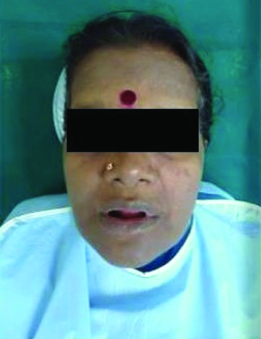
A 58-year-old female edentulous patient having microstomia presented in the Department of Prosthodontics, JSS Dental College with the chief complaint of inability to chew food due to missing teeth in the month of November 2011 with the history of last extraction done six months back. The maximum mouth opening of the patient (distance between upper and lower edentulous ridge) [1] was measured to be 23mm[Table/Fig-1]. The various causes of microstomia can be Cleft lip and Palate, Scleroderma, Temporomandibular disorder, mumps, space infections, burns, postsurgical and radiation therapy [2]. The patient’s medical and dental history was non-contributory. The cause of microstomia in this case seemed to be developmental in nature as all other causes were ruled out.
Microstomia patient with reduced mouth opening
Sectional complete denture was planned for this patient as conventional complete denture is difficult if not impossible for the patient to wear and for the prosthodontist to fabricate. The consent for above treatment was taken from the patient before hand.
Many techniques are used like hinged mandibular complete denture and sectional denture with hinge and stud attachments [3,4]. The technique described in this case report is a cheaper option as compared to other costly attachments since we used acrylic denture base to fabricate the sectional denture. The patient agreed to this treatment plan because it is a very cost effective technique.
Little modification in all phases of complete denture construction was done.
Step- Wise Procedure
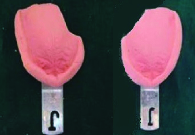
Sectional mandibular primary impression
Sectional maxillary primary impression
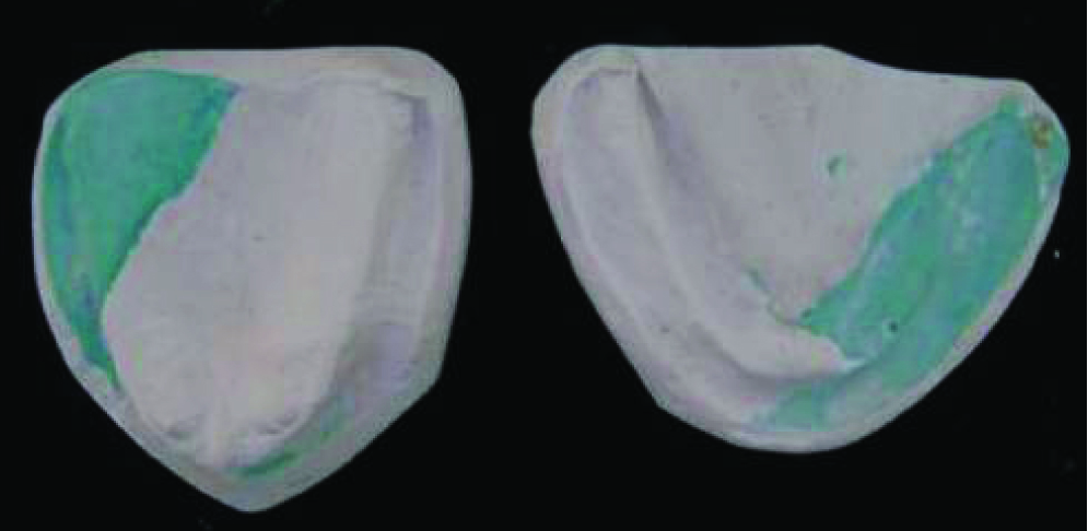
Preparation of diagnostic cast
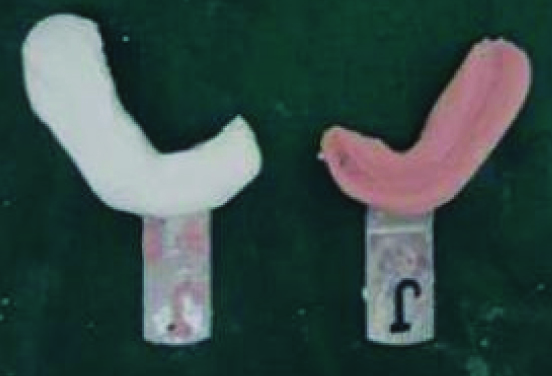
Two stock trays of similar size were cut more than half in opposite regions as shown in the [Table/Fig-2]. The first tray was used to make preliminary impression of one part of the ridge. It was then poured using dental plaster. Using the other tray, the impression of the remaining part of the ridge was made. The first retrieved cast was placed over the second impression and was secured with a rubber band.
The overlapping area of the cast and second impression works as a guide in the placement of the cast. Then the second sectional impression was also poured.
Finally a single diagnostic cast from two different sectional impressions was obtained.
Phase 2: Border Tracing and Secondary Impression
Before border moulding, a sectional custom tray was fabricated using die pins. Border tracing was done and a wash impression was made using a secondary impression paste of each half of the ridge separately.
After the wash impressions were made, the trays were assembled extra-orally using die pins and a master cast was prepared in a usual manner.
Phase 3: Fabrication of Permanent Denture Base and Preparation of the Denture Base
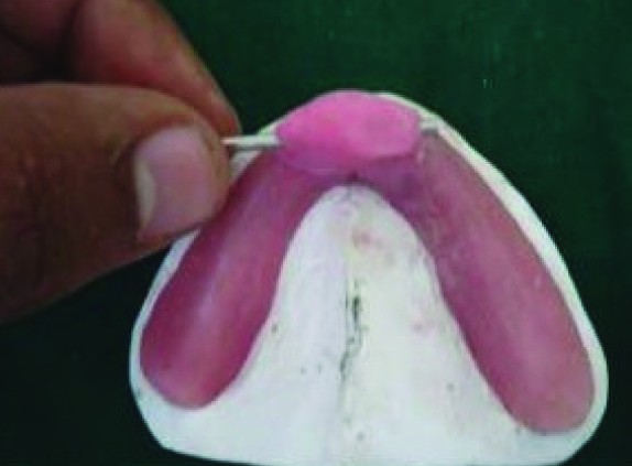
A usual procedure was carried out to fabricate heat cure acrylic permanent denture base. It was decided to fabricate buttoned sectional maxillary denture and cross pin, key-key way mechanism mandibular sectional denture.
The dentures bases were then modified as shown in the [Table/Fig-5,6].
Fabrication of permanent denture base and preparation of the denture base (maxillary)

Fabrication of permanent denture base and preparation of the denture base (mandibular)
Firstly, both record bases were sectioned into two halves.
Modification of maxillary record base:
Maxillary record base push buttons were attached to both halves and the anterior telescopic section was fabricated using autopolymerising resin to hold the two halves together by using buttons as shown in [Table/Fig-5].
Modification of mandibular record base:
To one half of the mandibular denture base, a denture key was attached as shown in the figure and to the other half a key way was attached which was covered with a housing. All these modification were made using autopolymerising resin as shown in the figure. Next, a hole was drilled through the key and the housing.
This assembly was reinforced by using a corresponding cross pin that conformed to the size of hole [Table/Fig-6].
Phase 4: Record Block Fabrication and Jaw Relation Recording
Record blocks were fabricated over modified record base and jaw relation record was carried out.
The record blocks were sectional in nature. They can be inserted in parts into the patient mouth and assembled to form one complete unit.
Teeth arrangement and try in

Teeth arrangement was done. A canine was attached to the cross pin present at one side of the mandibular denture base. The removal of this canine and thereby the cross pin helps in disassembling the parts of the lower sectional denture.
Phase 6: Denture Insertion and Follow Up [
8,
9]
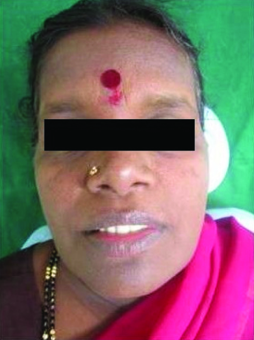
Extra oral view after insertion
Section wise denture ready for delivery

Finally the denture was inserted section wise, and post insertion follow up was done after one month. Section wise denture insertion is a comfortable way for such patients.
Discussion
Normal mouth opening is essential for the function of speech, nutritional needs, dental hygiene, facial expression and social interaction [5]. Limited mouth opening manifests as a consequence of certain medical conditions [3]. A maximal oral opening that is smaller than the size of final prosthesis can make prosthetic treatment challenging. We need to differ prosthetic treatment procedures accordingly in these patients [6].
Ansgar Cheng et al., fabricated collapsible complete denture in a case of limited mouth opening patients. As per Ansgar Cheng et al., collapsible hinged mandibular complete denture is kept stable by tongue pressure in lateral direction and resistance provided by ridge slopes. According to authors though the procedure was found to be time consuming, the result was outstanding. So, the goal of prosthodontic treatment include: providing lip support, improving articulation, reducing drooling and regaining favourable aesthetics [3].
In a similar case a sectional denture with Hinge and stud attachments were used for working of this denture design. The advantages of the design are ease of insertion and removal, Structural durability and maximum coverage for retention, stability, and support [4].
The disadvantages of this treatment plan are restricted tongue space, increased laboratory time and requirement of patient’s compliance.
Another group of authors fabricated sectional denture using Hinge and stud attachments for working of this denture design. Its advantages are custom made hinge is used which is more durable and less expensive, studs used in this method provide more rigid attachment than magnets and there is no fear of loss of magnetic effects when using studs [7].
In burns patients in particular various reconstructive and surgical techniques have been used to increase mouth opening [8,9].
This technique provides cheaper alternative to costly attachments, metal denture bases and other expensive equipments [Table/Fig-10]. The disadvantages of this type of technique are that the buttons should be regularly replaced when they lose their retention. The buttons resistance to corrosion in the patient mouth is questionable. Moreover, the autopolymerising resin used in this technique is not a long term solution due to its less strength and high monomer content.
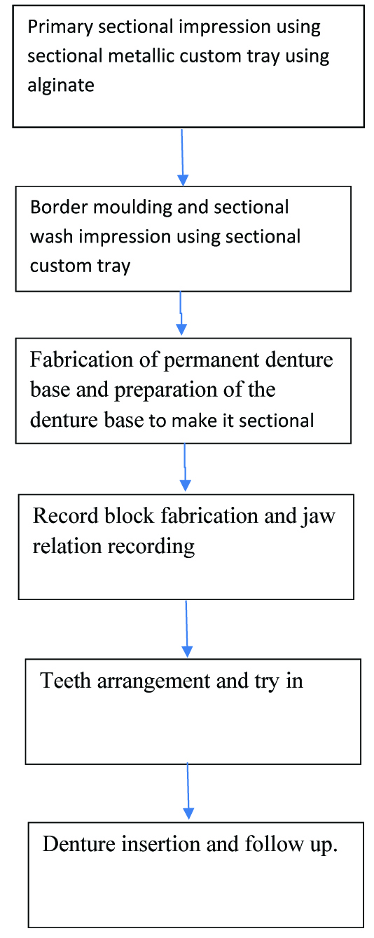
Flowchart simplifying the clinical procedure
Conclusion
Even though it is difficult to achieve all the aspects of prosthodontic treatment in a patient with microstomia, with improved working skills and technique we can still accomplish our goal. Other prosthodontic treatment like implants, obturators, and cast partial dentures can be done for this type of patients with slight modifications in all the procedures.
[1]. Wittenaar JH, Dijkstrea PU, Vissink A, Van Oort RP, Roodenburg JLN, Variation in repeated mouth opening measurements in head and neck cancer patients with or without trismusInt J Oral Maxillofacial Surg 2009 38(1):26-30. [Google Scholar]
[2]. Prasad R, Bhinde SV, Gandhi PV, Divekar NS, Madhav VNV, Prosthodontic management of a patient with limited mouth opening: a practical approachJIPS 2008 8:83-86. [Google Scholar]
[3]. Cheng AC, Wee AG, Morrison D, Maxymiw WG, Hinged mandibular removable complete denture for post-mandibulectomy patientsJ Prosthet Dent 1999 82(1):103-06. [Google Scholar]
[4]. Dikbas I, Koksal T, Kazazoglu E, Fabricating sectional-collapsible complete dentures for an edentulous patient with microstomia induced by sclerodermaQuintessence Int 2007 38(1):15-22. [Google Scholar]
[5]. Koymen R, Gulses A, Karacayli U, Aydintug YS, Treatment of microstomia with commissuroplasties and semidynamic acrylic splintsOral Surg Oral Med Oral Pathol Oral Radiol Endod 2009 107(4):503-07. [Google Scholar]
[6]. Baker PS, Brandt RL, Boyajian G, Impression procedure for patients with severely limited mouth openingJ Prosthet Dent 2000 84(2):241-44. [Google Scholar]
[7]. Cheng AC, Kwok-Seng L, Wee AG, Tee-Khin N, Prosthodontic management of edentulous patient with limited oral access using implant-supported prostheses: a clinical reportJ Prosthet Dent 2006 96(1):1-6. [Google Scholar]
[8]. Zweifel CJ, Guggenheim M, Jandali AR, Altintas MA, Kunzi W, Giovanoli P, Management of microstomia in adult burn patientsJournal of Plastic, Reconstructive & Aaesthetic Surgery 2010 63:e351-57. [Google Scholar]
[9]. Grishkevich VM, Post-burn microstomia: Anatomy and elimination with trapeze-flap plastyBurns 2011 37:484-89. [Google Scholar]