GA is an important parameter in obstetrics for management of pregnancy and evaluating foetal development [1]. High incidence of perinatal mortality has been noted in patients whose accurate gestational age is not known. Uncertain gestational age is associated with preterm delivery, low birth weight and post maturity.
Naegele’s rule, a well accepted method for estimating date of delivery, depends only on date of LMP, has some problems as some of women don’t recall LMP accurately [2].
Sonographic estimation of GA by using foetal biometric parameters such as BPD, FL, AC and HC assumed important role in management of pregnancy. There are some limitations with these parameters as BPD after 26 weeks becomes unreliable in conditions altering the shape of skull [3]. Femur length is shortened in cases of achondroplasia making it unreliable parameter in estimating GA.
Foetal cerebellum can be visualized as early as 10-11 weeks by USG. From second trimester onwards, it grows with a linear correlation with gestational age. TCD is least affected by external factors because it is surrounded by dense petrous bone which allows its use for assessing GA even in third trimester [5]. In cases of growth restriction, the cerebellum is the least affected parameter maintaining its size in case of foetal growth restriction hence accurate GA can be predicted with TCD [6]. With these advantages of TCD over other parameters, this study was conducted to evaluate the accuracy of TCD in estimation of gestational age and to compare it with other routine parameters in 15 to 40 weeks of gestation.
Materials and Methods
A prospective pilot study was conducted on the 100 pregnant women between 15 to 40 weeks of gestation with known LMP which had been confirmed by first trimester ultrasound, presented to radiology department for antenatal ultrasound scan during period of June 2013 to May 2014. Non anomalous singleton pregnancies without any risk factors between 15 to 40 weeks of gestation with normal liquor and without any growth disturbances were included in the study. Main purpose of this study was to demonstrate how accurately GA by TCD is correlating with that of GA by LMP so that TCD can be included in routine foetal biometry. To determine accuracy of TCD in estimating GA and by using GA by LMP as gold standard, we excluded many cases of irregular cycles and those with not known LMP, so the sample size considered for this study was less.
After explaining the procedure and the risks involved, written and informed consent was taken. Study was conducted after obtaining institutional ethical committee approval. With low frequency transducer (3.5 MHz), Trans cerebellar plane is identified by obtaining an oblique view through posterior fossa that included visualization of midline thalamus, cerebellar hemispheres and cisterna magna [5]. Single measurement is used for each pregnancy. Other parameters such as BPD, HC, AC and FL were also measured according to previously described standard planes. GA based on individual parameters was compared with GA by LMP.
The subjects were further divided into sub groups based on gestational age at the time of ultrasound scan into second trimester (15 to 28 weeks) which includes 50 members and third trimester (29 to 40 weeks) which includes 50 members. Subjects were also divided based on age and parity. The above ultrasound parameters were compared within the individual sub-groups to look for any confounding effects that these variables had on the study.
Statistical Analysis
Statistical analysis was performed using SPSS software version 17.0 Statistical analysis was done by using Karl Pearson’s coefficient of correlation (r) and regression analysis.
Results
There was no significant influence of age and parity on all parameters in estimation of gestational age in both second and third trimesters.
Mean GA based on LMP was 21.13 weeks in second trimester and 34.37 weeks in third trimester as depicted in [Table/Fig-1]. Total mean GA was 27.75 weeks. When mean GA based on BPD, HC, AC, FL and TCD were compared with that of LMP, all parameters in second trimester were showing GA which was near to that of LMP. TCD had mean GA of 21.12 in second trimester which was very near to GA by LMP. In third trimester TCD had mean GA of 34 weeks which was very close to GA by LMP. FL was having mean GA of 33.90 which was second close to GA by LMP. AC had mean GA which was far from mean GA by LMP among all parameters. When we compared overall mean GA, TCD showed mean GA which was closest to that of LMP.
Mean and standard deviations of all parameters with confidence intervals.
| Parameters | N | Mean | Std Deviation | 95% Confidence interval for Mean |
|---|
| Lower Bound | Upper Bound |
|---|
| LMP | Second trimesterThird trimesterTotal | 5050100 | 21.1334.3727.75 | 4.403.217.68 | 19.8833.4626.23 | 22.3835.2929.27 |
| HC | Second trimesterThird trimesterTotal | 5050100 | 21.0933.4527.27 | 4.482.797.24 | 19.8132.6525.83 | 22.3634.2428.70 |
| AC | Second trimesterThird trimesterTotal | 5050100 | 20.9633.3827.17 | 4.252.987.23 | 19.7532.5325.73 | 22.1634.2228.60 |
| FL | Second trimesterThird trimesterTotal | 5050100 | 21.1933.9027.59 | 4.493.087.48 | 19.9233.1126.10 | 22.4734.8629.07 |
| BPD | Second trimesterThird trimesterTotal | 5050100 | 21.2633.4827.37 | 4.492.797.18 | 19.9932.6925.95 | 22.5434.2728.79 |
| TCD | Second trimesterThird trimesterTotal | 5050100 | 21.1234.0027.60 | 4.452.907.42 | 19.7632.9425.92 | 22.2934.5928.87 |
| Average | Second trimesterThird trimesterTotal | 5050100 | 21.1533.7427.45 | 4.402.967.34 | 19.9032.9025.99 | 22.4034.5828.90 |
*LMP-Last menstrual period, HC-Head Circumference, AC-Abdominal Circumference, FL-Femur length, BPD-Biparietal diameter, TCD-Transcerebellar diameter.
[Table/Fig-2] shows intra-class correlation coefficient with confidence interval which shows the agreement between the parameters to evaluate gestational age with the gestational age by LMP. In second trimester all parameters showed nearly equal values and correlated with LMP. In third trimester TCD showed intra-class correlation of 0.982 which was higher than other parameters. FL was the next accurate parameter with correlation coefficient of 0.980. Of all the parameters, BPD showed the least correlation with the gestational age.
Intraclass correlation with confidence intervals and t-test p-values.
| Parameters | Intra-class correlation | 95% Confidence Interval | t test p value |
|---|
| Lower Bound | Upper Bound |
|---|
| Second trimester | HCACFLBPDTCDAVERAGE | 0.9970.9950.9970.9960.9980.997 | 0.9950.9910.9940.9920.9970.996 | 0.9980.9970.9980.9980.9990.999 | p 0.0001,HSp 0.0002,HSp 0.0001,HSp 0.0001,HSp 0.0001,HSp 0.0001,HS |
| Third trimester | HCACFLBPDTCDAVERAGE | 0.9590.9590.9800.9540.9820.977 | 0.6950.4460.9590.6940.8650.850 | 0.9820.9890.9940.9840.9930.992 | p 0.0007,HSp 0.0008,HSp 0.0005,HSp 0.0007,HSp 0.0006,HSp 0.0007,HS |
| Total | HCACFLBPDTCDAVERAGE | 0.9950.9960.9970.9950.9980.996 | 0.9890.9840.9970.9920.9950.996 | 0.9980.9980.9990.9970.9990.999 | p 0.0004,HSp 0.0005,HSp 0.0003,HSp 0.0004,HSp 0.0004,HSp 0.0004,HS |
*LMP-Last Menstrual Period, HC-Head Circumference, AC-Abdominal Circumference, FL-Femur Length, BPD-Biparietal Diameter, TCD-Transcerebellar Diameter.
[Table/Fig-3] shows the Pearson’s coefficient of correlation (r) of all parameters. In second trimester, all parameters had nearly equal r-values. TCD had highest correlation with value of 0.997. In third trimester, there was considerable difference in r-values; r-value was 0.982, when the GA by TCD was compared with the GA by LMP, which was more than the other parameters. The least correlation was seen with BPD, r=0.951. The second most accurate correlation was seen with the FL with r-value of 0.981.
Karl pearson correlation coefficients of Individual parameters compared with LMP.
| Parameters Trimester | Karl pearson correlation | p value | |
|---|
| LMP | With AC | Second trimesterThird trimester | 0.9910.972 | 0.00050.0006 | SigSig |
| With TCD | Second trimesterThird trimester | 0.9970.982 | 0.00020.0004 | SigSig |
| With BPD | Second trimesterThird trimester | 0.9920.951 | 0.00040.0008 | SigSig |
| With FL | Second trimesterThird trimester | 0.9940.981 | 0.00030.0004 | SigSig |
| With HC | Second trimesterThird trimester | 0.9940.954 | 0.00040.0008 | SigSig |
| With AVERAGE | Second trimesterThird trimester | 0.9960.979 | 0.00020.0007 | SigSig |
*LMP-Last Menstrual Period, HC-Head Circumference, AC-Abdominal Circumference, FL-Femur Length, BPD-Biparietal Diameter, TCD-Transcerebellar Diameter.
[Table/Fig-4] shows the regression analysis between LMP and other parameters in second and third trimesters. By using regression we can derive an equation to calculate the actual gestational age from TCD. The equation does not contain variables such as age and parity as these does not have statistically significant effect on estimation of GA by TCD.
Regression analysis between LMP and with other parameter depicting with p values and regression analysis values. aDependent Variable: LMP
| Trimester | Parameters | Unstandardized coefficients | Standardized coefficients | p value | r2 |
|---|
| B | Std Error | Beta |
|---|
| Second trimester | With AC | (Constant parameter) | -0.374 1.026 | 0.419 0.020 | 0.991 | 0.376 0.0004 | 0.983 |
| With TCD | (Constant parameter) | 0.418 0.985 | 0.254 0.012 | 0.997 | 0.106 0.0001 | 0.993 |
| With BPD | (Constant parameter) | 0.486 0.971 | 0.392 0.018 | 0.992 | 0.221 0.0003 | 0.984 |
| With FL | (Constant parameter) | 0.506 0.973 | 0.337 0.016 | 0.994 | 0.140 0.0002 | 0.988 |
| With HC | (Constant parameter) | 0.530 0.977 | 0.326 0.015 | 0.994 | 0.111 0.0002 | 0.989 |
| With AVERAGE | (Constant parameter) | 0.073 0.995 | 0.284 0.013 | 0.996 | 0.798 0.0002 | 0.992 |
| Third trimester | With AC | (Constant parameter) | -0.619 1.048 | 1.232 0.037 | 0.972 | 0.618 0.0008 | 0.944 |
| With TCD | (Constant parameter) | -2.327 1.087 | 1.043 0.031 | 0.981 | 0.030 0.0006 | 0.963 |
| With BPD | (Constant parameter) | -2.772 1.110 | 1.540 0.046 | 0.961 | 0.078 0.0009 | 0.924 |
| With FL | (Constant parameter) | -0.422 1.024 | 1.008 0.030 | 0.981 | 0.677 0.0006 | 0.962 |
| With HC | (Constant parameter) | -2.324 1.097 | 1.678 0.050 | 0.954 | 0.172 0.0009 | 0.908 |
| With AVERAGE | (Constant parameter) | -1.5257 1.064 | 1.085 0.032 | 0.979 | 0.166 0.0007 | 0.958 |
*LMP-Last Menstrual Period, HC-Head Circumference, AC-Abdominal Circumference, FL-Femur Length, BPD-Biparietal Diameter, TCD-Transcerebellar Diameter.
LMP = -0.221+1.015 (TCD)
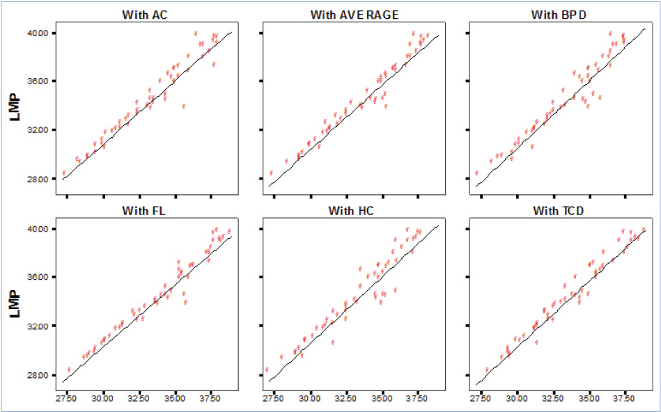
Graphical representation of regression analysis is done as depicted in [Table/Fig-5] (3rd trimester) and [Table/Fig-6] (2nd trimester).
Regression analysis graphs in third trimester.
*LMP-Last Menstrual Period, HC-Head Circumference, AC-Abdominal Circumference, FL-Femur Length, BPD-Biparietal Diameter, TCD-Transcerebellar Diameter.
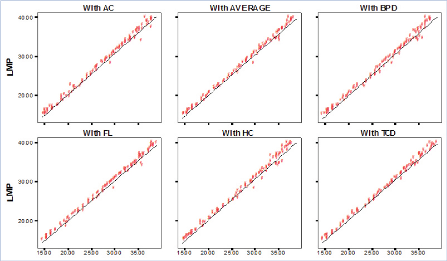
Regression analysis graphs in second trimester.
*LMP-Last Menstrual Period, HC-Head Circumference, AC-Abdominal Circumference, FL-Femur Length, BPD-Biparietal Diameter, TCD-Transcerebellar Diameter.
Discussion
Accurate knowledge of gestational age is required for proper management in obstetric care [1]. Professor Ian Donald of Glassgow was the first to use diagnostic ultrasonography to investigate gravid uterus and is considered as father of modern ultrasound. Along with Mac Vicar and Brown, he developed first 2D contact scanner in 1958 [7]. In modern era of advanced imaging, ultrasound is used in foetal biometry and LMP is used where early pregnancy scans are not available.
Routine biometric parameters for GA assessment such as BPD, HC, AC and FL have their own limitations like BPD and HC because of moulding of head in third trimester. Similarly, femur length is not reliable in cases of achondroplasia. Transcerebellar diameter was developed as an alternative parameter in foetal biometry. Since, cerebellum lies in the posterior cranial fossa covered by thick dura and bony calvarium, it is more resistant to deformation by extrinsic pressure. Foetal cerebellum is sonographically visualized as early as 10-11 weeks. TCD is least affected by factors affecting foetal growth allowing it to determine accurate gestational age even in third trimester [5] and cases of intrauterine growth restriction [6].
In a study done by Chavez MR, the concordance between the actual and predicted gestational age by TCD was high (r=0.92; p<.001). The agreement was superior in the second trimester (r=0.93) compared to third trimester (r=0.81; p<0.001) [8]. Chavez MR et al., studied TCD in twin pregnancies and concluded that the agreement between the actual gestational age and the predicted gestational age by TCD was comparable to that of singleton pregnancy (Pearson’s correlation coefficient r=0.997, p<0.001) [9].
In another study by Chavez MR et al., the concordance between the actual gestational age and the predicted gestational age by TCD was high for both IUGR and large fetus (r=0.98 and 0.95 respectively) [10]. Joshi BR et al., did a study measuring TCD in 594 singleton pregnancies in Nepalese population. They found that the gestational age and TCD (50th percentile in mm) coincided well till 20th week of gestation. They observed no significant clinical difference between the nomogram created by them and the previously published nomogram in gestational age between 21st and 28th weeks. But, they observed significant differences between their nomogram and the previously published nomograms in third trimester [11].
Gupta AD et al., from India, studied TCD in singleton pregnancies and observed that the gestational age of pregnant women not sure of their LMP can be reliably estimated by measuring the TCD which showed good correlation (r=+0.946, r2=89.6% and p<0.001). The increase in TCD throughout gestation helped in assessing the development of the cerebellum [12]. Naseem F et al., did a study in 228 patients with gestational age of 36 weeks measuring TCD and BPD by ultrasonography. They compared GA by TCD and BPD with LMP. In this study, they observed that in 228 patients, when compared with GA by LMP, TCD had given accurate gestational age in 209 patients and BPD had given accurate gestational age in 176 patients [13].
Rotmensch S et al., did a study measuring the cerebellar diameter in cases of down syndrome and found that cerebellar diameters in down syndrome fetuses were lesser than normal controls at all gestational age, (p<0.005) by an average of 0.67-0.87 mm. A ratio of 0.92 for observed/expected cerebellar diameter gave a sensitivity of 21%, specificity of 95% and PPV 1.66% and 0.50% in a population with risk of having down syndrome of 1 in 250 and 1 in 750 respectively. However, this difference in cerebellar size was too small to be used clinically [14].
In our study, when mean GA based all parameters were compared with that of LMP; all parameters in second trimester were showing GA which was near to that of LMP. TCD had mean GA of 21.12 in second trimester which was near to that GA by LMP. In third trimester, TCD showed mean GA which correlated better with GA by LMP. When we compared overall mean GA also, TCD showed better correlation with that of LMP. The intra-class correlation between the TCD and the actual gestational age showed excellent agreement. In second trimester, all parameters were showing nearly equal values. In third trimester, TCD had intra-class correlation which was higher than other parameters.
By comparing the gestational age by LMP and the gestational age by TCD, we obtained a Pearson’s coefficient of correlation (r) of all parameters. In second trimester all parameters were having nearly equal r-values. TCD had highest correlation among all. In third trimester, there was considerable difference in r-value with TCD being parameter having high correlation.
Limitation
Our study included small sample size (100 patients). TCD is not routinely done in foetal biometry. So, this study was done to demonstrate how accurately GA by TCD is correlating with that of GA by LMP so that TCD can be included in routine foetal biometry. To determine accuracy of TCD in estimating GA by using GA by LMP as gold standard, we excluded many cases of irregular cycles and those with not known LMP, decreasing our sample size. Hence, further studies with large sample size may be required to corroborate our findings and to establish TCD as accurate and more reliable parameter in estimation of gestational age in second and third trimesters.
Conclusion
This study showed that TCD is an accurate predictor of gestational age in the second and third trimester. The correlation between the LMP derived gestational age and the gestational age by TCD seems to decrease from second to third trimester. Even in the third trimester TCD is fairly accurate and better predictor of gestational age in comparison to the other ultrasound parameters such as BPD, HC, AC, FL. FL, though accurate, cannot be used as a single parameter for estimation of gestational age as it is the parameter that is most affected by IUGR. TCD is the parameter that is least affected by IUGR, therefore can be used as a single parameter for the estimation of gestational age. Our results also showed that TCD is not affected by age of the mother and parity also does not seem to have an effect on TCD.
*LMP-Last menstrual period, HC-Head Circumference, AC-Abdominal Circumference, FL-Femur length, BPD-Biparietal diameter, TCD-Transcerebellar diameter.
*LMP-Last Menstrual Period, HC-Head Circumference, AC-Abdominal Circumference, FL-Femur Length, BPD-Biparietal Diameter, TCD-Transcerebellar Diameter.
*LMP-Last Menstrual Period, HC-Head Circumference, AC-Abdominal Circumference, FL-Femur Length, BPD-Biparietal Diameter, TCD-Transcerebellar Diameter.
*LMP-Last Menstrual Period, HC-Head Circumference, AC-Abdominal Circumference, FL-Femur Length, BPD-Biparietal Diameter, TCD-Transcerebellar Diameter.