Recognition of the Lateral Pterygoid Muscle and Plate during Ultrasound-Guided Trigeminal Nerve Block
Ke-Vin Chang1, Chia-Shiang Lin2, Chih-Peng Lin3, Wei-Ting Wu4, Levent Özçakar5
1 Clinician, Department of Physical Medicine and Rehabilitation and Community and Geriatric Research Center, National Taiwan University Hospital, Bei-Hu Branch and National Taiwan University College of Medicine, Taipei, Taiwan.
2 Clinician, Department of Anesthesiology, Mackay Memorial Hospital, Mackay Medicine, Nursing and Management College, and Mackay Medical College, Taipei, Taiwan.
3 Clinician, Department of Anesthesiology, National Taiwan University Hospital, Taipei, Taiwan and National Taiwan University College of Medicine, Taipei, Taiwan.
4 Clinician, Department of Physical Medicine and Rehabilitation and Community and Geriatric Research Center, National Taiwan University Hospital, Bei-Hu Branch and National Taiwan University College of Medicine, Taipei, Taiwan.
5 Clinician, Department of Physical and Rehabilitation Medicine, Hacettepe University Medical School, Ankara, Turkey.
NAME, ADDRESS, E-MAIL ID OF THE CORRESPONDING AUTHOR: Dr. Ke-Vin Chang, Department of Physical Medicine and Rehabilitation, National Taiwan University Hospital, Bei-Hu Branch and National Taiwan University College of Medicine, Taipei, Taiwan.
E-mail: pattap@pchome.com.tw
Lateral pterygoid plate, Pterygopalatine fossa, Trigeminal neuralgia
To the Editor,
Trigeminal neuralgia is a specific facial pain syndrome, characterized by episodic, electric-like shock pain in the distribution of the face that is innervated by the fifth cranial nerve. The use of Ultrasound-guided (US) placement of local anaesthetic in the face has been demonstrated to provide a high degree of accuracy providing analgesia of the superficial branches of the trigeminal nerve and was validated in a cadaveric model [1]. In 2013, a new ultrasound-guided deep trigeminal nerve block technique via the pterygopalatine fossa was described to target the Gasserian ganglion i.e., sensory ganglion of the trigeminal nerve [2]. The authors’ demonstrated the sonoanatomy of the lateral pterygoid muscle and plate and described the needle trajectory using an in-plane approach to position the needle in the pterygopalatine fossa located between the maxilla anteriorly and the lateral pterygoid plate posteriorly.
Two case series have been published using the same radiation and magnetization free approach, resulting in treatment success using different regimens (local anaesthetic with corticosteroid in 2013 [3] and radiofrequency ablation in 2015) [4]. Recently, using Computed Tomography, Chuang YW et al., pointed out that the landmark muscle shown by the aforementioned method might be too superficial to be the lateral pterygoid muscle [5]. These studies used a linear transducer to describe the deep trigeminal nerve block via pterygopalatine fossa which may be limited by poor tissue penetration and narrowing scanning breadth. There is limited literature describing the course of the lateral pterygoid muscle.
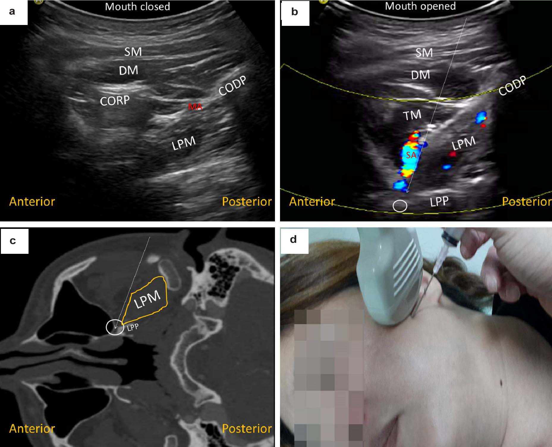
In this regard, using a curvilinear transducer, we aimed to explore the lateral pterygoid muscle and its surrounding anatomical sonoanatomy. The pterygoid muscle originates from the infratemporal crest of the sphenoid bone and the lateral pterygoid plate, and attaches to the condylar process of the mandible (inferior belly) and to the capsule of the temporomandibular joint (superior belly). To visualize the muscle, the ultrasound probe can first be placed along the zygomatic arch as previously described. The transducer is then relocated caudally in the horizontal plane, where a hypoechoic gap can be identified between the coronoid and condylar processes of the mandible [Table/Fig-1a], [Video-1]. Superficial to both mandibular processes is the masseter muscle and part of the temporalis muscle. The lateral pterygoid muscle, which appears quadrilateral in shape, is seen originating from the condyle and extending anteroinferiorly. The injectate should be placed deep to the superior head of the lateral pterygoid muscle along the pterygomaxillary fissure into the pterygopalatine fossa. To improve visualization of the injectate area between the zygomatic arch and the coronoid process, the clinician may ask the patient to open the mouth [Table/Fig-1b], [Video-1]. The pterygoid plate emerges as a straight hyperechoic structure that attaches to the anterior part of the lateral pterygoid muscle. Using colour Doppler, the maxillary artery can be visualized pulsating between the lateral pterygoid and temporalis muscles. Moreover, one of its branches, the sphenopalatine artery and/or its attributes, courses anteriorly into the pterygopalatine fossa where the maxillary branch (V2) of the Gasserian ganglion is located on its roof [Table/Fig-1b,c]. In addition to the in-plane technique, the needle can be introduced using an out-of-plane approach through a posterosuperior-to-anteroinferior direction to reach the anterior edge of the pterygoid plate and redirected forward towards the pterygopalatine fossa [Table/Fig-1d]. This out of plane technique requires further exploration in this anatomical area and is not recommended for the novice clinician due to the lack of needle tip visualization.
Ultrasound image of the lateral pterygoid muscle during mouth closing (a) and opening the circle shows the pterygopalatine fossa; the vessel enhanced in colour Doppler imaging is the sphenoid palatine artery and the dashed line illustrates the needle trajectory (b). Visualization of the lateral pterygoid muscle and pterygopalatine fossa by using computed tomography (c). Transducer and needle positioning during ultrasound-guided deep trigeminal nerve block. (d).
SM, superficial masseter muscle; DM, deep masseter muscle; TM, temporalis muscle
MA, maxillary artery; SA, sphenoid palatine artery; CORP, coronoid process
CODP, condylar process; LPM, lateral pterygoid muscle; LPP, lateral pterygoid plate.
The use of a curvilinear transducer evaluating the anatomical disposition of the lateral pterygoid muscle and pterygopalatine fossa may provide a better visualization of the previously described deep trigeminal ultrasound guided technique.
Declaration: The current research was supported by the research grant from National Taiwan University Hospital, Bei-Hu branch.
[1]. Spinner D, Kirschner JS, Accuracy of ultrasound-guided superficial trigeminal nerve blocks using methylene blue in cadaversPain Med 2012 13:1469-73. [Google Scholar]
[2]. Nader A, Schittek H, Kendall MC, Lateral pterygoid muscle and maxillary artery are key anatomical landmarks for ultrasound-guided trigeminal nerve blockAnaesthesiology 2013 118:957 [Google Scholar]
[3]. Nader A, Kendall MC, De Oliveria GS, Chen JQ, Vanderby B, Rosenow JM, Ultrasound-guided trigeminal nerve block via the pterygopalatine fossa: an effective treatment for trigeminal neuralgia and atypical facial painPain Physician 2013 16(5):E537-45. [Google Scholar]
[4]. Nader A, Bendok BR, Prine JJ, Kendall MC, Ultrasound-guided pulsed radiofrequency application via the pterygopalatine fossa: a practical approach to treat refractory trigeminal neuralgiaPain Physician 2015 18:E411-15. [Google Scholar]
[5]. Chuang YW, Chen CH, Landmark of ultrasound-guided trigeminal block: lateral pterygoid musclePain Physician 2015 18:E933-34. [Google Scholar]