Memory T Cells (CD45RO) Role and Evaluation in Pathogenesis of Lichen Planus and Lichenoid Mucositis
Mani Devi1, Dhanaraj Vijayalakshmi2, Kumar Dhivya3, Murali Janane4
1 Professor and Head, Department of Oral Pathology, Adhiparasakthi Dental College and Hospital, Chennai, Tamil Nadu, India.
2 Professor, Department of Oral Pathology, Adhiparasakthi Dental College and Hospital, Chennai, Tamil Nadu, India.
3 Reader, Department of Oral Pathology, Adhiparasakthi Dental College and Hospital, Chennai, Tamil Nadu, India.
4 Postgraduate Student, Department of Oral Pathology, Adhiparasakthi Dental College and Hospital, Chennai, Tamil Nadu, India.
NAME, ADDRESS, E-MAIL ID OF THE CORRESPONDING AUTHOR: Dr. Mani Devi, Adhiparasakthi Dental College and Hospital, Melmaruvathur-603319, Chennai, Tamil Nadu, India.
E-mail: devident@rediffmail.com
Introduction
Memory T cells have the ability to survive in a quiescent state for longer periods and are responsible for the rapid responses on subsequent exposure to antigen. Analyzing memory T cells in Oral Lichen planus (OLP) and Lichenoid Mucositis (LM) suggest that these cells may play a role in the immunopathogenic mechanisms.
Aim
To identify and evaluate Memory T cells in Lichen Planus (LP), Lichenoid Mucositis (LM) and Normal Mucosa (NM) using CD45RO monoclonal antibody immunohistochemically.
Materials and Methods
A total of 30 cases (15 cases of OLP and 15 cases of LM) clinically and histopathologically diagnosed, and 10 cases of NM were stained for CD45RO monoclonal antibody, immunohistochemically using Biotin Streptavidin method. Staining intensity of CD45RO expression was statistically analysed using Chi-square Test.
Results
The present study demonstrated a higher expression of CD45RO in connective tissue layer of OLP (53.3% intense staining) when compared to LM (20% intense staining) and no intense staining in NM. The difference in staining intensity pattern between the study groups was statistically significant (p=0.014).
Conclusion
This study demonstrates a statistically significant rise in memory T cells in LP than in LM, indicating the possible different immunopathogenic mechanisms.
Immune response, Immunohistochemistry, Naïve human T cells
Introduction
Human CD45 is a transmembrane tyrosine phosphatase present on all cells of haematopoietic origin, except RBCs. They exist in different isoforms that are expressed on a restricted group of cell types designated as CD45R [1]. Most naïve human T cells express a form of CD45R that is called CD45RA, whereas memory T cells express a different isoform called CD45RO [2].
Naive T cells are unprimed lymphocytes that have been stimulated by antigen to become immune competent lymphocytes. These cells have the ability to differentiate into Effector and Memory T cells [3]. Effector T cells include cytokine secreting CD4+ helper T cells and CD8+ cytotoxic T cells, which functions in protective immune response. Memory T cells have the ability to survive in a quiescent state for long periods and are responsible for the rapid responses on subsequent exposures to antigen [4].
OLP is a chronic mucocutaneous inflammatory disease characterised by basal keratinocyte damage and interface lymphocyte reaction. It causes bilateral white striations, papules or plaques on the buccal mucosa, tongue, and gingiva [5]. Lesions that appear clinically identical to LP caused by drugs and involving the oral mucous membrane is termed as LM [6].
Analysing memory T cells in OLP and LM suggest that these cells may play a role in the immunopathogenic mechanisms. With this background, we did this study to evaluate the presence of memory T cells in OLP and LM, by using monoclonal antibody CD45RO.
Materials and Methods
The study was conducted on the paraffin embedded tissue blocks retrieved from the archived files of Department of Oral and Maxillofacial Pathology, Ragas Dental College and Hospital, Chennai in April, 2006. A total of 30 cases (15 cases of OLP and 15 cases of LM) clinically and histopathologically diagnosed and 10 cases of NM were stained for CD45RO monoclonal antibody.
Immunohistochemistry Procedure [7]: For immunohistochemical evaluation 5-micron thick sections were cut from formalin fixed paraffin embedded tissue blocks mounted on Amino Propoxy Ethane Silane (APES) coated slides. The slides with tissue sections were deparaffinized using xylene and put in descending grades of alcohol and then rehydrated with water. Slides were then treated with 3% hydrogen peroxide for 20 minutes to quench endogenous peroxidase activity of cells that would otherwise result in non specific staining. The slides were then transferred to citrate buffer and autoclaved for antigen retrieval at 15 lbs pressure for 15 minutes. Primary antibody was added and incubated for one hour at room temperature (CD45RO monoclonal antibody). Then a drop of Biotinylated link from the secondary antibody kit (DAKO LSAB 2KIT) was added and incubated for 20 minutes. Then a drop of Streptavidin from the secondary antibody kit (DAKO LSAB 2KIT) was added and incubated for 20 minutes. Then a drop of freshly prepared DAB (3’– Diamino Benzidine tetra hydrochloride – a substrate chromogen) was added. Slides were counter stained with haematoxylin.
Evaluation of CD45RO expression: Analysis of CD45RO expression was done on the basis of staining intensity on cell membrane. Staining intensity was assessed for mild (+), moderate (++), intense (+++), or no expression (-). A couple of investigators compared CD45RO antibody staining intensity between the study groups using Kappa statistics. Kappa statistics was done for interpretation of the interobserver variation. Data entry and analysis was performed using SPSS version 10.0.5®. Percentage of intensity was calculated to assess CD45RO expression. A p value <0.05 was considered statistically significant.
Results
CD45RO antibody mean percentage staining intensity between the study groups [Table/Fig-1].
Table showing CD45RO antibody staining intensity of the connective tissue between the study groups.
| Lesion | Connective tissue n (%) | p-value |
|---|
| - | + | ++ | +++ |
|---|
| Lichen Planus (n 15) | - | 1 (6.7%) | 6 (40.0%) | 8 (53.3%) | .014* |
| Lichenoid Mucositis (n 15) | - | 5 (33.3%) | 7 (46.7%) | 3 (20.0%) |
| Normal Mucosa (n 10) | 1 (10.0%) | 6 (60.0%) | 3 (30.0%) | - |
Chi-square test was done to compare the percentage expression of CD45RO between Lichen Planus, Lichenoid Mucositis and Normal Mucosa
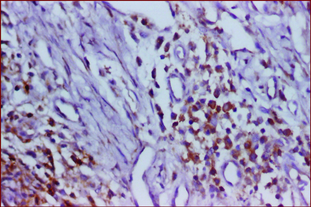
In the connective tissue of LP, one case (6.7%) showed mild staining, six cases (40%) moderate and eight cases (53.3%) showed intense staining [Table/Fig-2,3,4]. In LM, five cases (33.3%) showed mild staining, seven cases (46.7%) moderate staining and three cases (20%) were intensely stained [Table/Fig-5]. In NM, one case (10%) showed no staining, six cases (60%) mild staining and three cases (30%) were moderately stained. The difference in staining intensity pattern between the study groups was statistically significant (p=0.014).
Figure showing CD45RO antibody staining intensity in connective tissue between the study groups.
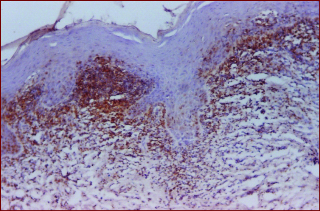
Photomicrograph of CD45RO expression in Lichen Planus (10X).
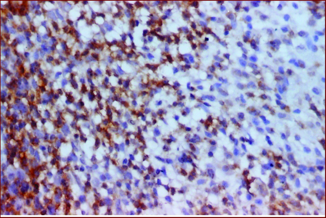
Photomicrograph of CD45RO expression in Lichen Planus (40X).
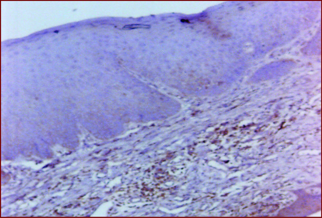
Photomicrograph of CD45RO expression in Lichenoid Mucositis (10X).
Discussion
LP is an interface reaction consisting typically of T lymphocytic infiltration in the upper layer of connective tissue in close apposition to the basal layer of epithelium [8]. Both CD4 and CD8 T cells are found in LP. The majority of the lymphocytes in the infiltrate are CD8 and CD45RO memory positive cells. The latter cell subtype is not normally found in healthy mucosa [9]. CD4 T cells, which express CD45RO antigen, are called memory cells and they proliferate in response to recall antigen. These cells show stronger helper function for the production of antibody. Naive T cell loses the CD45RA antigen after activation and begins to express CD45RO [7].
In our study, memory T cells were identified by using CD45RO antibody. The present study demonstrated a higher expression of CD45RO in connective tissue layer of LP (53.3% intense staining) when compared to LM (20% intense staining) and no intense staining in NM. However, NM showed 60% mild and 30% moderate staining. This difference was statistically significant. In LP, there are two populations of CD4 helper cell. One is CD45RA with suppressor inducer activity and the second is CD45RO, which serves as memory cell. The latter group predominates in OLP [10].
Porter SR et al., reported that in OLP, majority of T lymphocytes express ab TCR and are CD45RO positive memory T cells [11]. Walton LJ et al., reported that circulating memory CD45RO T cells were increased from 49.1% in controls to 65.5 % in OLP [12].
Rudrigues Nunez I et al., reported the mean proportion of CD45RO lymphocyte with higher expression in atrophic erosive OLP than in patients with reticular OLP [13]. Kirby AC et al., reported the presence of lymphocytes function associated antigen 3 (LFA-3) on Langerhans Cell (LC) [14]. Since LC are highly activated in OLP, this may be an important aspect of disease development. These cells also express Vascular Cell Adhesion Molecule-1 (VCAM-1) and Intercellular Adhesion Molecule-1 (ICAM-1) and present the antigen to memory T- cells (CD45RO), the predominant phenotype in OLP.
According to McCartan BE and Lamey PJ even though the T cell numbers are lower in LM than LP, the CD4/CD8 ratio is similar in both the conditions [8]. In OLP, lymphocytes are recruited into the mucosa by upregulation of adhesion molecule expression, possibly driven by cytokines. The damage to keratinocytes and the basement membrane is predominantly mediated via cytotoxic T lymphocytes. Furthermore, the activation of T lymphocytes and the resultant generation of cytokines are likely to perpetuate that OLP is a cell mediated immune response [11].
In OLP, the inflammatory infiltrate consists of a significant number of CD4 T cells in the lamina propria and CD8 T cells in close proximity to epithelial basement membrane. Accumulation of CD8 cells seems to increase gradually in disease progression [15]. CD4 helper T cells are stimulated to secrete Th1 cytokine IL2 and IFNg. Subsequently, CD8 cytotoxic T cells may be activated by the combination of antigen associated with MHC Class I on basal keratinocyte and by CD4 T cell derived IL2 and IFN g. Activated CD8 cytotoxic T cell trigger basal keratinocytes apoptosis in OLP [16]. In our study also the CD45RO staining intensity in OLP was higher than in LM and NM. These results were consistent with the previous studies cited above [16].
OLP is triggered by some events that cause CD4/CD45RA positive lymphocytes to differentiate into CD4/CD29 positive lymphocytes. From this point, immunosuppressive mechanisms are reduced, leading to an accumulation of memory CD45RO T cell [13].
The results of the present study demonstrate that there is a significant increase in the number and expression of memory T helper cells in OLP than in LM. This clearly indicates that the route of antigen presentation and penetration is different between these two lesions.
Limitation
The sample size was small and future studies with larger sample size are needed for more conclusive results.
Conclusion
Increased CD45RO expression was observed in the connective tissue in the sequence of OLP>LM>NM and this difference in staining intensity was statistically significant. The present study demonstrates a statistically significant increase in number and expression of memory T cells in OLP than in LM, indicating the possible different immunopathogenic mechanisms. Based on the results of our study, we conclude that memory T cells have a significant role in the pathogenesis of OLP and LM.
*Chi-square test was done to compare the percentage expression of CD45RO between Lichen Planus, Lichenoid Mucositis and Normal Mucosa
[1]. Leitenberg D, Novak TJ, Farber D, Smith BR, Bottomly K, The extracellular domain of CD45 controls association with the CD4-T cell receptor complex and the response to antigen-specific stimulationThe Journal of experimental medicine 1996 183(1):249-59. [Google Scholar]
[2]. Arlettaz L, Barbey C, Dumont-Girard F, Helg C, Chapuis B, Roux E, CD 45 isoform phenotypes of human T cells: CD 4+CD 45 RARO+memory T cells re-acquire CD 45 RA without losing CD 45 ROEuropean journal of immunology 1999 29(12):3987-94. [Google Scholar]
[3]. Abbas AK, Lichtman AH, Cellular and Molecular immunology 2000 5th edWB Saunders company publication:110-115. [Google Scholar]
[4]. Berard M, Tough DF, Qualitative differences between naive and memory T cellsImmunology 2002 106(2):127-38. [Google Scholar]
[5]. Jungell P, Oral Lichen planus reviewInternational Journal of Oral and Maxillofacial Surgery 1991 20:129-31. [Google Scholar]
[6]. Müller S, Oral manifestations of dermatologic disease: A focus on lichenoid lesionsHead and neck pathology 2011 5(1):36-40. [Google Scholar]
[7]. Yamazaki K, Nakajima T, Aoyagi T, Hara K, Immunohistological analysis of memory T lymphocytes and activated B lymphocytes in tissues with periodontal diseaseJournal of periodontal research 1993 28(5):324-34. [Google Scholar]
[8]. McCartan BE, Lamey PJ, Expression of CD1 and HLA-DR by Langerhans cells (LC) in oral lichenoid drug eruptions (IDE) and idiopathic oral lichen planus (LP)Journal of oral pathology and medicine 1997 26(4):176-80. [Google Scholar]
[9]. Fitzpatrick Dermatology in General Medicine 1999 15th edMc Graw – Hill Publication:463-477. [Google Scholar]
[10]. Boyd AS, Update on the diagnosis of lichenoid dermatitisAdvances in dermatology 1996 11:287 [Google Scholar]
[11]. Porter SR, Kirby A, Olsen I, Barrett W, Immunologic aspects of dermal and oral lichen planus: A reviewOral Surgery, Oral Medicine, Oral Pathology, Oral Radiology, and Endodontology 1997 83(3):358-66. [Google Scholar]
[12]. Walton LJ, Macey MG, Thornhill MH, Farthing PM, Intra-epithelial subpopulations of T lymphocytes and Langerhans cells in oral lichen planusJournal of oral pathology and medicine 1998 27(3):116-23. [Google Scholar]
[13]. Rodríguez-Núñez I, Blanco-Carrión A, García AG, Rey JG, Peripheral T-cell subsets in patients with reticular and atrophic-erosive oral lichen planusOral Surgery, Oral Medicine, Oral Pathology, Oral Radiology, and Endodontology 2001 91(2):180-88. [Google Scholar]
[14]. Kirby AC, OIsen I, Farthing PM, Porter SR, Expression of lymphocyte function-associated antigen 3 in oral lichen planusOral diseases 1995 1(4):193-97. [Google Scholar]
[15]. DeRossi SS, Ciarrocca KN, Lichen planus, lichenoid drug reactions, and lichenoid mucositisDental Clinics of North America 2005 49(1):77-89. [Google Scholar]
[16]. Khan A, Farah CS, Savage NW, Walsh LJ, Harbrow DJ, Sugerman PB, Th1 cytokines in oral lichen planusJournal Of Oral Pathology & Medicine 2003 32(2):77-83. [Google Scholar]