All Premolars with Three Roots Confirmed with Cone Beam Computed Tomography: A Rare Anatomical Variation
Tushar Krishna Yadav1, Nidhi Anup Gupta2, Vikesh Sisodia3
1 Associate Professor, Department of Paediatric and Preventive Dentistry, Y.M.T. Dental College and Hospital, Navi Mumbai, Maharashtra, India.
2 Associate Professor, Department of Paediatric and Preventive Dentistry, Y.M.T. Dental College and Hospital, Navi Mumbai, Maharashtra, India.
3 Associate Professor, Department of Paediatric and Preventive Dentistry, Y.M.T. Dental College and Hospital, Navi Mumbai, Maharashtra, India.
NAME, ADDRESS, E-MAIL ID OF THE CORRESPONDING AUTHOR: Dr. Nidhi Anup Gupta, S-504, Jal Vayu Vihar, Phase-II, Sector-20, Kharghar, Navi Mumbai-410210, Maharashtra, India.
E-mail: dr.nidhig2910@gmail.com
Bicuspids, C-shaped canal, Imaging technology, Number of roots
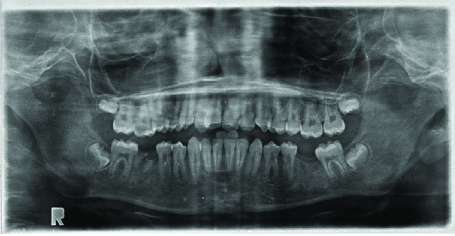
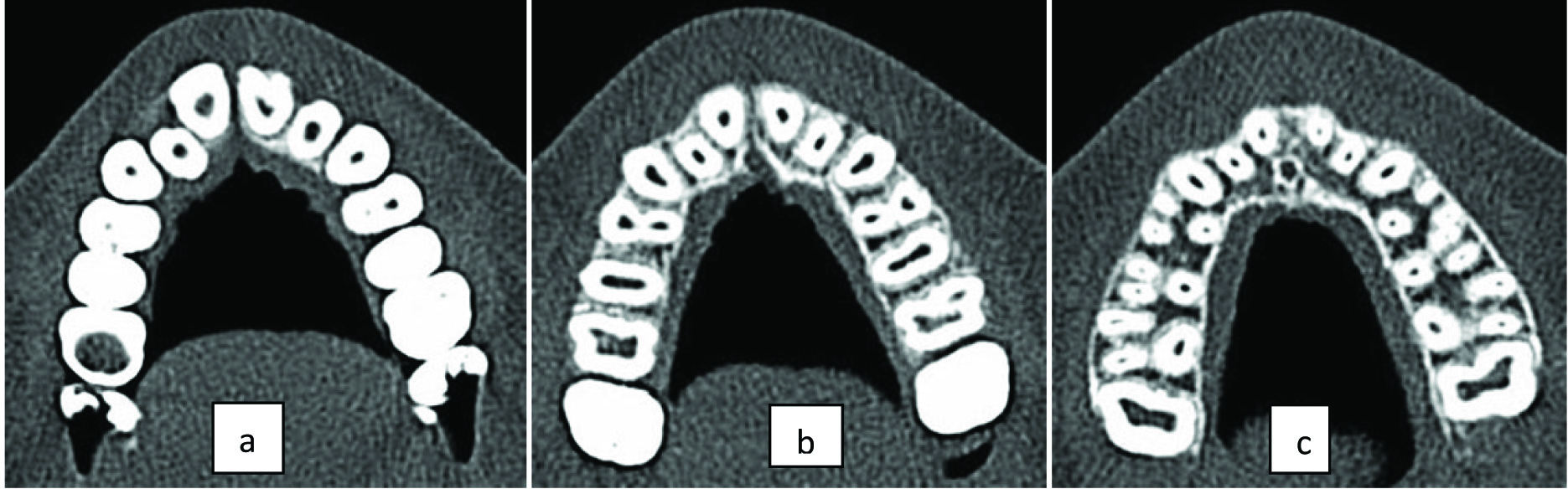
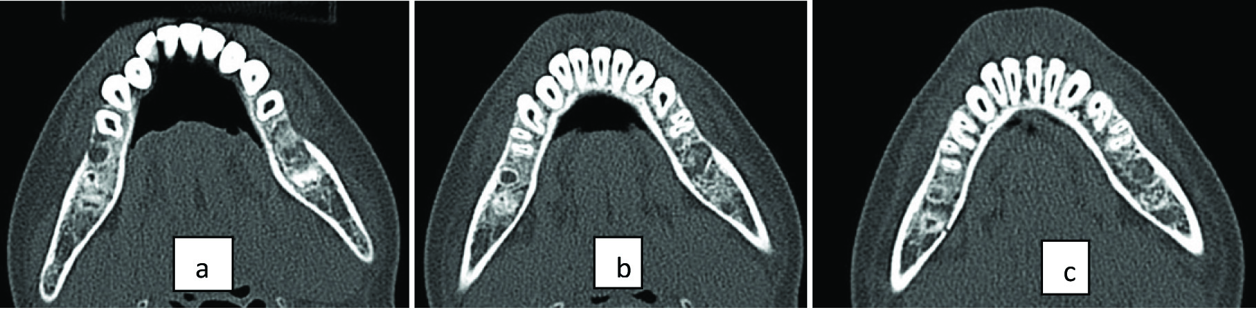
A 13-year-old boy with a non contributory medical history reported with a chief complaint of pain in relation to his lower right jaw region. Clinical examination showed a grossly carious tooth with evidence of intraoral swelling and sinus tract formation. The Orthopantomograph (OPG) revealed root stumps in relation to 46 which were advised for extraction. While examining the OPG the adjacent premolars showed two roots. Being an unusual finding and also the patient was being considered for early placement of implants in 36 and 46 region was sent for Cone Beam Computed Tomography (CBCT). The OPG showed all eight premolars with two roots and the lower left first premolar appeared to have three roots [Table/Fig-1]. The CBCT showed three roots in all the premolars [Table/Fig-2,3], c-shaped canal in the mandibular right and left first premolar [Table/Fig-3].
Preoperative OPG showing root stumps (46) and three roots in all premolars.
CBCT axial view of maxilla showing: (a) coronal third section showing pulp chamber; (b) middle third section showing two roots and two canals; (c) apical third section showing three rooted bilateral maxillary first and second premolars.
CBCT axial view of mandible showing: (a) coronal third section showing pulp chamber; (b) middle third section showing two roots and two canals; (c) apical third section showing three rooted bilateral mandibular first and second premolars.
Discussion
While travelling, road maps are helpful to reach our destination; similarly a map can be created in the mind with the help of the CBCT for predictable and successful endodontic treatment, which will lead to less endodontic failures to missed and untreated canal spaces [1].
The bifurcation of mandibular first premolar appearing bilaterally is 59.6% and maxillary first premolar with trifurcation appearing bilaterally is 4% [2]. Therefore, if a particular anomaly is rare like the three rooted premolars, there is 90% chance that the anomaly would be bilateral [2]. This fact stands true in our case report.
The presence of three roots is rare [Table/Fig-4] [3-6]. It is a remarkable finding that in our study all the premolars have three roots [Table/Fig-5].
Shows various case reports and studies done on premolars with three roots [3-6].
| S. No. | Tooth type | Author | Type of study | Inference |
|---|
| 1. | Maxillary first premolar | Gupta S et al., [3] | In vitro study (clearing technique) in North Indian population | Prevalence of three roots is 0.4% |
| 2. | Maxillary second premolar | Khreisat ASA et al., [4] | Case report | Three roots |
| 3. | Mandibular first premolar | Cleghorn BN et al., [5] | Case report showing 3D microcomputed tomography of extracted first premolar for orthodontic purposes. | Three rooted tooth with three canals |
| 4. | Mandibular first premolar | Parekh V et al., [6] | Clearing technique | Type 1 (50%) according to vertucci classifiation was most common and type 6 (2.5%) vertucci was least common. Type 6 and type 8 were not seen |
| 5. | Mandibular second premolar | Parekh V et al., [6] | Clearing technique | Type 1 (80%) according to vertucci classifiation was most common and type 4 (2.5%) vertucci was least common. Type 2, 3, 6, 7, 8 were not seen |
| 6. | Mandibular second premolar | Cleghorn BM et al., [5] | Case report showing 3D microcomputed tomography of extracted first premolar for orthodontic purposes | C shaped canal |
Shows variations in root canal morphology in the premolars of our case reports.
| Tooth number | No of roots | No of root canals | Vertuccis classification |
|---|
| 14 | 3 | 3 | Buccal- type VPalatal- type I |
| 15 | 3 | 3 | Buccal-type VPalatal- type V |
| 24 | 3 | 3 | Mesiobuccal- type IDistobuccal- type IPalatal- type I |
| 25 | 3 | 3 | buccal- type VPalatal- type I |
| 34 | 3 | 3, c- shaped canal in distal root | Mesiobuccal- type IDistobuccal- type IPalatal- type I |
| 35 | 3 | 3 | mesial- type Idistal- type I |
| 44 | 3 | 3, c- shaped canal in distal root | mesial- type Idistal- type I |
| 45 | 3 | 3 | mesial- type Idistal- type V |
The exact reason for presence of three roots is not known but it could be related to the epithelium diaphragm of the hertwigs root sheath. The differential growth of the epithelial diaphragm causes the division of the root trunk into two or three roots. When the enamel organ is grown if three tongue like extensions are developed from the horizontal epithelial diaphragm, three roots are formed [7].
CBCT can be a useful adjunct but the disadvantage of using CBCT in paediatric patients, uncooperative patients, and those who have neuromuscular disorders often cannot remain stable throughout the scanning procedure [8] and therefore, the images obtained are not clear, as seen in our case report. This was also a limitation of our case report.
Conclusion
Variations of this grade as seen in our case is extremely rare but is possible, so it is necessary that before starting any endodontic treatment, the clinician should take into consideration the variability of the root canal morphology and CBCT can be of great help to the clinician to achieve the same.
[1]. Cantatore G, Berutti E, Castellucci A, Missed anatomy: frequency and clinical impactEndodontic Topics 2009 15:3-31. [Google Scholar]
[2]. Sabala CL, Benenati FW, Neas BR, Bilateral root or root canal aberrations in a dental school patient populationJournal of Endodontics 1994 20(1):38-42. [Google Scholar]
[3]. Gupta S, Joysinha D, Gowhar O, Tyagi SP, Singh N, Gupta S, Root and canal morphology of maxillary first premolar teeth in north Indian population using clearing technique: An invitro studyJ Conserv Dent 2015 18(3):232-36. [Google Scholar]
[4]. Khreisat ASA, Khreisat IS, Khzouz W, Three rooted maxillary second premolar- a case reportPakistan Oral & Dental Journal 2013 33(3):547-49. [Google Scholar]
[5]. Cleghorn BM, Christie WH, Dong CCS, Anomalous mandibular premolars: a mandibular first premolar with three roots and a mandibular second premolar with a c-shaped canal systemInternational Endodontic Journal 2008 41:1005-14. [Google Scholar]
[6]. Parekh V, Shah N, Joshi H, Root canal morphology and variations of mandibular premolars by clearing technique: An in vitro StudyJ Contemp Dent Pract 2011 12(4):318-21. [Google Scholar]
[7]. Kumar GS, Orban’s oral histology and embryology 2014 13th edIndiaElsevier Health Sciences:35 [Google Scholar]
[8]. Thomas SL, Application of cone-beam CT in the office settingDent Clin N Am 2008 52:753-59. [Google Scholar]