Accessory Grooves on the Diaphragmatic Surface of the Liver: A Cadaveric Study
Satheesha B. Nayak1, Ashwini Aithal Padur2, Naveen Kumar3, Bincy M. George4
1 Professor, Department of Anatomy, Melaka Manipal Medical College (Manipal Campus), Madhav Nagar, Manipal, Karnataka, India.
2 Senior Grade Lecturer, Department of Anatomy, Melaka Manipal Medical College (Manipal Campus), Madhav Nagar, Manipal, Karnataka, India.
3 Assistant Professor, Department of Anatomy, Melaka Manipal Medical College (Manipal Campus), Madhav Nagar, Manipal, Karnataka, India.
4 Associate Professor, Department of Anatomy, Melaka Manipal Medical College (Manipal Campus), Madhav Nagar, Manipal, Karnataka, India.
NAME, ADDRESS, E-MAIL ID OF THE CORRESPONDING AUTHOR: Dr. Satheesha B Nayak, Professor, Department of Anatomy, Melaka Manipal Medical College (Manipal Campus), Madhav Nagar, Manipal-576104, Karnataka, India.
E-mail: nayaksathish@gmail.com
Introduction
Usually liver does not possess any grooves on its diaphragmatic surface, but there are reports on the presence of grooves on this surface.
Aim
The present study was conducted to study the gross features of diaphragmatic surface of the liver and document the presence of any grooves on it and also to correlate our study with the previous studies.
Materials and Methods
Ninety seven formalin embalmed livers stored in 10% formaldehyde were observed for the presence of grooves on diaphragmatic surface.
Results
Fifteen (15.46%) livers had at least one groove on the diaphragmatic surface. Left lobe did not reveal the presence of any abnormal grooves. Presence of a single groove was observed in six (6.18%) livers; Double grooves were found in five (5.15%) livers; Triple grooves were found in three (3.09%) livers and four grooves were found in one (1.03%) liver. The deepest among all the grooves was measured 1.5 cm.
Conclusion
The knowledge of these grooves is of importance to radiologists and surgeons during their routine procedures.
Abdomen, Hepatic, Laparoscopy, Sonography, Surgery
Introduction
Liver is the largest gland, situated in the upper right quadrant of the abdomen. It is wedge shaped and resembles a five sided pyramid. It has right, anterior, posterior, superior and inferior surfaces. The right, anterior, superior and posterior surface are smoothly continuous with each other and form a diaphragmatic surface, which is related to the diaphragm [1].
Normally, there is no other structure between the liver and diaphragm that can cause grooves on the diaphragmatic surface. There is no developmental explanation for the formation of the grooves on the diaphragmatic surface as well. However, some studies in the past have reported the presence of grooves on the diaphragmatic surface. These grooves are named as “cough furrows” appearing due to chronic cough or due to a condition called “corset liver” occurring due to application of a tight corset to correct scoliosis or to get an attractive figure [2,3].
Yadav GD et al., have reported such a case found during laparotomy [4]. Kanchan T et al., have also reported the presence of grooves as found during autopsy [5]. Though, there are reports from many studies regarding the occurrence of unusual grooves/notches/furrows on different aspects of liver, most of them are from surgical or radiological observations. There are not many cadaveric studies in this regard. In the current study, we aimed to document the incidence of grooves on the diaphragmatic surface of the liver and study their orientation and also aimed to do the relevant literature survey to compare and contrast our findings with the previous studies.
Materials and Methods
The study was conducted in the Melaka Manipal Medical College (Manipal Campus), Manipal, Karnataka, India on the cadavers donated for teaching and research purpose. During our routine demonstration classes for medical undergraduate students, we observed a few livers with abnormal grooves/notches/fissures on the diaphragmatic surface. Later, we observed 97 livers stored in 10% formaldehyde specifically for the presence of any grooves on the diaphragmatic surface. All the livers stored in the formalin tanks in the past 20 years were included in the study. The observations were completed in the year 2016.
The age of the cadavers from which the livers were obtained ranged between 40 and 90 years. We could not make gender difference of the livers since they were in store for a long time. But from our cadaver stock, it is evident that approximately 10% livers belonged to females. The damaged livers and livers with obvious diseased appearance were excluded from the study. The anterior, posterior and superior surfaces were carefully observed for presence of abnormal grooves and the observational data was documented [Table/Fig-1]. Relevant photographs were taken from the specimens with abnormal grooves. The data was converted to percentage occurrence of grooves. A thorough review of literature was conducted and our observations were compared with the previous studies.
Table showing the number and percentage of grooves seen on the diaphragmatic surface of the liver.
| Total Number of livers included in the study = 97 |
|---|
| No. of grooves | No. of Livers | Percentage |
|---|
| Presence of at least one abnormal groove | 15 | 15.46 |
| Presence of single groove | 6 | 6.18 |
| Presence of two grooves | 5 | 5.15 |
| Presence of three grooves | 3 | 3.09 |
| Presence of four grooves | 1 | 1.03 |
Results
Total number of livers observed was 97. Among them 15 (15.46%) livers had at least one abnormal groove on the diaphragmatic surface. All the abnormal grooves were confined to the right anatomical lobe of the liver and they extended from anterior surface to posterior surface across the superior surface.
It was interesting to observe that all the grooves were parallel to each other and were parallel to the sagittal plane.
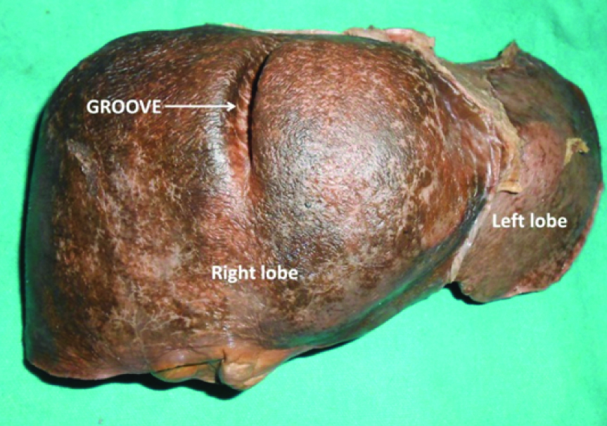
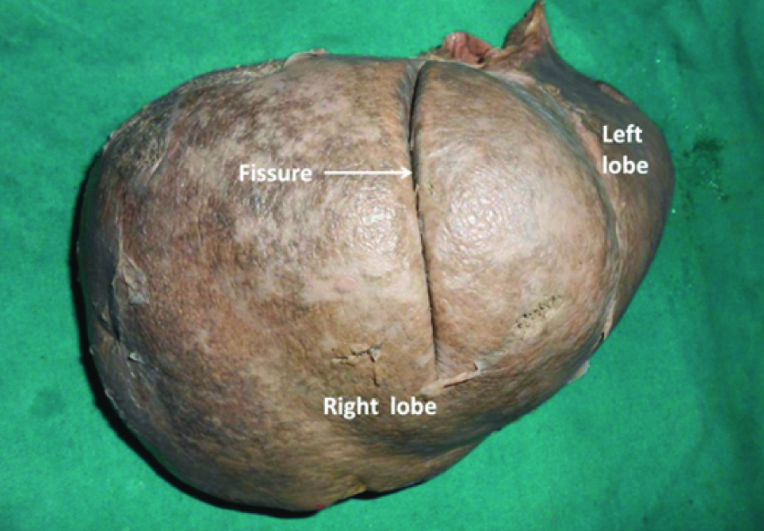
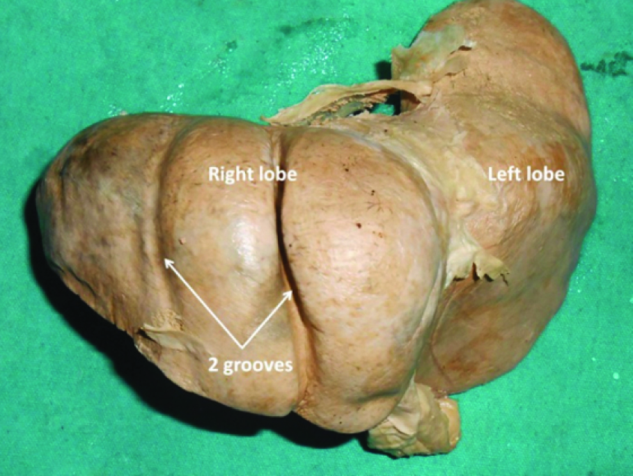
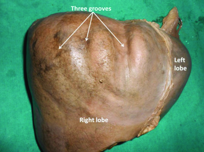
Left lobe did not reveal the presence of any abnormal grooves. Presence of a single groove was observed in six (6.18%) livers [Table/Fig-2] and [Table/Fig-3]; Double grooves were found in five (5.15%) livers [Table/Fig-4]; Triple grooves were found in three (3.09%) livers [Table/Fig-5] and four grooves were found in one (1.03%) liver [Table/Fig-6]. The deepest among the groove was 1.5 cm and looked like a deep fissure [Table/Fig-3].
Anterosuperior view of the liver showing the accessory groove extending from anterior surface to superior and posterior surfaces.
Superior view of the right lobe of the liver showing the accessory fissure extending from anterior surface to superior and posterior surfaces.
Anterosuperior view of the liver showing the two accessory grooves extending from anterior surface to superior and posterior surfaces.
Anterosuperior view of the right lobe of liver showing three accessory grooves extending from anterior surface to superior and posterior surfaces.
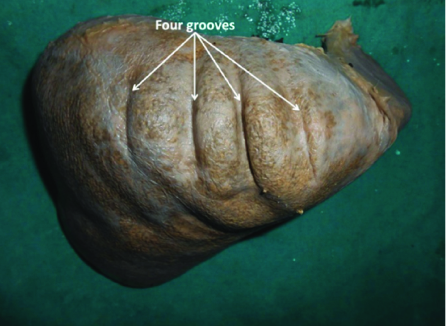
Superior view of the liver showing four accessory grooves extending from anterior surface to superior and posterior surfaces.
Discussion
In the medical literature, there are reports about the occurrence of unusual grooves on the liver. Most of them are surgical or radiological findings and were observed in general irrespective of the surfaces of the liver. Othman FB et al., studied 40 livers for the presence of accessory fissures. They could find accessory fissure in only two (5%) livers [6]. These fissures were found on posterior and inferior surface of the liver. In another cadaveric study on Afro-Caribbean population, the surface grooves were observed in 12% of cases inclusive of all surfaces [7]. In another cadaveric study, additional fissures on the inferior surface were found in 1.81% of cases [8]. Lim JH et al., have reported sonographic observation of a parasagittal fissure through the liver parenchyma on the posterior aspect of the right lobe among 15 out of their 2000 patients [9]. In an autopsy study by Machhi V et al., 40% of livers had diaphragmatic sulci and in 47% of cases they were located on the right lobe and were multiple [10]. In contrast to this, in the current study whatever grooves were found, were located on right lobe only (100%). Also, prevalence of sulci on diaphragmatic surface was low in our study (15.46%) when compared to the above study (40%). From another cadaveric study, Ono ML et al., have reported the presence of 79 sulci found in 50 livers out of the total 420 livers studied [11]. They opine that the grooves are not formed during development of the liver. According to them, formation of grooves is a postnatal event. Some sonographic studies have documented the presence of accessory grooves on the liver. Martinoli C et al., have observed a fissure on the left lobe in 5% of cases, caused by the infolding of the lesser omentum [12]. In another study, Auh YH et al., found that the accessory fissures are found more on the superior surface of the right lobe, incidence averaging up to 25%. They opine that the frequency of the grooves increases with advancing age. They have found the depth of the sulci reaching up to 2 cm [Table/Fig-7] [13]. Presence of many accessory fissures may mimic pathological liver nodules in CT scans. When only some parts of these fissures are observed sonographically, there is a high chance of getting them mistaken for echogenic liver lesions.
Table of comparison between previous studies and the current study about the occurrence of accessory fissures on the liver [6-13].
| Authors (reference) | Mode of study | Occurance of grooves in percentage |
|---|
| Othman FB et al., [6] | Cadaveric | 5% |
| Gardner MT et al., [7] | Cadaveric | 12% |
| Nayak BS [8] | Cadaveric | 1.81% |
| Lim JH et al., [9] | Sonographic | 0.0075% |
| Machhi V et al., [10] | Autopsy | 40% |
| Ono ML et al., [11] | Cadaveric | 0.42% |
| Martinoli C et al., [12] | Sonographic | 5% |
| Auh YH et al., [13] | Sonographic | 25% |
| Current study | Cadaveric | 15.4% |
Clinically, the hepatic grooves cannot be ignored because they are likely derived from the Chilaiditi sign [14]. Knowledge of these minor or major grooves may be very important to radiologists and surgeons to understand the liver segmentation [15].
Limitation
One of the limitations of the current study is the sample size. The study involved 97 livers. The study can be extended to a larger sample size. Other limitation of the study is that we are not able to explain the exact causative factor behind the presence of these accessory sulci.
Conclusion
Very few cadaveric studies have focused on accessory grooves on the diaphragmatic surface of the liver. In that respect, this study is unique. One more unique feature of this study is that all the grooves found were parallel to the sagittal plane and were parallel to each other. They are definitely not the “cough furrows” because they are not parallel to the ribs. They may be anatomically and physiologically normal but knowledge of their presence may avoid radiologic misdiagnosis and confusions. The knowledge of the grooves may be useful to operating surgeons during surgical resections and liver transplant surgeries.
[1]. Standring S, Ellis H, Healy JC, Standring S, LiverGray’s Anatomy: The Anatomical Basis of Clinical Practice 2005 39th edLondonElsevier Churchill Livingstone:1213-lp25. [Google Scholar]
[2]. Sherlock S, Dooley J, Disease of the liver and biliary system 1993 9th editionOxfordBlackwell:4 [Google Scholar]
[3]. Philips DM, LaBrecque DR, Shirazi SS, Corset liverJ Clin Gastroenterol 1985 7(4):361-68. [Google Scholar]
[4]. Yadav GD, Deka P, Accessory sulcus of the liver - An incidental laparotomy findingIndian J Surg 2008 70(2):92-93. [Google Scholar]
[5]. Kanchan T, Acharya J, Naik R, Grooves on the hepatic surfaceJ Forensic Leg Med 2014 21:24-25. [Google Scholar]
[6]. Othman FB, Latiff AA, Suhaimi FH, Das S, Accessory sulci of the liver. An anatomical study with clinical implicationsSaudi Med J 2008 29(9):1247-49. [Google Scholar]
[7]. Gardner MT, Cawich SO, Shetty R, Pearce NW, Naraynsingh V, Hepatic surface grooves in an Afro-Caribbean population: A cadaver studyItalian Journal of Anatomy and Embryology 2015 120(2):117-26. [Google Scholar]
[8]. Nayak BS, Study on the anomalies of liver in the south Indian cadaversInt J Morphol 2013 31(2):658-61. [Google Scholar]
[9]. Lim JH, Ko YT, Han MC, Kim CW, Choi BI, Im JG, The inferior accessory hepatic fissure: Sonographic appearanceAJR Am J Roentgenol 1987 149(3):495-97. [Google Scholar]
[10]. Macchi V, Feltrin G, Parenti A, De Caro R, Diaphragmatic sulci and portal fissuresJ Anat 2003 202(Pt 3):303-08. [Google Scholar]
[11]. Ono ML, Murakami G, Sato TJ, Sawada K, Hepatic grooves and portal segmentationKaibogaku Zasshi 2000 75(6):517-23. [Google Scholar]
[12]. Martinoli C, Cittadini G, Conzi R, Pastorino C, Neumaier CE, Rollandi G, Sonographic characterization of an accessory fissure of the left hepatic lobe determined by omental infoldingJ Ultrasound Med 1992 11(2):103-07. [Google Scholar]
[13]. Auh YH, Rubenstein WA, Zirinsky K, Kneeland JB, Pardes JC, Engel IA, Accessory fissures of the liver: CT and sonographic appearanceAJR Am J Roentgenol 1981 43(3):565-72. [Google Scholar]
[14]. Yavuz A, Batur A, Bulut MD, Bora A, Göya C, Andic C, Anterior hepatic grooves accompanied by Chilaiditi sign: A retrospective radiological analysis of a neglected anatomical factSurg Radiol Anat 2015 37(5):483-92. [Google Scholar]
[15]. Lee JH, Lee SY, Hwang DW, Park KM, Lee YJ, Practical usefulness of minor grooves of the hepatic surface for liver resectionHepatogastroenterology 2012 59(114):458-60. [Google Scholar]