Effective cleaning and shaping of the root canal and creation of an apical seal is the essential goal for successful endodontic treatment. In root canal cleaning and shaping, mechanical instrumentation clubbed with irrigating solutions play a vital role in removing the pre-existing debris, bacteria, toxic products from the inaccessible areas of the root canal, which otherwise would result in persistent irritation to the periapical tissues and impair the healing process [1].
NaOCl (0.5% to 6%) is the commonly recommended irrigating solution because of its pronounced antimicrobial activity and the ability to dissolve organic matter [2]. However, it is toxic if extruded beyond the root apex into the periapical areas [3]. NaOCl has no effect on inorganic part of dentin and smear layer; therefore use of 17% EDTA is recommended. But the application of EDTA for more than one minute led to the erosion and change in biomechanical properties of dentin of the root canal wall [4]. Furthermore, NaOCl breaks into sodium chloride and oxygen, which interfere with resin sealers polymerization and hence decreasing bond strength [5]. Therefore, the emphasis on finding an alternative root canal irrigant which is less toxic but equally effective still continues.
Epoxy resin-based AH Plus sealer (DentsplyDeTrey GmbH, Konstanz, Germany) has been extensively used with gutta percha because of its potential for better wettability of dentin and gutta percha, reduced solubility, very low shrinkage while setting, sealability, bonding and micro retention to the root dentin and adequate biological performance [8]. According to Dogan H et al., adhesion of resin based sealer materials to the dentinal surfaces is affected by different irrigation regimens due to change in permeability and solubility characteristics of dentin caused by the usage of irrigants [9].
Literature is scarce on the effect of chlorine dioxide irrigation on the resin sealer-dentin bond strength. Hence, the purpose of this study was to evaluate and compare 5% ClO2 with or without EDTA with 3% NaOCl and EDTA combination as endodontic irrigants on the adhesion of AH Plus sealer to radicular dentin using micro- push out bond strength test. Also, to compare micro- push out bond strength at coronal and mid-root levels within the group and to evaluate the mode of failure of the specimens under stereomicroscope. The null hypothesis is that ClO2 with or without EDTA reduced bond strength of AH Plus sealer to radicular dentin.
Materials and Methods
In the recent in vitro study, 40 permanent intact central incisors with complete root formation, straight root canals and extracted for mainly periodontal reasons were collected from the Department of Oral Surgery, Dayanand Sagar College of Dental Sciences, Bengaluru, Karnataka, India. Teeth were thoroughly cleaned using curettes and stored in saline solution at 4°C until it was used for the study.
Preparation of test solutions: The commercial two bottle preparation -solution A and solution B of chlorine dioxide 13.8% (BioClenz, Frontier Pharmaceuticals, Melville, New York, USA) were freshly mixed as per the manufacturer’s instruction and diluted to desired concentration with distilled water using the formula C1V1 = C2V2, where C1 = concentration of stock solution, V1 = volume of stock solution, C2= concentration of required solution, V2 = final volume of solution of desired concentration [10]. Therefore, 3.62 ml of 13.8% ClO2 was mixed with 6.38 ml of distilled water to prepare 10 ml of 5% ClO2 solution. The pH of ClO2 was 4.67, as measured by a pH meter (ELICO, pH meter Ll 120).
Preparation of specimens: Each tooth was decoronated at the cemento enamel junction to a standardize root length of 15±1mm. Working length was calculated by inserting size 15 K-file (Dentsply, Mailleffer, Ballaigues, Switzerland) 1mm short of the root length. Exterior surface of apical 3rd of all roots was covered with sticky wax to prevent flow of irrigants through apical foramen and to allow an effective reverse flow of the irrigant, simulating clinical conditions. The samples were then divided randomly into four groups (n=10) to receive different irrigants during instrumentation.
Group I: 5 ml 3% NaOCl (Deorcare, Kochi, Kerala, India) followed by 5 ml 17% EDTA (Smear off –Deorcare, Kochi, Kerala, India) final rinse for one minute;
Group II: 5 ml 5% ClO2 followed by 5 ml 17% EDTA final rinse for one minute;
Group III: 5 ml 5% ClO2 - during instrumentation and final rinse;
Group IV: 5 ml Saline -during instrumentation and final rinse.
Canals were enlarged using rotary file system (Dentsply, Mailleffer, Ballaigues, Switzerland) up to size F3 (30, 0.09 taper). According to Khadami A et al., root canal needs to be enlarged to an apical 30 size of 0.06 coronal taper, to allow penetration of irrigants [11]. Throughout the preparation, the canals were irrigated according to the sequence mentioned with 30-gauge side vented needle (Indsurgical Inc., Bengaluru) between each file size change. Finally the canals were rinsed with 5 ml of deionized water to remove any precipitate if formed. Canals were dried using paper points. AH Plus sealer was applied along the walls of canals using lentulospiral (Dentsply, India) and obturated with Protaper Gutta-percha master cone F3 (Dentsply, Mailleffer, Ballaigues, Switzerland). All the samples were coronally sealed with CavitTM–G (3M ESPE). The roots were placed in 100% humidity for 48 hours to ensure complete setting of sealers and incubated for two weeks at 37°C to simulate clinical conditions. Six horizontal sections of 1±0.05 mm were obtained from the coronal and middle thirds of each root using diamond disk (Minitom, Struer, Denmark) at low speed with constant cooling water. Apical 5 mm sections were discarded due to small size of filling material. The best specimens of each third were mounted in cold cured acrylic resin in the custom made copper mould. Each section was coded and measured for the apical and coronal diameter obturated area using a digital caliper (INSIZE, India).
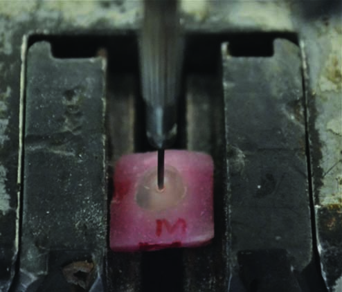
Push-out testing: Each root sections were then subjected to a compressive load via a Universal Testing Machine (UTM-LIoyd LR 50 K;LIoyd Instruments Ltd, Fareham, UK) at a cross head speed of 1 mm/minute using a 0.8 mm diameter stainless steel cylindrical plunger. The plunger tip was positioned to contact only the filling material, to avoid formation of cracks and erroneous reading [Table/Fig-1]. The push out force was applied in an apicocoronal direction until bond failure occurred, which was manifested by extrusion of the obturation material and a sudden drop along the load deflection curve recorded by computer software. The maximum failure load was recorded in Newton (N) and converted into Mega Pascal (MPa). The bond strength was calculated from the recorded peak load divided by the computed surface area that was obtained by the following formula: PBS (MPa) =N/A; where, N=Maximun load, A=Adhesion area of root canal filling (mm2). The adhesion (bonding) surface area of each section was calculated as:
Push out bond strength test.
A=πL (r1+r2) where, L= (r1-r2)1/2 +h2, π is the constant 3.14, r1 is apical radius, r2 is coronal radius and h is the thickness of sample in millimeters [12].
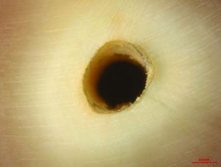
Analysis of failure modes: All debonded specimens were examined under a stereomicroscope (Discovery-V 20, CarlZeis, Germany) at 25X magnification to determine failure pattern: Adhesive -at the filling material/dentin interface, Cohesive- within filling material and mixed failure.
Statistical Analysis
The data were collected, tabulated and the mean and standard deviation of PBS were calculated at each root level for each group. The data were statistical analysed by using SPSS software version 17 with the significance set at p<0.05, using One way analysis of variance (ANOVA), Bonferroni and t-test.
Results
µPBS (MPa) in the groups [Table/Fig-2]:
Mean Micro Push Bond Strength (MPa) recorded in the groups according to ANOVA, p>0.05
| Group | Mean | Std. Deviation | SE of Mean | 95% CI for Mean | Min | Max | F- value | p- value |
|---|
| Lower Bound | Upper Bound |
|---|
| Group I | 2.22 | 0.52 | 0.12 | 1.98 | 2.47 | 1.59 | 3.55 | 0.013* | 3.815 |
| Group II | 2.16 | 0.77 | 0.17 | 1.80 | 2.52 | 1.01 | 3.73 |
| Group III | 2.06 | 0.76 | 0.17 | 1.70 | 2.41 | 1.26 | 3.40 |
| Group IV | 1.27 | 0.31 | 0.07 | 1.13 | 1.42 | 0.78 | 2.15 |
denotes significant difference
Higher mean µPBS was recorded in Group I followed by Group II, Group III and Group IV respectively. The difference in mean µPBS among the groups was found to be statistically significant (p<0.05) [Table/Fig-2].
In order to find out which pair of groups exhibited a significant difference, multiple comparisons test was done using Bonferroni test [Table/Fig-3]. The difference in mean µPBS was found to be statistically significant (p<0.001) between (Group I and Group IV) and (Group II and Group IV) as well as between (Group III and Group IV).
Multiple comparisons using Bonferroni test.
| (I) Group | (J) Group | Mean Differ- ence (I-J) | p-value | 95% CI for Mean Diff |
|---|
| Lower Bound | Upper Bound |
|---|
| Group 1 | Group 2 | 0.060 | 1.000 | -0.47 | 0.59 |
| Group 3 | 0.164 | 1.000 | -0.37 | 0.70 |
| Group 4 | 0.599 | 0.019* | 0.07 | 1.13 |
| Group 2 | Group 3 | 0.104 | 1.000 | -0.43 | 0.63 |
| Group 4 | 0.539 | 0.045* | 0.01 | 1.07 |
| Group 3 | Group 4 | 0.436 | 0.017* | -0.10 | 0.97 |
denotes significant difference
Comparison of µPBS between the sub groups within each group (t-test) [Table/Fig-4]:
Comparison of µPBS between the sub-groups within each group
t-test; p>0.05 Not significant.
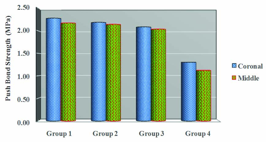
No significant difference was observed between coronal and middle region in any of the groups with respect to mean µPBS (p>0.05).
Failure patterns of tested groups [Table/Fig-5,6 and 7]:
Specimen showing adhesive failure.
Specimen showing cohesive failure.
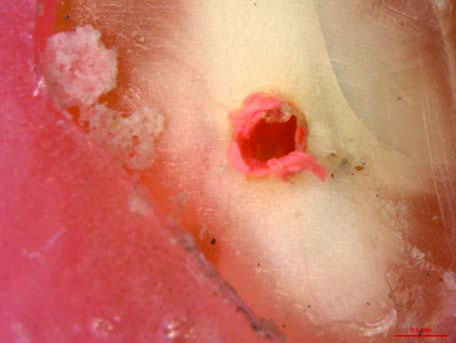
Specimen showing mixed failure.
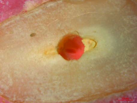
The saline group exhibited adhesive type of bond failure. Other experimental specimens exhibited predominance of mixed failure.
Discussion
ClO2 has recently come under consideration as a possible root canal irrigant, as it has the ability to dissolve organic and inorganic tissue [13,14]. The effective components of sodium hypochlorite, hypochlorous acid (HOCl), hypochlorite ions (OCl-) are negatively charged and might be repelled, when it comes in contact with negatively charged bacterial cell wall. But, ClO2 dissolves well in the apolar lipid phase of the cell membrane, remains as gas in water, hence better bacterial penetrability than NaOCl and bring about its destruction at a pH from 3 to 9 [6].
ClO2 has antiviral, antifungal and antibacterial properties. Its powerful oxidizing properties enable it to kill bacteria by disrupting the transport of nutrients across the cell wall. [15,16]. The recent detection of Cytomegalo virus and Epstein-Barr virus associated with periradicular lesions may promote the use of ClO2, which kills both enveloped and non enveloped viruses [15]. ClO2 damages Candida albicans (ATCC10231) by permeating into plasma membranes with minimal fungicidal concentration of 20 mmol/l [17].
A unique property of ClO2 is, it reacts with four essential amino acids (cysteine, methionine, tyrosine and tryptophan) present in bacteria, which is required for its survival hence bacteria do not develop resistance against it. So, a promising finding is that, due to all salient properties of ClO2, it powerfully eradicates the experimental E. faecalis biofilm from the mechanically prepared canal system and is more active against the reinfection than the currently widely used conventional irrigants [7].
Barnhart BD et al., concluded that ClO2 is less cytotoxic and produces little or no trihalomethanesas compared to sodium hypochlorite. [18]. Hyper pure ClO2 solution is volatile and quickly evaporates without any remnants and does not impede the healing process hence it can be regarded as a safe irrigant [7].
Since, ClO2 is unstable above the concentration of 9.5% 1{(ClO2)/ (air)} and is pungent at higher concentrations [15], an experimental concentration of 5% ClO2 was used as it shows antimicrobial properties even at a concentration of 0.12% [7,13,14].
The micro push out test was used in this study to evaluate the interfacial strength and dislocation resistance between the root filling material and the radicular dentin as smaller test specimens are ‘stronger’ than larger ones due to the lower probability of presence of the critical sized defects. In µPBS tests, higher apparent ‘strength’ can be measured with more failures at the interface [19].
Group I had highest bond strength values where dentin was treated with 3% NaOCl followed by 17% EDTA. The effective removal of the smear layer and smear plugs formed during biomechanical preparation allows the extension of the resin sealer tags into the open dentinal tubules, creating efficient micro retention [20]. Attal JP et al., and Dogen Buzoglu H et al., reported that EDTA significantly decreased the wetting ability of dentin making it hydrophobic [21,22] which provide adhesion with hydrophobic resinous AH plus. The irrigation regime followed is in agreement with current protocol, including the use of 5 ml EDTA final rinse as proposed by Mello I et al., [23].
Although the bond strength in Group II was marginally lower than Group I, it was slightly higher than Group III. This difference could be due to the additive effect of EDTA in smear layer removal, thus enhancing the demineralization effect of chlorine dioxide on root dentin and hence improving the push out bond strength of Group II (5%ClO2+17%EDTA) over Group III (5% ClO2). The capacity of ClO2 in removal of smear layer could be because of its low pH i.e., 4.67. Earlier studies have confirmed that pH of an irrigant is indirectly proportional to the amount of demineralization of root canal dentin [24]. Various studies have also suggested an inverse relationship between pH of a solution and time taken by the solution to dissolve the tissue [25]. In the current study, the pH of ClO2 was 4.67, much lower than the pH of NaOCl (pH = 12), this might be one of the reasons for ClO2 being less effective in dissolving pulp tissue; resulting in surface with remnants of organic components in smear layer, thus lowering the bond strength of Group II and III than Group I. In a similar study by Nishikiori R et al., the pH of ClO2 was raised up to 12 by using sodium hydroxide and it was concluded that ClO2 was similar to NaOCl in dissolving the bovine pulp tissue [26]. But the addition of sodium hydroxide to increase the pH might affect the smear layer removing capacity of ClO2 that can affect the bond strength to root dentin.
Based on evidences from previous studies, though ClO2 has lesser tissue dissolution and smear layer removal properties than NaOCl and EDTAC combination [13,14] still the bond strength values of Group III specimens, did not show statistically significant difference in this study. The reason could be that, 5% ClO2 being a weak acid, it is likely that it will affect the dentin similar to other organic acids such as maleic acid or lactic acid and it may cause a mineral gradient that could yield itself better to resin infiltration [27]. Also, adhesion of AH Plus to root dentin is related to covalent bonds between epoxide rings and the exposed amino groups in the collagen network [28]. ClO2 could have exposed collagen network and preserved it to give comparable bond strength values. However, this needs to be investigated further. In the saline group, there would have been heavy organic and inorganic remnants in the smear layer, which resulted in low bond strength to root dentin.
Another notable finding regarding bond strength value was that the µ-PBS was more in coronal third than middle third in all the specimens although there was no statistical significant difference. This is probably due to better volume and penetration of the irrigant and the more organized structure of tubular dentin in coronal third, which apparently yields better infiltration of epoxy resin. This can also be explained by the apical structure of dentin which has low number of dentinal tubules and irregular structure of secondary dentin that might have resulted in reduced penetration of adhesives into the middle root dentin compared to coronal dentin [27], though the differences were not statistically significant in the present study.
In the assessment of failure pattern under stereomicroscope, it was found that the saline group exhibited adhesive type of bond failure correlating to the results of the study, indicating decreased bond strength values. Other test specimens exhibited predominance of mixed failure. The result of Group I and II is partially in agreement with Aranda Garcia AJ et al., study who observed that the mixed failure mode was predominant for both NaOCl/EDTA when the AH Plus/Gutta percha was used [29]. In addition, in the present study even the Group III specimens showed predominantly mixed failures, comparable to that of Group I and Group II suggesting smear layer removal and pulp dissolution action of ClO2.
Based on results of our study, it can be concluded that bond strength of epoxy sealers to root dentin after using various irrigants and irrigation protocol used in this study is not significantly different from one another (except with Group IV). Hence, the null hypothesis was rejected.
Limitation
A limitation of this study is that it determines adhesion only on basis of PBS test. Further studies regarding penetration of the sealer into root dentin; its ultra structural changes are needed.
Conclusion
In the present study, the bond strength values of 5% ClO2 with or without EDTA are comparable with conventional NaOCl and EDTA combination. Hence, ClO2 can be considered as an effective alternative endodontic irrigant with its tissue dissolution and smear layer removal properties.
Further studies, on variable parameters such as volume, concentration of chlorine dioxide, effects of various exposure times, and effects on other endodontic sealers is needed, in order to enable its routine use in endodontics.
*denotes significant difference
*denotes significant difference