In Endodontics eradication of microorganisms is the primary goal and also serves as predictor for the long-term success of the endodontic therapy. Root canal irrigation plays an important role in the debridement and disinfection of root canal systems. Many clinical approaches have been evaluated for disinfection and control of the root canal biofilm during endodontic treatment [1]. Even then the presence of bacteria in root canals has been considered to be responsible for endodontic treatment failure. Location, harbouring, and multiplication of bacteria within root canals are the factors most crucial for disinfection. Therefore, the disinfection in this anatomical structure poses to be a clinical problem. Mechanical instrumentation approach is limited in its effectiveness by the bacterial colonisation and survival in dentinal tubules, lateral canal ramifications, canal isthmuses, and other irregularities in the root canal [1].
Even the root canal systems found in primary teeth frequently contain many ramifications and deltas between canals making thorough debridement quite difficult by instrumentation therefore profuse irrigation plays a vital role in achieving effective removal of debris and necrotic tissue [2].
Antimicrobial irrigants, especially sodium hypochlorite (NaOCl), are very effective in reducing bacteria populations, as they have a proteolytic effect. Although NaOCl acts directly on target bacteria, several factors—such as the anatomical complexities of root canals, deep invasion of microorganisms into dentinal tubules, and formation of biofilm on the surface of the root apex—make it difficult to completely eliminate microorganisms from root canals and periapical lesions [3].
Different techniques have been proposed to improve the efficacy of irrigating solutions, including changes of concentration, temperature, surfactant, and agitation [4]. Activation of irrigants appears to be an important method of increasing antibacterial and antibiofilm activity of root canal irrigants, not only within the root canal, but also within the anatomical complexities of the root canal system and dentinal tubules. Though traditional chemomechanical cleansing measures have shown acceptable results in endodontic outcomes, several literature reports have suggested that the additional use of lasers in removing bacterial load in areas where traditional methods may fail to succeed can give superior outcomes [1]. Laser light can reach the areas of root canal that are impossible to reach by the conventional techniques of irrigation [1]. LAI by means of a diode laser has proven to be more effective in enhancing the decontaminating action of NaOCl [5]. Also, the use of another technique PDT with the lasers seems to be a promising alternative to reduce microbial bacteria in the infected root canals of teeth and has been recommended as an alternative or supplement to currently used disinfection methods [6]. Enterococcus faecalis is the pathogenic microbe responsible for failure of endodontic treatment both in permanent as well as primary teeth since it is the most resistant strain found especially in the biofilm community [7]. The effectiveness of the latest advancements in disinfection of root canal systems hence if are proven to give significant disinfection ability against this pathogen, will obviously be effective against most other species of root canal microbiota.
Hence, this study was designed to compare the efficacy of latest advancements in disinfection techniques using a high power diode laser namely direct laser irradiation, photodynamic therapy and laser activated irrigation using sodium hypochlorite.
Materials and Methods
The study was an in vitro, intergroup, experimental study conducted in the Department of Pedodontics and Preventive Dentistry, Bapuji Dental College and Hospital, Davangere, Karnataka, India.
Ethical clearance was obtained for conducting the study by the ethical review board of Bapuji Dental college and Hospital, Davanagere, Karnataka, India.
Sample Size Determination
The sample size estimation for the evaluation of amount of disinfection through the microbiologic analysis was done using a formula wherein the mean and standard deviation values from the previous studies were used. Sample size was determined to be 20 per group [6].
Source of Specimens
Sixty primary teeth both single rooted and multi-rooted teeth with two third of their root length intact [8] extracted for therapeutic reasons like over-retained primary teeth causing ectopic eruption of permanent successors, cases referred for serial extraction, in case patients are not willing for pulpectomy procedures or resorption on lingual aspect of roots were collected for the purpose of this study. Primary teeth of same type were included in all groups that is comparison between same type of primary teeth was ensured in all the three groups.
Preparation of Teeth Specimens
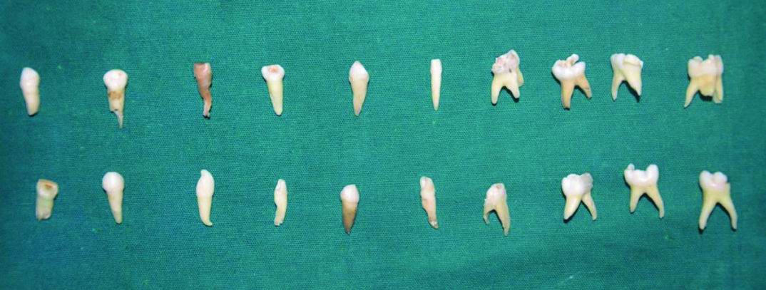
Scaling was done for all teeth specimens to remove any calculus and periodontal tissues [9] [Table/Fig-1]. Working length was determined radiographically to 2 mm short of radiographic apex.
Teeth samples with two thirds of root length for all groups.
Instrumentation was completed to size 30 No H files (Mani files). Samples were irrigated using 10 ml 2.5% sodium hypochlorite during canal preparation, and canal patency was maintained [9]. The pulp chamber was flooded with sodium hypochlorite and replenished with 1 ml of 2.5% sodium hypochlorite irrigant after each instrument. The teeth were transferred to a flask with deionized water for sterilization by autoclaving for 30 minutes at 121°C with 15 lb. pressure [10]. The sterilised specimens were tested for adequate sterilisation using spore test. Teeth were randomly divided into three groups [Table/Fig-2].
Group distribution for the study.
| Group number | Group type | Number of specimens per group |
|---|
| Group I | Direct Laser- irradiation | 20 |
| Group II | Photodynamic therapy | 20 |
| Group III | Laser activated irrigation with 2.5 NaOCl | 20 |
Bacteria and Culture Conditions
The sterilized tooth specimens were inoculated with E.faecalis American Type Culture Collection (ATCC) 29212 that was cultured in Brain-Heart Infusion (BHI) broth (Difco, Detroit, MI) at 37°C for 18 hours in an atmosphere of 5% CO2 using inoculating tips. The organisms were harvested by centrifugation at 10,000×g for five minutes then suspended in saline and adjusted to 3×10⁶ cells/ml using a spectrophotometer. Specimens were kept immersed in broth at 37°C to allow bacterial growth. The medium was replaced once a week for four consecutive weeks. A period of four weeks was chosen for inoculation of bacteria as recognisable and appreciable bacterial colonies has shown to be produced after it. Thereafter, teeth were removed from the bacterial culture. The root apices were covered with Cavit™. Each tooth was ends wiped with 2.5% sodium hypochlorite to disinfect the outside of the tooth before further treatment. Cavit was removed and experimental irrigation procedure was completed for all the three groups [1].
Bacterial Colony Count Before the Experimental Intervention [Table/Fig-3]
Colony count using digital colony count meter.
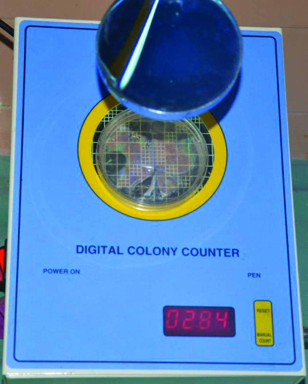
Sterile ringer solution was added in all canals and paper points were inserted into canal terminus and left for 60 seconds to soak up the contents in the canals. Here the sterile ringer solution acts as a medium for carrying the bacteria from canal to agar plate. The paper points were used to inoculate the bacteria on blood agar plates and these plates were incubated at 37°C in a CO2 chamber for 48 hours. The bacterial colonies were counted in CFU (Colony Forming Units)/mm3 using a digital colony counter [10].
Experimental Procedure [Table/Fig-4]:
Laser irradiation for all groups.(Images from left to right).
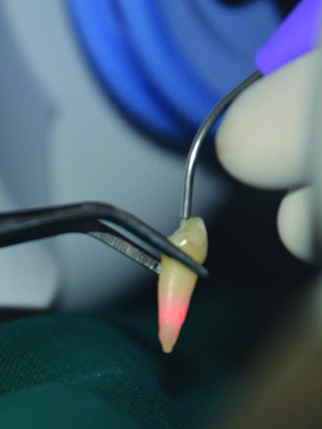
Group 1: Direct laser Irradiation
The physical parameters of it are: wavelength – 810 nm, output power- 5 watts, output energy – 20%. The optical fibre was introduced 1 mm short of the working length and was withdrawn from apical to coronal according to the recommendations of Gutknecht N et al., and the root canals were irradiated for a period 20 seconds, repeated three times at intervals of 10 seconds between each one [11].
Group 2: Photodynamic therapy
Light source and photosensitizer; the irradiation source was a diode laser (AMD Picasso diode laser). Its wavelength, output power, and duty was 810 nm, 1.5 watts (W), and 20%, respectively, while the diameter of quartz optical fiber was 400 μm. Indocyanine green (12.5 mg/ml) a commonly used fluorescent fundus contrast medium, was used as the photosensitizer [10].
Laser Irradiation; the optical fibre of the diode laser was inserted into the root canal to reach its apex and laser irradiation was performed for a period of 60 seconds [10].
Group 3: Laser Activated irrigation with 2.5% NaOCl: The physical parameters; wavelength – 810 nm, output power- 1.5 watts, output energy – 20%.
Laser activated irrigation with 2.5% NaOCl; the canal chamber was filled with 2.5% NaOCl and the optical fibre was introduced 1 mm short of the working length and laser irradiation was performed with the irrigant in the canal for a period 20 seconds, repeated three times at intervals of 10 seconds between each one [12].
For evaluation of disinfection after the intervention [Table/Fig-2]: The samples from the root canals were then obtained in a similar manner as obtained preoperatively and plated on blood agar plates, incubated at 37oC and bacterial colony count using a digital colony count meter was performed.
Statistical Analysis
The data obtained was subjected to statistical analysis using SPSS software (Version 20.0). One way Analysis was applied for intergroup comparison. Tukey’s post-hoc test was applied for pairwise comparison between groups. Paired t-test was applied for intragroup comparison.
Results
[Table/Fig-5] shows the preoperative comparison of mean values of bacterial colony count of all the three groups. No statistically significant difference in preoperative colony count values of all groups was found. [Table/Fig-6] depicts a highly significant difference in mean bacterial count for all the three groups postoperatively. [Table/Fig-7] shows Tukey’s post-hoc test results for comparison between the three groups. On interpreting the table it was found that there was statistically significant difference between results of group 1 and Group 2 and also between Group 1 and Group 3. However, no statistical difference between Group 2 and Group 3 was found. [Table/Fig-8] summarises the comparison of mean preoperative and postoperative values for all groups and shows that there is statistically significant decrease in postoperative bacterial count for all the three groups.
Preoperative comparison mean CFU/mm3 of all the three groups and One-way ANOVA Test performed at baseline.
| Groups | I(Laser irradiation) | II(Photodynamic therapy) | III(Laser activated irrigation) |
|---|
| (Mean CFU/mm3 ± SD) |
|---|
| Bacterial count | 176±70 | 171±58 | 172±30 |
| F-value | 0.040 |
| p-value | 0.961 |
One-way ANOVA test applied and bacterial colony count postoperatively.
| Groups | I(Laser irradiation) | II(Photodynamic therapy) | III(Laser activated irrigation) |
|---|
| (Mean CFU/mm3 ± SD) |
|---|
| Bacterial count | 104.45±50 | 32.60 ±17 | 32.90±24.77 |
| F-value | 29.32 |
| P-value | < 0.001** |
Tukey’s post-hoc test results for comparison between the three groups.
| Post opbacterialcount | Group | Group | p-value |
|---|
| Laser irradiation | PDT | 0.001 |
| LAI | 0.001 |
| PDT | LAI | 0.96 |
PDT- Photodynamic therapy LAI – laser activated irrigation using sodium hypochlorite
Comparison of preoperative and postoperative bacterial count for all the three groups.
| Groups | Mean difference | Std. Deviation | T-value | Significance |
|---|
| Pair 1 preop I– postop 1 | 71.60000 | 34.13657 | 9.380 | <0.001 |
| Pair 2 preop II– postop II | 138.60000 | 57.42950 | 10.793 | <0.001 |
| Pair 3 preop III– postop III | 139.75000 | 40.40958 | 15.466 | <0.001 |
Paired T-test is used for preoperative and postoperative mean difference (CFU/mm3) for all the three groups.
Discussion
Endodontic treatment of primary teeth is commonly practiced in dentistry. Control of infection is important because the medullary bone spaces favour dissemination of infection and also due to close approximation of developing permanent tooth germ to the roots of the primary teeth [7]. The anatomic complexities in root canals of primary teeth root canals make complete debridement difficult. In such cases chances of failure of endodontic treatment are high. Therefore, disinfection plays an important role in pulpectomy procedures even in primary teeth [13].
Da Silva LA et al., concluded from their study that the success of endodontic treatment depends on several factors, the most important of which is the reduction or elimination of bacterial infection. Because the microbiota of root canals of primary teeth with necrotic pulp and periapical lesions is similar to that found in permanent teeth, endodontic treatment should be similar [7]. Hence, pulpectomy procedures should include the neutralization of necrotic content, instrumentation and intracanal dressings as essential steps [7]. Thus, laser assisted disinfection techniques in primary teeth were evaluated in the present study.
It is a well-known fact that Enterococcus faecalis is the most resistant strains in root canals of permanent teeth as well as primary teeth [7]. Therefore promising results obtained through use of laser assisted disinfection techniques for this species of bacteria, could possibly give better results with other root canal microbiota too.
Lasers are an alternative to conventional or advanced means of disinfection of root canals using endoactivator. Use of diode laser with methods like laser activated irrigation using sodium hypochlorite, or photodyanamic therapy are used for disinfecting the areas that are impossible to reach with the traditional techniques. High power diode lasers are bactericidal due to dose dependent heat generation. Its antimicrobial effectiveness against diverse microorganisms has already been demonstrated in a study by Gutknecht N et al., [11]. Laser-induced bacterial killing is because of thermal heating of the environment above the lethal values and local heating inside bacteria [6]. According to some authors lasers activate the irrigation solutions via the transfer of pulsed energy [9]. In case of laser activated irrigation using sodium hypochloride, Groot SD et al., observed that the laser-induced bubble grew larger when NaOCl was used as an irrigant solution. It was also found that a higher amount of smaller bubbles were present after laser activation when using sodium hypochlorite as the irrigant solution. This was contributory to the activation of sodium hypochlorite and enhancement in its effectiveness as an irrigant [14].
Laser energy is used to activate a nontoxic photosensitizer in presence of oxygen, and free oxygen radical released from these dyes causes damage to the membrane and DNA of microorganisms without affecting host cell viability in photoactivated disinfection [6]. The results of a study by Nagayoshi M et al., suggest that use of a diode laser in combination with photosensitizer dye like, indocyanine green may be useful for clinical treatment of periapical lesions [10]. Likewise, Bago I et al., has found that PDT and endo activator system were more successful disinfection of root canals than the diode laser and NaOCl syringe irrigation alone [6]. Similarly, Neelakantan P et al., has concluded that diode laser and Er:YAG laser activation were superior to ultrasonics in dentinal tubule disinfection [12].
The results of our study have shown that although a significant decrease in bacterial count was obtained with all the three groups, the need of an additional disinfection media which may be either an irrigant or a photosensitive dye is required for better disinfection as both laser activated irrigation using NaOCl, and photo activated irrigation showed better results than direct laser irradiation. Direct laser irradiation of the canals though gave significant decrease in bacterial count, the lack of an active disinfecting media could be the reason contributing to its inferiority when compared to the other two techniques.
This could also be attributed to survival of E. faecalis or its high resistance to heat, because of its cell-wall structure [6]. The activation of the disinfecting solution by the laser via the transfer of pulsed energy which accentuates the efficacy of disinfecting solutions.
However, in the present study the effectiveness of was found similar as there was presence of an active disinfectant medium in both groups and only the efficacy of both solutions was increased by the use of laser. Whereas, with a variety of these lasers assisted disinfection methods the clinician may often be in a dilemma as to which one to choose. Therefore, in an attempt to resolve and throw some light on this issue, the present study compared the effectiveness of each group in laboratory settings.
A comparison of the preoperative bacterial count for all the three groups was found to be insignificant which allowed us to be able to compare the three groups for their postoperative values.
Moreover, high-power lasers have the potential to cause dentine charring, ankylosis, root resorption and periradicular necrosis [6]. Therefore, for the second and third groups as there was an assisting media for disinfection, the parameters of laser settings were kept at a power output of 1.5 watt.
Hence, the use of diode lasers in endodontic disinfection especially in necrotic primary teeth is recommended as it could improve the success of pulpectomy procedures.
Limitation
However, the present study being an in vitro study the effectiveness of these lasers assisted disinfection strategies need to be proved clinically through randomised controlled trials and longer term follow ups in pulpectomised primary teeth for accurately and precisely giving credit to lasers for their success.
Conclusion
Disinfection strategies using a high power diode laser by techniques like direct laser irradiation, laser activated irrigation or photoactivated disinfection give significant results in eliminating E. fecalis. Also, laser activated irrigation and photoactivated disinfection were found to be similar in their effectiveness while direct laser irradiation was found to give least results among the three.
PDT- Photodynamic therapy LAI – laser activated irrigation using sodium hypochlorite
Paired T-test is used for preoperative and postoperative mean difference (CFU/mm3) for all the three groups.