Introduction
Over the past many years various root end filling materials have been used which have been tested for their physical properties but each of them had certain limitations. In clinical practice, root end filling materials are exposed to oral tissue fluids which may compromise their longevity.
Aim
The aim of this study was to investigate the effects of oral tissue fluids on compressive strength of Mineral Trioxide Aggregate (MTA) and biodentine.
Materials and Methods
MTA and biodentine cylinders measuring 6 mm × 4 mm were prepared using acrylic blocks. They were divided into six groups; (Group 1) (MTA) (n=3), (Group 2) MTA contaminated with saliva, (MTA-S) (n=3), Group 3: MTA contaminated with blood, MTA-B (n=3), Group 4: Biodentine (BD), Group 5: Biodentine contaminated with saliva (BD-S) (n=5), Group 6: Biodentine contaminated with blood (BD-B) (n=5). The mould was contaminated with saliva and blood and incubated at 37°C at 100% humidity for three days and compressive strength (MPa) was measured using universal testing machine and the data was analyzed statistically using one-way ANOVA test.
Results
There was no significant difference in the compressive strength between the three groups i.e., MTA, MTA-S, MTA-B (p > 0.05). However, there was higher compressive strength in the MTA-B group when compared to MTA and MTA-S. Also, there was no statistical significant difference between BD, BD-S, BD-B (p>0.05).
Conclusion
This study showed that the compressive strength of MTA and biodentine was not adversely affected by contamination with oral tissue fluids like blood and saliva.
Introduction
The ultimate goal of the root canal treatment is to seal the communicating pathways between the pulp and periradicular tissues which prevents microleakage of irritants and microbes from infected root canals [1]. The success of root-end filling material lies in providing a complete fluid tight apical seal [2]. The failure of surgical endodontic treatment is due to inadequate apical seal [3]. The ideal root end filling material should be biocompatible with tissue fluids, non toxic and non carcinogenic, dimensionally stable, radiopaque, have antibacterial property, good handling characteristics, should be able to set in wet conditions, and should have physical properties like adequate compressive strength and bond strength, hardness and adhere to the root canal dentine to achieve a good apical seal [4]. During surgical endodontic therapy the presence of moisture or contact with tissue fluids like blood and saliva may affect its sealing ability [5]. Over the past many years various root end filling materials have been used like amalgam, guttapercha, zinc-oxide eugenol cements (IRM, Super-EBA), glass ionomer cements, composite resins, compomers, MTA, biodentine, bioaggregate, endosequence, etc., which have been tested for their sealing ability and physical properties but each of them have shown limitations [1,6].
MTA is a hydrophillic, tricalcium silicate-based material which has excellent biocompatibility and sealing property. It is used for various procedures such as pulp capping, pulpotomy, pulpectomy, apexogenesis, apexification in teeth with open apex, repair of root perforations, and as a root canal and root-end filling material (Vosoughhosseini et al., 2008) [7]. The main disadvantage is the difficulty in handling and condensing the cement inside the root canal and its longer setting time (two hours 45 minutes) [8]. The study by Vanderweele RA et al., reported that the retention of MTA decreased on contamination with blood [9] and study by Nekoofar MH et al., demonstrated that blood contamination decreased the compressive strength of MTA [10].
Biodentine (Septodont, Saint-Maur-des-Fosse’s, France) is used as a dentine replacement material similar to that of MTA. It is available in powder-liquid formulation where the powder is composed of tricalcium silicate, dicalcium silicate, calcium carbonate, calcium oxide, iron oxide, and zirconium oxide as a radiopacifier and the liquid constitutes of calcium chloride as an accelerator and hydrosoluble polymer as a water reducing agent. The setting time of Biodentine is nine to12 minutes [11].
Various studies have evaluated the compressive strength of MTA contaminated with blood, but there is a lack of evidence in the literature regarding the effect of oral tissue fluids like saliva on compressive strength of calcium silicate based cements. Therefore, it is necessary to determine the strength of these cements on exposure to the oral fluids which may come in contact during setting reaction and interfere with the setting mechanism. This study was aimed to evaluate the effect of oral tissue fluids like saliva and blood on compressive strength of MTA and biodentine.
Materials and Methods
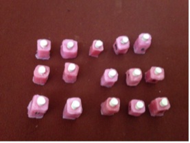
Ethical approval and clearance for this in vitro study was obtained from Scientific Review Board, Saveetha Dental College, Chennai, India. A total of 24 cylindrical samples were prepared with internal diameter of 4 mm width and 6 mm length using acrylic blocks [Table/Fig-1]. They were divided into six groups:
MTA and biodentine samples.
Group 1-MTA (n=3),
Group 2-MTA-S (n=3) Contaminated with saliva,
Group 3-MTA-B (n=3) Contaminated with blood,
Group 4-(BD) Biodentine (n=5),
Group 5-(BD-S) Biodentine contaminated with saliva (n=5),
Group 6-(BD-B) Biodentine contaminated with blood (n=5).
Both MTA and biodentine were manipulated based on manufacturer’s instructions. The moulds were coated with saliva or blood according to the group before placement of the material and a glass slab was placed at the base of the material to generate a smooth surfaced base. Samples were incubated at 37°C at 100% humidity for three days [12]. The force needed to break the samples (in N/mm2) was tested by universal testing machine (Instron model 1011, UK) at a crosshead speed of 1 mm/s and compressive strength (MPa) was calculated using the following formula:
RC = F x 9.807 / A [13]
RC = compressive strength (MPa), F = force/unit area (kg), 9.807 (gravity) = constant and A = base area (7.06 mm2).
Statistical Analysis
The means and standard deviations for all the groups were calculated and the data was analyzed statistically using one-way ANOVA test with a p-value set at 0.05 [Table/Fig-2].
Compressive strength (MPa) of MTA and biodentine contaminated with blood and saliva with (p-value > 0.05) for MTA and biodentine groups.
| S. No | Sample | CompressiveStrength (MPA)Mean ± Sd | p-value |
|---|
| 1 | MTA | 157.87 ± 76.30 | 0.52 |
| 2 | MTA-S | 114.70 ±47.36 |
| 3 | MTA-B | 176.44 ± 67.47 |
| 4 | BD | 205.41 ± 21.59 | 0.18 |
| 5 | BD-S | 156.64 ± 57.31 |
| 6 | BD-B | 176.93 ± 28.41 |
*one-way ANOVA
Results
After three days of incubation, samples were tested for the compressive strength (MPa). On contamination with oral tissue fluids like saliva and blood, there was no statistical significant difference in the compressive strength between the three groups i.e., MTA, MTA-S, MTA-B (p>0.05) [Table/Fig-2]. However, there was higher compressive strength in the MTA-B group when compared to MTA and MTA-S. Also, there was no statistical significant difference between BD, BD-S, BD-B (p>0.05). Among these three groups BD showed a higher compressive strength than BD-S, BD-B [Table/Fig-2].
Discussion
The success of root end filling material lies in sealing the mechanical or pathological communication between the periapical environment and root canal system. It should have the capacity to withstand the mechanical forces of condensation during perforation repair or when used as a retrograde filling material [14]. The most common cause for failure of endodontic therapy is apical microleakage [15].
During clinical applications like perforation repair or in apexification the materials may come in contact with tissue fluids like saliva or blood which may penetrate the MTA and inturn may affect the strength by altering the setting mechanism leading to failure of the set cement [16]. Also, compressive strength is an indicator of the setting and hydration processes [17]. In clinical situations, root end filling materials come into direct contact or even mix with blood during or after placement. In addition, it has been stated that the ‘air entrapment’ features of blood proteins affect the microstructure of cements and increases their porosity [18]. When used as a restorative material, it may be subjected to masticatory stress, so adequate knowledge about the compressive strength is necessary to prevent the failure of the set cement [19]. Compressive strength is one of the main physical properties of hydraulic cements. When used in vital pulp therapies, the cement should have the capacity to withstand masticatory stress [20]. In this study, freshly drawn blood was used because the presence of an anticoagulant may decrease the bond strength [21].
In this study, MTA and biodentine were used because of their excellent sealing ability and less microleakage when compared to amalgam, intermediate restorative material, glass ionomer cement and zinc oxide eugenol [22].
MTA was introduced by Torabijenad and was later approved by FDA and became commercially available as ProRoot MTA in 1998 [23]. MTA has excellent sealing ability, biocompatibility, good compressive strength, insoluble in body fluids once set [24]. ProRoot MTA also has the favourable ability to set under moist conditions [25]. MTA was prepared by mixing powder with sterile distilled water in a 3:1 ratio. On hydration the particles in the powder forms a colloidal gel that solidifies to form hard barrier [26]. Setting time is affected by factors like moisture and air entrapped during trituration [27,28].
MTA contains fine hydrophilic particles like calcium hydroxide and silicon which has the ability to set in the presence of wet environment [29]. There are various studies on the influence of blood on properties of MTA. An in vitro study by Salem MA et al., reported that exposure to blood during setting has an adverse effect on marginal adaptation and the surface microstructure of MTA [30]. Therefore, by addition of the accelerators reduces the infusion of the blood into the material thereby protecting it from deleterious effect by improving its initial strength [31]. Recently, Biodentine has been used as a dentine replacement material in large carious lesions [32]. On hydration reaction, tricalcium silicate produces calcium silicate gel and calcium hydroxide and they may precipitate at the surface. So, the unreacted tricalcium silicate grains are surrounded by hydrated calcium silicate gel, which are impermeable to water and decreases the setting reactions [32].
Biodentine, due to its high porosity has higher capacity for ion exchange [33]. Biodentine improves in compressive strength with time over several hours [34].
A study by Grech L et al., reported that biodentine had low fluid uptake and sorption values, low setting time and superior mechanical properties. The fluid uptake and setting time was the highest for MTA compared to biodentine [34]. This was supported by Camilleri J et al., 2013 who stated that biodentine is more dense and less porous when compared to MTA which explains its less fluid uptake [35]. The lower the porosity, higher will be the mechanical strength [36].
To the best to our knowledge on the available literature, this is the first study evaluating the compressive strength of MTA and biodentine on contamination with blood and saliva. Charland T et al., reported that the exposure to blood did not have a significant difference on the setting time of MTA [37]. Kim Y et al., stated that the on exposure of MTA to foetal bovine serum affected the setting reaction of MTA [38]. Though both MTA and biodentine are tricalcium based cements, the shorter setting time of biodentine makes it a demanding material for apical surgeries [39].
Thomas B et al., compared the effect of pH on compressive strength of MTA and biodentine and stated that biodentine showed a higher compressive strength in acidic and in alkaline environment compared to that of MTA [40].
Poplai G et al., reported that in the presence of acidic conditions surface hardness of biodentine was affected [41].
Limitation
The limitation of this study includes limited sample size for evaluating the material properties. Even though the oral scenario is recreated, it was not done under controlled oral conditions. Further more clinical studies are warranted for evaluating the effect of oral tissue fluids on compressive strength under well controlled oral conditions.
Conclusion
In this study, the compressive strength of MTA and biodentine was similar in comparison, both under normal as well as in contaminated conditions. This study concludes that compressive strength of MTA and biodentine was not significantly affected by contamination with oral tissue fluids like blood and saliva.
*one-way ANOVA
[1]. Priyanka SR, Veronica A, Literature review of root-end filling materials.iosr journal of dental and medical sciencesIOSR-JDMS 2013 9(4):20-25. [Google Scholar]
[2]. Fogel HM, Peikoff MD, Microleakage of root-end filling materialsJ Endod 2001 27:456-58. [Google Scholar]
[3]. Elemam RF, Pretty I, Comparison of the success rate of endodontic treatment and implant treatmentISRN Dentistry 2011 2011:640509 [Google Scholar]
[4]. Torabinejad M, Pitt Ford TR, Root end filling materials: A reviewEndod Dent Traumatol 1996 12(4):161-78. [Google Scholar]
[5]. Mathew LA, Kini S, Acharya SR, Kamath S, Menezes ND, Rao S, A comparative evaluation of the microleakage of blood-contaminated mineral trioxide aggregate and biodentine as root-end filling materials: An in vitro studyJ Interdiscip Dentistry 2016 6(1):19-24. [Google Scholar]
[6]. Vasudev SK, Goel BR, Tyagi S, Root end filling materials - A reviewEndodontology 2013 15:12-18. [Google Scholar]
[7]. Vosoughhosseini S, Lotfi M, Shahi S, Baloo H, Mesgariabbasi M, Saghiri MA, Influence of white versus gray mineral trioxide aggregate on inflammatory cellsJ Endod 2008 34:715-17. [Google Scholar]
[8]. Camilleri J, Montesin FE, Di Silvio L, Pitt Ford TR, The chemical constitution and biocompatibility of accelerated Portland cement for endodontic useInt Endod J 2005 38:834-42. [Google Scholar]
[9]. Vanderweele RA, Schwartz SA, Beeson TJ, Effect of blood contamination on retention characteristics of MTA when mixed with different liquidsJ Endod 2006 32:421-24. [Google Scholar]
[10]. Nekoofar MH, Stone DF, Dummer PM, The effect of blood contamination on the compressive strength and surface microstructure of mineral trioxide aggregateInt Endod J 2010 43:782-91. [Google Scholar]
[11]. Vipin A, Vineeta N, Namrata S, Pooja A, Bioactive dentin replacementIOSR-JDMS 2013 12(4):51-57. [Google Scholar]
[12]. Ramazani N, Sadeghi P, Bacterial leakage of mineral trioxide aggregate, calcium-enriched mixture and biodentine as furcation perforation repair materials in primary molarsIranian Endodontic Journal 2016 11(3):214-18. [Google Scholar]
[13]. Umesh V, Hambire UV, Tripathi VK, Atmaram GM, Improvement in the compressive strength and flexural strength of dental compositeARPN Journal of Engineering and Applied Sciences 2012 7(8):1066-69. [Google Scholar]
[14]. Aggarwal V, Singla M, Miglani S, Kohli S, Comparative evaluation of push-out bond strength of ProRoot MTA, Biodentine, and MTA Plus in furcation perforation repairJ Conserv Dent 2013 16(5):462-65. [Google Scholar]
[15]. Muliyar S, Shameem KA, Thankachan RP, Francis PG, Jayapalan CS, Hafiz KAA, Microleakage in endodonticsJ Int Oral Health 2014 6(6):99-104. [Google Scholar]
[16]. Oloomi K, Saberi E, Mokhtari H, Mokhtari Zonouzi HR, Nosrat A, Nekoofar MH, Evaluation of the effect of blood contamination on the compressive strength of MTA modified with hydration acceleratorsRestorative Dentistry & Endodontics 2013 38(3):128-33. [Google Scholar]
[17]. Nekoofar MH, Oloomi K, Sheykhrezae MS, Tabor R, Stone DF, Dummer PM, An evaluation of the effect of blood and human serum on the surface microhardness and surface microstructure of mineral trioxide aggregateInt Endod J 2010 43:849-58. [Google Scholar]
[18]. Nekoofar MH, Stone DF, Dummer PM, The effect of blood contamination on the compressive strength and surface microstructure of mineral trioxide aggregateInt Endod J 2010 43:782-91. [Google Scholar]
[19]. Kaup M, Dammann CH, Schäfer E, Dammaschke T, Shear bond strength of Biodentine, ProRoot MTA, glass ionomer cement and composite resin on human dentine ex vivoHead and Face Medicine 2015 11:14 [Google Scholar]
[20]. Kayahan MB, Nekoofar MH, McCann A, Sunay H, Kaptan RF, Meraji N, Effect of acid etching procedures on the compressive strength of 4 calcium silicate-based endodontic cementsJournal of Endodontics 2013 39(12):1646-48. [Google Scholar]
[21]. De Carvalho Mendonça EC, Vieira SN, Kawaguchi FA, Powers J, Matos AB, Influence of blood contamination on bond strength of a self-etching systemEuropean Journal of Dentistry 2010 4(3):280-86. [Google Scholar]
[22]. El Sayed M A, Saeed M H, In vitro comparative study of sealing ability of Diadent BioAggregate and other root-end filling materialsJ Conserv Dent 2012 15:249-52. [Google Scholar]
[23]. Schmitt D, Lee J, Bogen G, Multifaceted use of ProRoot MTA root canal repair materialPediatr Dent 2000 23:326-30. [Google Scholar]
[24]. Ankita Khandelwal, Karthik J, Roopa R, Nadig Jain Arpit, Sealing ability of mineral trioxide aggregate and Biodentine as root end filling material, using two different retro preparation techniques - An in vitro studyInt J Contemp Dent Med Rev, vol 2015 [Google Scholar]
[25]. Torabinejad M, Hong CU, McDonald F, Pitt Ford TR, Physical and chemical properties of a new root-end filling materialJ Endod 1995 21:349-53. [Google Scholar]
[26]. Prasad A, Pushpa S, Arunagiri D, Sawhny A, Misra A, Sujatha R, A comparative evaluation of the effect of various additives on selected physical properties of white mineral trioxide aggregateJ Conserv Dent 2015 18(3):237-41. [Google Scholar]
[27]. Parirokh M, Torabinejad M, Mineral trioxide aggregate: a comprehensive literature review--Part I: chemical, physical, and antibacterial propertiesJ Endod 2010 36(1):16-27. [Google Scholar]
[28]. Chogle S, Mickel AK, Chan DM, Huffaker K, Jones JJ, Intracanal assessment of mineral trioxide aggregate setting and sealing propertiesGen Dent 2007 55:306-11. [Google Scholar]
[29]. Akcay H, Arslan H, Akcay M, Mese M, Sahin NN, Evaluation of the bond strength of root-end placed mineral trioxide aggregate and biodentine in the absence/presence of blood contaminationEuropean Journal of Dentistry 2016 10(3):370-75. [Google Scholar]
[30]. Salem Milani A, Rahimi S, Froughreyhani M, Vahid Pakdel M, Effect of blood contamination on marginal adaptation and surface microstructure of mineral trioxide aggregate: A SEM studyJournal of Dental Research, Dental Clinics, Dental Prospects 2013 7(3):157-63. [Google Scholar]
[31]. Oloomi K, Saberi E, Mokhtari H, Mokhtari Zonouzi HR, Nosrat A, Nekoofar MH, Evaluation of the effect of blood contamination on the compressive strength of MTA modified with hydration acceleratorsRestor Dent Endod 2013 Aug 38(3):128-33. [Google Scholar]
[32]. Biodentine Active Biosilicate Technology Scientific File, Septodont, Paris, France [Google Scholar]
[33]. Villat C, Tran V. X, Pradelle-Plasse N, Impedance methodology: a new way to characterize the setting reaction of dental cements,”Dental Materials 2010 26(12):1127-32. [Google Scholar]
[34]. Grech L, Mallia B, Camilleri J, Investigation of the physical properties of tricalcium silicate cement-based root-end filling materialsDent Materials 2013 29(2):20-28. [Google Scholar]
[35]. Camilleri J, Sorrentino F, Damidot D, Investigation of the hydration and bioactivity of radiopacifiedtricalcium silicate cement, Biodentine and MTA angelusDental materials: official publication of the Academy of Dental Materials 2013 29(5):580-93. [Google Scholar]
[36]. Anshu Gupta, Sohez Makani, Kiran Vachhani, Hitesh Sonigra, Kailash Attur, Rimil Nayak, Biodentine: an effective pulp capping material 2016 3(1):15-19. [Google Scholar]
[37]. Charland T, Hartwell GR, Hirschberg C, Patel R, An evaluation of setting time of mineral trioxide aggregate and endo sequence root repair material in the presence of human blood and minimal essential mediaJ Endod 2013 39:1072-2. [Google Scholar]
[38]. Kim Y, Kim S, Shin YS, Jung IY, Lee SJ, Failure of setting of mineral trioxide aggregate in the presence of fetal bovine serum and its preventionJ Endod 2012 38(4):536-40. [Google Scholar]
[39]. Caron G, Azerad J, Faure MO, Machtou P, Boucher Y, Use of a new retrograde filling material (Biodentine) for endodontic surgery: two case reportsInt J Oral Sci 2014 6(4):250-53. [Google Scholar]
[40]. Thomas B, Chandak M, Patidar A, Kothai H, Effect of pH on the compressive strength of grey mineral trioxide aggregate and biodentinIOSR Journal of Dental and Medical Sciences 2014 Oct 13(10):50-53. [Google Scholar]
[41]. Poplai G, Jadhav SK, Hegde V, Effect of acidic environment on the surface microhardness of biodentineWorld J Dent 2013 4(2):100-102. [Google Scholar]