Incidence of Natal Teeth in Newborns in Government Medical College and Hospital, Chengalpattu: A Pilot Study
Vidya Albert Yen1, Niraimathy Kuppuswami2
1 Associate Professor, Department of Dental Surgery, Chengalpattu Medical College and Hospital, Chennai, Tamil Nadu, India.
2 Associate Professor, Department of Dental Surgery, Chengalpattu Medical College and Hospital, Chennai, Tamil Nadu, India.
NAME, ADDRESS, E-MAIL ID OF THE CORRESPONDING AUTHOR: Dr. Vidya Albert Yen, 2/3, East Road, West C.I.T. Nagar, Chennai-600035, Tamil Nadu, India.
E-mail: vidyaannie@yahoo.com
Introduction
Natal teeth are teeth which are seen at birth and neonatal teeth erupt during the first thirty days after birth. Exact cause is however unknown but trauma, malnutrition, infection or environmental factor can be the causitive factor. Different studies have shown different incident rates.
Aim
To find the incidence of natal teeth in neonates born in the hospital over a period of seven months and also to find out the most commonly occurring natal teeth among them.
Materials and Methods
The present study was conducted in the Government Medical College and Hospital, Chengalpattu, Chennai, Tamil Nadu, India, for a period of seven months. A questionnaire was given to the mothers for gaining relevant information regarding the age, gender of neonate, type of delivery, etc. The cohort of neonates delivered in the hospital was examined clinically to note the presence of natal teeth.
Results
Out of 4,341 children four neonates were born with natal teeth. Our study showed female preponderance and most commonly erupted teeth were mandibular anteriors. The type of delivery had no significant relation with the presence or absence of natal teeth.
Conclusion
The presence of natal teeth was very rare in our study group. The neonates with natal teeth should be thoroughly examined and parent counselling is also important in these cases.
Neonates, Predecidous teeth, Precocious dentition
Introduction
Natal teeth are teeth present at birth and neonatal teeth are teeth erupted within the first month of life [1]. In 59 BC Titus Livius was the first person who documented natal and neonatal teeth [2]. Natal teeth are three times more common than neonatal teeth [3]. The exact aetiology however is unknown, but certain types of infection, febrile status, trauma, malnutrition, superficial position of tooth germ, hormonal stimulation and maternal exposure to environmental toxins have been implicated as causative factors [4]. Few studies states that natal/neonatal teeth are more common in Muslim children as compared to Hindu children [5].
Dentitia praecox, dens connatalis, congenital teeth, foetal teeth, infancy teeth, predeciduous teeth, and precocious dentition are some of the terminologies used to describe these teeth [3]. Several superstitions are associated with these teeth. In 23 BC Gaius Plinius Secundus believed that a splendid future awaited for male infants with natal teeth. In different countries like India, Poland and Africa superstition existed generally in regard to natal teeth and in African folks children born with teeth were murdered soon after birth because it was believed to bring misfortune. In China, children with natal teeth consider as bad omen [2].
Incidence of natal teeth is 1:3000 live births. Most common natal teeth are lower primary central incisors [6]. Incidence of natal and neonatal teeth is 85% in mandibular incisors, 11% in maxillary incisors, 3% in mandibular canine and molars and only 1% in maxillary posterior region [7]. Of these 90% of teeth are primary and only 10% are supernumerary [6]. Predilication for females was cited by some authors with Kates et al., reporting a 66% proportion for female against a 31% proportion for male [8]. The presence of natal teeth can cause feeding problems, loosening and risk of aspiration, ulceration of ventral part of the tongue and frenulum [6].
The incidence of natal teeth has been already investigated by Baskar S et al. from March 2015 to April 2015 and did not report presence of natal and neonatal teeth among infants born from normal and assisted reproductive technology treatment in a Tertiary Care Centre at Chennai, Tamil Nadu, India [9].
Natal teeth may be associated with some syndromes like Hallermann-Steriff syndrome, Elllis-van creveld syndrome, steatocystoma, multiplex, congential pancyhonchia, Wiedemann-Rauten strauch, cleft lip and palate [10].
Therefore, the present study was conducted to find out the incidence of natal teeth among neonates in the region of Chengalpattu and see which is the most commonly occuring natal teeth among them. We also wanted to see if the type of delivery also had any influence on the incidence of natal teeth.
Materials and Methods
This pilot study was conducted at Government Medical College and Hospital, Chengalpattu for a period of seven months. The study was approved by the Ethical Committee of the Institute and informed consent was taken from the parent of each child after giving proper knowledge about the natal teeth and procedure of the study. All the neonates who were born during the study period were included. The mothers were asked to fill a questionnaire that included information regarding age of the mother, religion, gender of neonate, birth order, mother’s physical condition before delivery and type of delivery. All the neonates underwent dental examination to find out the location, number and clinical appearance of natal teeth. No treatment was given to the children at that point of time as the data was recorded immediately after the child was born.
The materials used during dental examination were mouth mirror, pen torch and gloves. Photograph of each and every child with natal teeth was taken for record. No radiograph was done as it was a preliminary pilot study. The data were recorded in an excel sheet.
Results
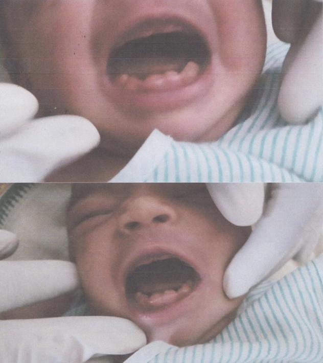
The present study was conducted for a period of seven months and all the children who were born during this period were evaluated. In total 4,341 children were born, out of these only four children were born with natal teeth. From our results we found a female preponderance as all the four neonates were girl child. Type of delivery had no significant relation with the presence of natal teeth as two children were born by caesarean section and two by normal delivery. The most commonly present natal teeth were mandibular anteriors which presented as oedema of the gingival tissue with an unerupted but palpable tooth [Table/Fig-1-b]. Three neonates had only two teeth present but one of them had six teeth at the time of birth [Table/Fig-2]. Most of the children born with the natal teeth were first born child of their parents. All the children born were Hindu [3].
Neonates with mandibular anterior teeth present at the birth.
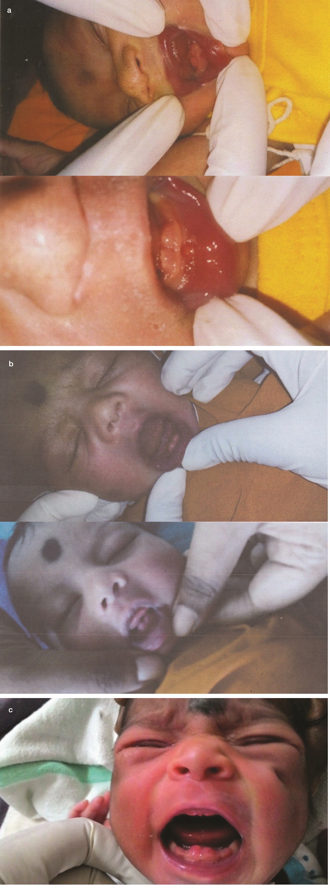
A neonate with six natal teeth.
Discussion
From so many years cases of infant born with natal and neonatal teeth have been reported in the dental literature. The presence of natal teeth is a very rare condition. The aetiology of neonatal teeth is unknown but can be related to several factors such as superposition of germ [11,12], infection or malnutrition [13], eruption accelerated by febrile incidents or hormonal stimulation [14], hereditary transmission of a dominant autosomal gene [15], osteoblastic activity inside the germ area related to the remodeling phenomenon [16] and hypovitaminosis [17]. The natal teeth are present at the time of birth and neonatal teeth erupt 30 days after delivery [1]. The incidence of natal and neonatal is 1:2000 to 1: 3500 and the prevalence is 1:700 to 1:30,000 depending on the type of study [5]. The results of the present study are similar to other reported studies. The incidence of natal teeth in our study was 1:1085.2 which is almost similar to the studies published in past. There is no difference in prevalence between males and females however, in our study we found a female preponderance [18]. The most commonly present teeth were mandibular incisors which are also reported in other studies [7,19]. As described earlier the type of delivery has no relationship with the presence or absence of natal teeth. Clinically, the natal teeth are poorly developed and are small and cone shaped [20]. Rao RS et al., stated that natal teeth are more common in Muslim children which is inconsistent with the findings of the present study [5].
A major complication from natal/neonatal teeth is ulceration on the ventral surface of the tongue caused by the tooth’s sharp incisal edge. This condition is also known as Riga-Fede disease or syndrome [21,22]. The constant ulceration may cause interference in proper suckling and feeding and there are chances of nutritional deficiencies in neonate and cause infants’ failure to gain weight [23]. One of the major complications associated with natal teeth is possibility of swallowing and aspiration which has already been described previously. Other complications are injury to mother’s breast and inconvenience during suckling [3]. Natal teeth may be associated with syndromes like Ellis-van creveld syndrome, Jackson-Lawler, Hallermann-streiff, Steatocystoma multiplex with natal teeth [24].
The treatment includes extraction with topical anaesthetic cream as there is very poor root development. In neonates under the age of 10 days vitamin K levels should be evaluated or prophylactic vitamin K injection should be given before extracting the tooth. The extraction can be done if the tooth is supernumerary, loose or associated with a cleft lip. Most of the time, the doctors prefer conservative management that includes grinding/smoothening of sharp edges of the tooth, changes in feeding technique, composite resin to form a dome shape over the edge of the tooth so the tongue glides over the tooth [24]. Knowing how to manage natal teeth is important for proper well being of a child [19].
Limitation
The results of the present study cannot be generalised as it included only neonates born in that hospital during those seven months. As this was just a pilot study, no follow up was done. Further, studies with large group of children at different places should be conducted in future.
Conclusion
Presence or absence of natal teeth does not depend on the type of delivery or the birth order of the child. Three children out of four were females and the mandibular anteriors were the most commonly present. The children with teeth at the time of birth should be thoroughly examined and proper treatment should be provided at the appropriate age. The parents should be counselled properly in these cases.
[1]. Massler M, Savara BS, Natal and neonatal teeth: A review of 24 cases reported in the literatureJ Pediatr 1950 36:349-59. [Google Scholar]
[2]. Ardeshana A, Bargale S, Karri A, Dave B, Dentitia praecox - natal teeth: a case report and reviewJournal of Applied Dental and Medical Sciences 2016 2(1) [Google Scholar]
[3]. Mhaske S, Yuwanati MB, Mhaske A, Raghavendra R, Kamath K, Saawarn S, Natal and neonatal teeth-an overview of the literature, hindawani publishing corporation ISRN Paediatrics 2013 2013:956269 [Google Scholar]
[4]. Leung AKC, Robson WLM, Natal teeth: a reviewJournal of the National Medical Association 2006 98(2):226-28. [Google Scholar]
[5]. Rao RS, Mathad SV, Natal teeth: Case report and review of literatureJ Oral Maxillofac Pathol 2009 13(1):41-46. [Google Scholar]
[6]. Venkatesh C, Adhisivam B, Natal teeth in an infant with congenital hypothyroidismIndian Journal of Dental Research 2011 22(3):498 [Google Scholar]
[7]. Chowdhary S, Tandon S, Congenital teeth: superstition and reality – a case report and review of literatureInternational Journal of Scientific Study 2014 1(5):53-56. [Google Scholar]
[8]. Kates GA, Needleman HL, Holmes LB, Natal and neonatal teeth: a clinical studyJ Am Dent Assoc 1984 109:441-43. [Google Scholar]
[9]. Baskar S, Deepa G, Incidence of natal and neonatal teeth in infants born from assisted reproductive technology treatments and normal reproductive patterns at tertiary care centre, Chennai. A pilot studyInternational Journal of Pharmacy Practice and Pharmaceutical Sciences 2015 2(11):01-03. [Google Scholar]
[10]. Cunha RF, Boer FAC, Torriani DD, Frossard WTG, Natal and neonatal teeth: review of the literatureAmerican Academy of Pediatric Dentistry 2001 23:2 [Google Scholar]
[11]. Boyd JD, Miles AE, Erupted teeth in ciclops faetusBr Dent J 1951 91:173 [Google Scholar]
[12]. Shafer WG, Hine MK, Levy BM, Distúrbios do desenvolvimento das estruturas bucais e parabucaisIn: Tratado de Patologia Bucal 1985 4a EdRio de JaneiroGuanabara:2-79. [Google Scholar]
[13]. Leung AKC, Natal teethAm J Dis Child 1986 140:249-51. [Google Scholar]
[14]. Bigeard L, Hemmerle J, Sommermater JI, Clinical and ultrastructural study of the natal tooth: enamel and dentin assessmentsJ Dent Child 1996 63:23-31. [Google Scholar]
[15]. Shafer WG, Hine MK, Levy BM, Distúrbios do desenvolvimento das estruturas bucais e parabucaisIn: Tratado de Patologia Bucal 1985 4a edRio de JaneiroGuanabara:02-79. [Google Scholar]
[16]. Jasmin JR, Clergeau-Guerithalt A scanning electron microscopic study of the enamel of neonatal teethJ Biol Buccale 1991 19:309-14. [Google Scholar]
[17]. Anderson RA, Natal and neonatal teeth: histologic investigation of two black femalesJ Dent Child 1982 49:300-03. [Google Scholar]
[18]. Gorlin RJ, Goldman HM, Thoma K, In: Patologia Oral 1973 4th edBarcelonaSalvatore:163-166. [Google Scholar]
[19]. Wang CH, Lin YT, Lin YJ, A survey of natal and neonatal teeth in newborn infantsJournal of the Formosan Medical Association 2016 :pii:S0929-6646(16)30038-39. [Google Scholar]
[20]. Basavanthappa NN, Kagathur U, Basavanthappa RN, Suryaprakash ST, Natal and neonatal teeth: A retrospective study of 15 casesEur J Dent 2011 5(2):168-72. [Google Scholar]
[21]. Buchanan S, Jenkins CR, Riga-Fedes syndrome: natal or neonatal teeth associated with tongue ulceration: Case reportAust Dent J 1997 42:225-27. [Google Scholar]
[22]. Tomizawa M, Yamada Y, Tonouchi K, Watanabe H, Noda T, Treatment of Riga-Fede’s disease by resin-coverage of the incisal edges and seven cases of natal and neonatal teethShoni Shikagaku Zasshi 1989 27:182-90. [Google Scholar]
[23]. Hedge RJ, Sublingual traumatic ulceration due to neonatal teeth (Riga-Fede disease)J Indian Soc Pedod Prev Dent 2005 3:51-52. [Google Scholar]
[24]. Smith DD. Natal and neonatal teeth. Available at: http://www.dermnetnz.org/topics/natal-and-neonatal-teeth [Google Scholar]