The rationale behind the root canal therapy in infected teeth is the elimination of debris, toxins and microorganisms by chemomechanical preparation. Conversely, even after cleaning and shaping, total sterilization of the root canal system remains questionable. It has been known that root canal instrumentation produces a smear layer that covers the surfaces of root canal walls containing both inorganic and organic materials [1]. Keeping or removing the smear layer is a highly controversial issue, as presence of smear layer itself may be infected and could harbor bacteria within the dentinal tubules [2]. This is significant in teeth with infected root canal system where the outcome of the endodontic treatment depends on the elimination of bacteria and their byproducts from the root canal system [3].
Traditionally, myriad of compounds in aqueous solutions have been suggested as root canal irrigants for removal of the smear layer including inert substances such as saline or acids like citric acid, lactic acid, tannic, polyacrylic acid or chelator solutions like bis-dequalinium acetate, EDTA; natural polysaccharide like 0.2% chitosan; broad spectrum antibiotics like tetracyclines and chlorine compounds like sodium hypochlorite [4].
NaOCl (2.25% - 5.25%) is a most commonly used irrigant in endodontic therapy which has not effectively removed the smear layer but effectively dissolves organic tissue [5]. Chelating agents like citric acid and EDTA are highly biocompatible and safe to use but they have little or no antibacterial effect. Natural polysaccharide like 0.2% chitosan has high chelating capacity for metallic ions that might be probably responsible for the depletion of the inorganic portion of the smear layer [6].
Researchers have proved the efficacy of various auxiliary chemical substances on permanent teeth, but their effects on primary dentition are not widely known. Therefore, the present study was done to evaluate the efficacy of four root canal irrigants individually on smear layer removal in primary tooth root canals after hand instrumentation.
Materials and Methods
Prior to the commencement of this in vitro study, Institutional Ethical Committee approval was obtained. A total number of 40 retained extracted primary anteriors with intact roots or with at least 2/3rd roots were collected for the study. Teeth with curved roots, less than 1/3rd roots and those that are endodontically treated were excluded from the study. Following extraction, teeth were cleaned by removing the remaining soft tissue and stored in 0.9% saline at 4°C till further use.
Superficial grooves were placed in a mesiodistal direction using diamond disk along the longitudinal axis of tooth in cementum which were not extending to the root canal. Profuse and constant irrigation was done with saline to facilitate smooth split. Endodontic access was obtained and a size 10 K-file was placed into the root canal until the tip was just visible at the apical foramen and the working length was determined by reducing 1 mm from the length recorded when the file was visible at the apex. Further instrumentation was done according to the conventional step back preparation from size 15 – 45 K files. During instrumentation, the canals were irrigated with 3 ml saline between every instrument change using 25 gauge needles kept at a depth of 2 mm from the working length.
After completion of preparation, four subgroups were made and had ten teeth each, as Group I: 5.25% NaOCl (Asian Acrylates, Mumbai, India), Group II: 6% citric acid solution (freshly prepared), Group III: smear clear (Sybron Endo, CA, USA) and Group IV: 0.2% chitosan (Yaizu Suisankagaku Industry Co. Ltd. Japan) and a final irrigation was done with 10 ml of respective irrigating solutions for 1 min and dried with paper points. Then the roots were split along the longitudinal axis using a chisel through the grooves placed previously and only one undamaged half from each sample with full root length were transferred to the testing lab in a sterile plastic container.
The exposed surfaces were then mounted on a metallic stub, gold sputtered and examined at cervical, middle and apical regions under Scanning Electron Microscope (SEM) at a magnification of X1000 and the representative digital photomicrographs from each root third were taken. These photomicrographs were evaluated individually by two examiners (kappa value of 0.86) who were blind to the irrigation regimens and attributed scores according to the criteria given by Rome WJ et al., [7]. in 1985 [Table/Fig-1].
Scoring criteria given by Rome WJ et al., [7].
| Score | Criteria |
|---|
| 0 | No smear layer, all dentinal tubules open and no erosion of tubules. |
| 1 | No smear layer, all dentinal tubules open and erosion of tubules. |
| 2 | Minimum smear layer; > 50% dentinal tubules visible. |
| 3 | Moderate smear layer; < 50% of dentinal tubules open. |
| 4 | Heavy smear layer; outline of dentinal tubules obliterated. |
Statistical Analysis
The data obtained were subjected to statistical analysis using Kruskal Wallis Anova test and Post-hoc Mann Whitney U test.
Results
The results obtained from this study are summarized in [Table/Fig-2,3,4,5,6 and 7] showing the scanning electron photomicrographs of the tested irrigants. Complete removal of the intracanal smear layer was not found with any of the tested root canal irrigants. According to this study, Group II exhibited better efficacy in removing smear layer without altering the normal dentinal structures with lowest mean scores (p<0.001) followed by Group III, Group IV and Group I. There was no stastistically significant difference (p<0.05) between the scores at each root third (cervical, middle, apical) for all groups, though apical third scores were less than the other root thirds.
Comparison of efficacy by four irrigants on removal of smear layer.
| Root third | Groups | Mean | SD | f-value | p-value |
|---|
| Cervical third | I | 2.60 | 0.52 | 31.467 | 0.001* |
| II | 0.40 | 0.52 |
| III | 1.30 | 0.67 |
| IV | 2.40 | 0.52 |
| Middle third | I | 2.60 | 0.52 | 21.87 | 0.001* |
| II | 0.90 | 0.32 |
| III | 2.20 | 0.42 |
| IV | 2.30 | 0.48 |
| Apical third | I | 3.20 | 0.79 | 21.72 | 0.001* |
| II | 1.20 | 0.42 |
| III | 2.30 | 0.48 |
| IV | 3.10 | 0.57 |
Kruskal Wallis Anova test. p>0.05 (not significant), p<0.05 (significant), p<0.001 (highly significant)* SD-Standard deviation
Multiple pair wise comparisons between groups at cervical third, middle third and apical third.
| Group | Mean at cervicalthird (S.D) | p-value | Mean atmiddlethird (S.D) | p-value | Mean atapical third(S.D) | p-value |
|---|
| I | 2.60 (0.52) | 0.001* | 2.60 (0.52) | 0.001* | 3.20 (0.79) | 0.001* |
| II | 0.40 (0.52) | 0.92 (0.32) | 1.20 (0.42) |
| I | 2.60 (0.52) | 0.001* | 2.60 (0.52) | >0.05 | 3.20 (0.79) | 0.001* |
| III | 1.30 (0.67) | 2.20 (0.42) | 2.30 (0.48) |
| I | 2.60 (0.52) | >0.05 | 2.60 (0.52) | >0.05 | 3.20 (0.79) | >0.05 |
| IV | 2.40 (0.52) | 2.30 (0.48) | 3.10 (0.57) |
| II | 0.40 (052) | 0.001* | 0.92 (0.32) | 0.001* | 1.20 (0.42) | 0.001* |
| III | 1.30 (0.67) | 2.20 (0.42) | 2.30 (0.48) |
| II | 0.40 (0.52) | 0.001* | 0.92 (0.32) | 0.001* | 1.20 (0.42) | 0.001* |
| IV | 2.40 (0.52) | 2.30 (0.48) | 3.10 (0.57) |
| III | 1.30 (0.67) | 0.001* | 2.20 (0.42) | >0.05 | 2.30 (0.48) | 0.001* |
| IV | 2.40 (0.52) | 2.30 (0.48) | 3.10 (0.57) |
Post-hoc Mann Whitney U test (p≤0.05 significant)
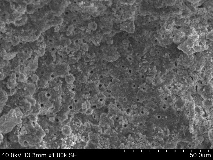
SEM view after sodium hypochlorite irrigation;
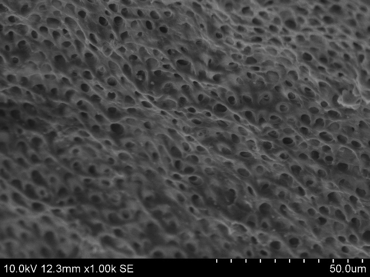
SEM view after citric acid irrigation.
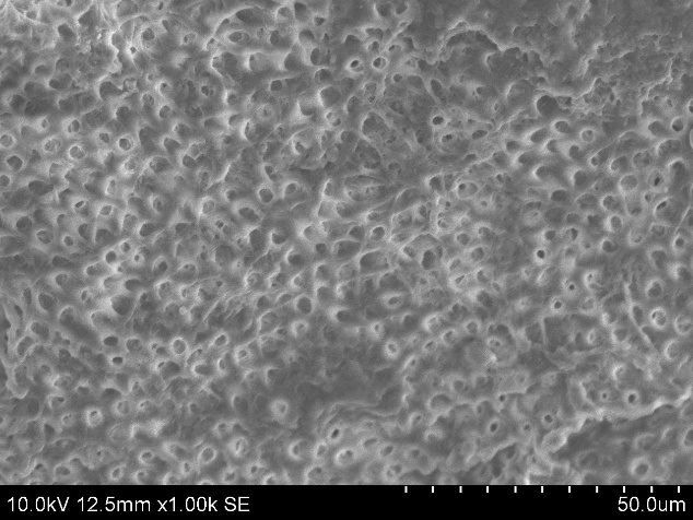
SEM view after smear clear irrigation;
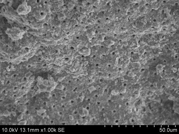
SEM view after chitosan irrigation.
Discussion
Cleaning, decontamination, shaping and enlarging the root canal system of the primary teeth is essential as the canal needs to be filled with the nonsetting pastes. To minimize the bacterial contamination and prevent the reinfection of the root canal system, these pastes must penetrate the tubules. Smear layer presence may compromise the quality of the root canal filling as it may delay or prevent the penetration of endodontic irrigants and intracanal medications and also interfere with the adhesion of root canal sealers to the root canal walls [2,8]. Therefore, removal of smear layer is often inevitable for success of endodontic treatment and is achieved with use of various chemical irrigants during root preparation.
The purpose behind irrigation of a root canal is to dissolve the organic component, the debris and demineralise the inorganic component [9]. Apart from type of irrigant used other factors that can influence the process of irrigation include extent of instrumentation, quantity and temperature of irrigation solution, canal diameter, length of time of contact, type and gauge of irrigating needle and depth of penetration of irrigating needle [10]. With so many variables affecting its function, till to date no single irrigant is effective in removing both organic and inorganic material. Thus, the present study intended to find out efficacy of the four root canal irrigants on removal of smear layer.
NaOCl is a popular irrigant for the excellent lubricant action and broad spectrum of antibacterial activity and its capacity to dissolve organic tissue. It has also been suggested that higher the concentration, the better the antibacterial and tissue dissolution properties, hence, 5.25% NaOCl was used for this study [11]. However, the scanning electron microscopic pictures of NaOCl in the present study showed the absence of superficial debris with the presence of smear layer at all root thirds, signifying the inability of 5.25% NaOCl in complete removal the smear layer. These results were similar with Yamada RS et al., and Baumgartner JC et al., suggesting that 5.25% NaOCl was competent in removing organic and loose superficial debris, leaving exposed inorganic component of smear layer preventing its further removal [12,13].
Citric acid is a weak organic acid belonging to the chelate agents. It is used in periodontal therapy for conditioning dentin and restorative dentistry. Decalcified capacity of citric acid was due to chelation of Ca2+ ions and acidity of the solution. Citric acid solutions are endorsed for endodontic use at larger concentrations (25% and 50%), whereas, the latest researches bring more data about the efficient performance of the weaker solutions of citric acid (6-19%) and that’s the reason for cosidering 6% citric acid solution in this study [14]. In the present study, 6% citric acid used was found to be efficient in significant removal of smear layer exposing the dentin tubules than other irrigant groups, except some debris similar to crystals that are spread over the dentinal surface in the apical root third of few samples. Inability of the irrigant to penetrate deep into the apical part of the root canal might be because of its high surface tension. These results were in accordance with Hariharan VS, et al., showing the superior efficacy of 6% citric acid than saline, 5.25% NaOCl, 10% EDTA and 2% chlorehexidine on removing the smear layer in primary teeth root canals [15] and Yamaguchi M et al., stating that the solutions of 0.5, 1, 2 M citric acid showed antimicrobial effects against the facultative and obligative anaerobes suggesting citric acid may possibly be used as irrigating solutions for root canals [16].
Smear clear is composed of 17% EDTA, cetrimide and two additional proprietary surfactants. To improve the efficacy of irrigant, a quaternary ammonium salt cetrimide (cetyltrimethylammonium bromide) and a cationic detergent were added [17]. Cationic surfactants are known to increase the penetration of irrigating solutions into the dentinal tubules, as it reduces the surface tension and fluid viscosity, thus enabling the chelating solution to be carried more easily to the full depth of the canal and it also has a bactericidal and fungicidal properties. Therefore, smear clear was chosen in this study [18]. In the present study, scanning electron microscopic pictures of smear clear showed efficient smear layer removal in the cervical third but the middle and apical third elicited higher quantity of smear layer covering the dentin walls and lesser number of exposed dentin tubules. Study conducted by Seddigheh K et al., [19]. also revealed that irrigation with smear clear efficiently removed smear layer from coronal thirds of the canals than middle and apical thirds. Zehnder M et al., stated that reducing the surface tension of endodontic chelator solutions did not improve their calcium chelating ability [20].
Chitosan is a natural polysaccharide prepared by the deacetylation of chitin obtained from the shells of crabs and shrimps and endowed with properties of biocompatibility, biodegradibility, bioadhesion and atoxicity to human cells [21,22]. High chelating capacity for different metallic ions and its low cost, made it preferred as an irrigant for the study [23]. Under the Scanning electron microscopic view, 0.2 % chitosan was found to be ineffective in removing the smear layer in all the three regions of the root surface, but found to be effective than 5.25% NaOCl. It is well reported that the effectiveness of a chelating agent depends on numerous factors including pH concentration, application time and quantity of the solution [24]. Darrag AM and Silva PVet al., revealed that application of the 0.2% of chitosan solution for 3-5 min was the most viable combination for use on the root dentin whereas, less application time might be as certained for the ineffectiveness of chitosan in the present study [25,26].
Analysis of the dentinal walls of all the specimens demonstrated that cleaning have been more effective on coronal and middle thirds than on the apical third, this may be due to reason that size of the canals in these thirds, allowed better circulation and action of irrigating solution. Outcomes may vary while using root canal irrigants to remove smear layer in in vivo, as the root canals are usually wet and the surface tension of the endodontic solutions may not play a role in their penetration ability. Nevertheless, these in vitro results cannot be extrapolated completely to invivo situations. Hence, further research is required and more in vivo studies need to be done to evaluate these root canal irrigants in detail regarding its physical, chemical, biological and antimicrobial properties in order to verify the benefits and consequences to humans.
Conclusion
In conclusion, the present in vitro results of scanning electron microscopic view demonstrated that 6% citric acid can potentially remove smear layer in primary root canals when used as final irrigant after hand instrumentation.
Kruskal Wallis Anova test. p>0.05 (not significant), p<0.05 (significant), p<0.001 (highly significant)* SD-Standard deviation
Post-hoc Mann Whitney U test (p≤0.05 significant)