Microleakage at the tooth/restoration interface is considered to be a major factor influencing the longevity of the restoration. Adequate adhesion and endurance (longevity) of dentin bonding have always been the topic of interest for dentistry. Microleakage from a restoration can act as a seed sower for the secondary caries to develop and gradually leading to treatment failure. This allows bacteria and oral fluids to invade the resin dentin border hence deteriorating the bond area causing teeth sensitivity, secondary caries or pulp inflammation. So an impervious sealing of the cavity is utmost mandatory for durable composite restoration [1,2].
Cervical restorations are ever challenging because of difficulties in moisture control, caries access, and proximity to gingival margin. Both mechanical and the non mechanical factors act to hinder the longevity of the cervical restoration like microshear forces exerted during mastication and the extent of caries. Newer materials are readily introduced in the market with improved chemicomechanical properties, longevity, patient safety and comfort [3].
Adhesive systems are classified as “total etch” or “self etch” depending on their procedure of application and mechanism of adhesion. Several studies have shown that water trees, water bubbles, phase separation and incomplete polymerization of monomer occurred in the adhesive interface of all-in-one and one-bottle self etch adhesives [4]. These occurrences are not typical for traditional hydrophobic adhesives (i.e., total etch or self etch primer adhesives). Furthermore, the amount of nanoleakage within the bonding resin layer is large for one step self etching adhesives due to their increased concentrations of water and solvent (acetone or ethanol) [5].
Increase in the number of geriatric patients has contributed to an increase incidence of cervical caries, and microleakage is more critical in class V cavities because of high C factor [6,7]. An array of research has lead to advent of newer material which promises to have added advantages and minimized disadvantages in contrast to total etch and self etch. Hence, aim of this in vitro study was to evaluate the sealing ability of total etch, self etch and universal adhesive systems in class V restorations at occlusal and gingival margin using dye penetration method. The research hypothesis was that there exists a significant difference in the extent of microleakage between tooth and restoration interface in class V composite resin restorations using different bonding agents.
Materials and Methods
In this in vitro study 120 caries free human maxillary and mandibular premolar teeth indicated for orthodontic extraction, from 30 subjects of age group 14 to 20 years were collected from outpatient department of Oral and Maxillofacial Surgery, Rungta College of Dental Sciences and Research, Bhilai, Chattisgarh, India. Selected teeth were stored in sodium hypochlorite for one week for disinfection and then stored in normal saline to prevent dehydration. Calculus and stains on specimen were removed by ultrasonic scaling and then they were cleaned with pumice using rubber cups to remove any residual tissue tags. Each tooth was then mounted on plaster of paris. Class V cavity was prepared on the facial surface of each tooth using high speed airotor handpiece and straight fissure diamond bur, under water spray coolant. Standardized preparations were obtained by making cavity preparations that were approximately 3 mm wide, 1.5 mm deep and 2.5 mm high parallel to the Cemento-Enamel Junction (CEJ). The gingival half of the preparation was extended 1 mm below the CEJ. No bevels were used in the preparation.
Each preparation was rinsed with distilled water for 20 seconds and dried using filter paper. The teeth were then divided into four groups of 30 teeth each and bonding agent was applied [Table/Fig-1].
Experimental groups included in the study.
| Groups | Sample size(Teeth) | DentinBonding Agent | Generation | Application |
|---|
| A | 30 | AdperTM Single Bond 2 (3M ESPE) | 5th | Etchant was applied with a syringe, after waiting for 15 seconds it was rinsed with water and cavity was blot dried with filter paper. Single coat of Adper Single Bond (fifth generation) bonding agent was applied with applicator tip. Gently air was blown followed by second coat of bond. Light curing was done for 40 seconds. Application of Tetric N Ceram composite with composite handling instrument in horizontal increments of 2 mm each. Each increment was light cured for 40 seconds. |
| B | 30 | AdperTM SE Plus (3M ESPE) | 6th | Self Etch Primer was applied with a microbrush for 15 seconds followed by gentle air dispersion. And then light cured for 20 seconds. Adhesive Adper SE Plus (sixth generation) was applied with a microbrush followed by gentle air dispersion. And then light cured for 20 seconds. Tetric N Ceram composite was applied using same technique. |
| C | 30 | AdperTM Easy One (3M ESPE) | 7th | Adhesive (seventh generation) was applied with a microbrush followed by gentle air dispersion. And then light cured for 20 seconds. Tetric N Ceram composite was applied using same technique. |
| D | 30 | AdperTM Single Bond Universal (3M ESPE) | 8th | Adhesive (eighth generation) was applied with a microbrush followed by gentle air dispersion. And then light cured for 20 seconds. Tetric N Ceram composite was applied using same technique. |
Polishing was done with disks for 20 second each for coarse, medium, fine and ultrafine disks. The specimens were stored for 24 hours in distilled water. Thermocycling of 200 cycles was carried out at 5°C to 55°C, 60 second dwell time and five second transfer time at Low (LG Make) and high (Mahavir Make) Temperature Chamber respectively.
After thermocycling, apical 2 mm of teeth were sealed with a layer of sticky wax and all tooth surfaces were covered with two coats of nail polish with the exception of 1 mm around the tooth restoration interface. The teeth were then immersed in 2% methylene blue dye for 24 hours.
After staining, the teeth were rinsed with distilled water in order to remove any residual stain and the radicular parts of the teeth were cut horizontally 4.5 mm below the CEJ. Coronal parts were sectioned buccolingually in the approximate center of the restoration with a low speed diamond disc mounted in mandrill and jig.
Microleakage was assessed for both occlusal (enamel) and gingival (cementum) margins, using a stereomicroscope at a magnification of 16X and images were taken using a digital camera. The depth of dye penetration was observed according to the following criteria [8]:
0: No evidence of dye penetration;
1: Dye penetration along interface to half of cavity depth;
2: Penetration greater than half, not including axial wall;
3: Penetration involving axial wall but not pulp;
4: Penetration involving pulp.
Statistical Analysis
A non parametric analysis of ANOVA i.e., Kruskal-Wallis was used to determine whether there were significant differences among the groups. Intragroup comparison was made using Dunn’s procedure for non parametric data. Occlusal and gingival margins within the treatment groups were compared using the Mann-Whitney test. The level of significance was established as p<0.05 for all tests.
Results
Microleakage Scores at the occlusal margin [Table/Fig-2,3]: When comparison was made between mean microleakage scores of different materials at occlusal margin, AdperTM Easy exhibited the least microleakage (mean=1.23) whereas AdperTM SE Plus exhibited more microleakage (mean=2.10). Intragroup comparison revealed that there was a statisticaliy significant difference in microleakage among AdperTM SE Plus and AdperTM Easy One adhesive systems. (p=0.03 S, p<0.05)
Distribution of microleakage scores at the occlusal margins.
| Groups | Mean | SD | % of nomicroleakage |
|---|
| Single Bond | 1.36 | 1.35 | 40 |
| Adper SE | 2.10 | 1.18 | 13.3 |
| Adper Easy | 1.23 | 1.04 | 23.3 |
| Universal | 1.40 | 1.19 | 26.7 |
Kruskal-Wallis test applied: χ2-value=8.62, p-value=0.035, Significant
Post-Hoc Kruskal Wallis test (Dunn’s Procedure) applied for intragroup comparison of microleakage score at occlusal margin.
| Groups | p-value | 95% Confidence Interval |
|---|
| Lower Bound | Upper Bound |
|---|
| Single Bond | Adper SE | 0.088 | -1.53 | 0.07 |
| Adper Easy | 0.973 | -0.67 | 0.93 |
| Universal | 1.000 | -0.83 | 0.77 |
| Adper SE | Adper Easy | 0.030 * | 0.06 | 1.67 |
| Universal | 0.112 | -0.10 | 1.50 |
| Adper Easy | Universal | 0.949 | -0.97 | 0.63 |
p-value < 0.05 = Statistically significant*.
Microleakage scores at the gingival margin [Table/Fig-4,5]: There was no statistically significant difference observed between all the groups (p-value=0.97 NS, p>0.05). When comparison was made between mean microleakage scores of different material at gingival margin AdperTM SE Plus exhibited the least microleakage (mean=2.6) whereas, AdperTM Easy One exhibited the more microleakage (mean=2.66).
Distribution of microleakage scores at the gingival margins.
| Groups | Mean | SD | % of no microleakage |
|---|
| Single Bond | 2.63 | 0.92 | 3.3 |
| Adper SE | 2.60 | 1.07 | 10 |
| Adper Easy | 2.66 | 1.06 | 6.7 |
| Universal | 2.60 | 1.00 | 10 |
Kruskal-Wallis test applied: χ2-value=0.23, p-value=0.97, Not significant.
Post-Hoc Kruskal Wallis test (Dunn’s Procedure) applied for intragroup comparison of microleakage score at gingival margin.
| Groups | p-value | 95% Confidence Interval |
|---|
| Lower Bound | Upper Bound |
|---|
| SingleBond | Adper SE | 0.999 NS | -0.65 | 0.71 |
| Adper Easy | 0.999 NS | -0.71 | 0.65 |
| Universal | 0.999 NS | -0.65 | 0.71 |
| Adper SE | Adper Easy | 0.994 NS | -0.75 | 0.61 |
| Universal | 1.000 NS | -0.68 | 0.68 |
| Adper Easy | Universal | 0.994 NS | -0.61 | 0.75 |
p-value < 0.05 = Statistically significant*.
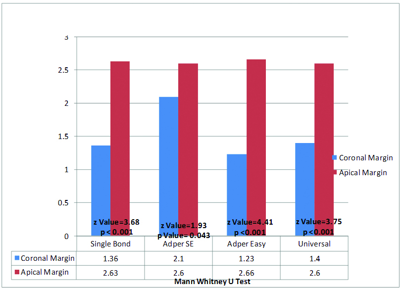
Comparison between microleakage scores at the occlusal and gingival margin [Table/Fig-6]: On intergroup comparison of mean microleakage score between occlusal and gingival margin statistically highly significant difference was observed with AdperTM Single Bond 2, AdperTM Easy One and AdperTM Single Bond Universal (p<0.001) whereas, in Group AdperTM SE Plus, the difference was statistically significant (p<0.05) which suggests that, microleakage at the gingival margin was more than the occlusal margin for all the materials.
Comparision between microleakage scores at occlusal and gingival margins.
Discussion
In the era of evolving biomedical sciences and that too when restorative dentistry is emphasized upon, the bonding agents have evolved in a major manner with advances being introduced every now and then. The factor which definitely triggered this evolution was the “need” be it on the patient’s perspective like aesthetics or on the dentist perspective like longevity, high bond strength and “minimally invasive” or “minimum intervention” care [9].
Sealing is the most concerned property when bonding agent is to be used. Adhesion (sealing) is merely proportional to microleakage. The literature has proved that three main factors could affect the sealing. One factor is the composite polymerization shrinkage that induces stress at the bonding interface. Second factor is that the substrate is a biological tissue, which makes adhesion difficult. Third factor is chemical composition of adhesive itself [8].
According to the results obtained in present study, it was observed that Group C showed least microleakage, followed Group A and Group D and Group B showed highest microleakage score at occlusal margin. The results were in accordance with studies by Nair M et al., Tabari M et al., and Kambale S et al., [10-12].
In Self Etch adhesives the acidic characteristics of the active monomers are responsible for dissolving the smear layer and demineralizing the underlying dentin. This demineralization is self-limiting because the acidity of the monomers is gradually buffered by the mineral content of the dentin. This implies that the resultant morphological aspect of the bonded interface is largely dependent on the characteristics of the dentin to which the adhesive is being applied and on the aggressiveness of the acidic monomers. Recently, ultramild Self Etch adhesives (Clearfil S3 Bond, Kuraray Inc., Japan: AdperTM Easy One and AdperTM Single Bond Universal, 3M ESPE, USA) have become available that present lower acidity (pH>2.5). Strong Self Etch systems dissolve the smear layer completely similar to etch and rinse while ultramild leaves the tubules intact with smear plug. The partial demineralization resulting from mild and ultramild Self Etch systems has reported to be an advantage because of the possibility of chemical interaction between some functional monomers (such as MDP and 4-META) and the remaining hydroxyapatite crystals along the collagen fibrils. It has been claimed that this chemical bonding results in the improved bond durability reported for these systems [13]. Thus, the success of Self Etch adhesives is largely related to their simplicity of use and to the theoretical ability to etch and infiltrate simultaneously, thus preventing discrepancies between demineralization and infiltration [14].
Self Etch agents consist of acidic monomers, which get activated in presence of water and starts demineralization process. Strong Self Etch agents contain higher amounts of water that remains within the adhesive interface, and is difficult to remove. The poor performance of AdperTM SE Plus may be due to the water/acidic monomer ratio in an aqueous mixture, which might not be stable for each application. Therefore, the increase or decrease in water concentration may affect the degree of ionization of the acidic monomer [10].
Moreover, AdperTM SE Plus has only water as solvent and contains Tri Ethylene Glycol Dimethacrylate (TEGDMA), which absorbed more amount of water after polymerization than Bisphenol-Glycidyl Methacrylate (BIS-GMA), which might explain its high microleakage score due to residual water, which can cause phase separation, osmotic blistering, polymerization inhibition, and reduced shelf life. Also, water is a poor solvent for organic compounds such as monomers. AdperTM Easy One and AdperTM Single Bond Universal have secondary solvent ethanol, which due to its high solubility parameter and osmotic pressure helps in displacement of residual water and carry the polymerizable monomers into the opened dentin tubules [15].
In our study, Total Etch adhesive (AdperTM Single Bond 2) showed more microleakage than Single Step Self Etch (AdperTM Easy One). This finding was in accordance with studies by Tabari M et al., Pushpa R et al., and Pontes DG et al., [11,16,17].
Water is necessary to maintain collagen fibril expansion in etch and rinse for resin infiltration but on contrary it plays antagonist role in hybrid layer formation. This reduces the mechanical properties of the interface and reduces the durability of the bonded surface. Thus, uneven stress distribution along the components of the hybridized zone causes enzymatic degradation of collagen fibrils that were left exposed, and the hydrolysis of the poorly formed adhesive polymer [18,19]. Ethanol wet bonding technique, has been proposed to avoid problems of traditional wet bonding technique, which removes excess water and maintains the interfibrillar spaces for resin infiltration [13].
The adhesives used, sealed the occlusal margin far better than the gingival margin, reason for this may be attributed to the presence of higher organic component, tubular configuration, fluid pressure and the lower surface energy of dentin which make bonding relatively difficult than enamel [20,21]. Another factor is great magnitude of polymerization shrinkage which cannot get compensated by water sorption and stress relaxation [22].
Limitation
Like all researches pertaining to biomedical sciences the present study also comes out with certain limitations. As per our assessment scanning electron microscopy and conofocal laser scanning microscopy rather than stereomicroscopy would yield more authentications. As conditions in the oral cavity are always different from outside environment much attention was employed to duplicate the same in vitro, however it is impossible to create such environment outside the oral cavity in-toto, which could in turn have lead to certain unavoidable bias.
But for an important and significant research pertaining to microleakage of bonding agents in composite restorations the above stated limitations can be overlooked upon when considered for the cutting edge conclusion provided by the present study. The research clearly indicates outstanding superior and efficient property of AdperTM Easy One and AdperTM Single Bond Universal over the other materials in controlling microleakage, which thereby significantly adds to the longevity and strength of restorations which is the cornerstone principle of restorative dentistry. We strongly recommend further in vivo studies to be conducted to add more insight into this research and to evaluate other clinical parameters which cannot be judged in vitro for example postoperative sensitivity and marginal discolouration occurring in composite restorations.
Conclusion
Under the umbrella of strong results and future recommendations this study makes a point to prove some valid conclusions. Still all adhesives under investigation exhibited significant microleakage to some or the other extent. Higher degree of microleakage was observed at gingival margin compared to occlusal margin. Self Etch AdperTM Easy One excelled among the other at the occlusal margin whereas, AdperTM SE Plus farewell at the gingival margin.
Kruskal-Wallis test applied: χ2-value=8.62, p-value=0.035, Significant
p-value < 0.05 = Statistically significant*.
Kruskal-Wallis test applied: χ2-value=0.23, p-value=0.97, Not significant.
p-value < 0.05 = Statistically significant*.