Auxanographic Carbohydrate Assimilation Method for Large Scale Yeast Identification
Suganthi Martena Devadas1, Mamatha Ballal2, Peralam Yegneswaran Prakash3, Manjunath H Hande4, Geetha V Bhat5, Vinitha Mohandas6
1 Research Scholar, Department of Enteric Diseases Division, KMC, Manipal University, Manipal, Karnataka, India.
2 Professor, Department of Enteric Diseases Division, KMC, Manipal University, Manipal, Karnataka, India.
3 Associate Professor and Incharge-Medical Mycology Laboratory, Department of Microbiology, KMC, Manipal University, Manipal, Karnataka, India.
4 Professor and Head, Department of Medicine, KMC, Manipal University, Manipal, Karnataka, India.
5 Microbiologist and Infection Control Officer, Department of Microbiology, Bhagwan Mahaveer Jain Hospital, Bengaluru, Karnataka, India.
6 Professor, Department of Microbiology, Bangalore Institute of Dental Sciences, Bengaluru, Karnataka, India.
NAME, ADDRESS, E-MAIL ID OF THE CORRESPONDING AUTHOR: Dr. Mamatha Ballal, Professor, Department of Microbiology, Enteric Diseases Division – Incharge, Central Research Lab, Kasturba Medical College - Manipal, Manipal University, Madhav Nagar, Manipal-576104, Karnataka, India.
E-mail: mamatha.ballal08@gmail.com
Introduction
The auxanographic carbohydrate assimilation had been an important method for differentiation of yeasts. Prevailing methods described in the literature for carbohydrate assimilation has limited scope for use in large scale yeast identification.
Aim
To optimize the large scale auxanographic carbohydrate assimilation method for yeast identification.
Materials and Methods
A modified auxanographic carbohydrate assimilation method was developed and a total of 35 isolates of Candida species comprising of four ATCC (American Type Culture Collection) Candida strains (Candida albicans ATCC 90028, Candida tropicalis ATCC 90018, Candida parapsilosis ATCC 750, Candida krusei ATCC 6258) and 31 clinical isolates of Candida tropicalis (n=13), Candida krusei (n=7), Candida glabrata (n=3), Candida kefyr (n=3), Candida albicans (n=5) were validated. The carbohydrates tested were Glucose, Sucrose, Maltose, Lactose, Cellubiose, Raffinose, Trehalose, Xylose, Galactose and Dulcitol.
Results
A total of 35 Candida species were tested for their carbohydrate assimilative property and the results were consistent with the existing standard protocols. A well circumscribed opaque yeast growth indicated assimilation of the test carbohydrate and translucent to opalescent growth with the outline of initial inoculum alone indicated lack of assimilation. The control plate indicated no growth of the Candida species.
Conclusion
The carbohydrate assimilation tests finds utility for yeast diversity studies exploring novel ecological niches. The technique described here facilitates testing of an extended range of carbohydrates and yeasts in a cost effective manner.
Candida, Control plate, Ecological niche
Introduction
Yeasts comprise of a diverse group of fungi with immense industrial significance and pathogenic implication. Primary identification of yeasts is achieved by an assortment of morphological, biochemical and physiological methods [1-3]. Yeast carbohydrate assimilation technique remains one of the most common and widely used for definitive identification of yeasts in resource limited diagnostic laboratories and research settings. It relies on the aerobic utilization of carbon or nitrogen as the sole source for the metabolic process. The results are evident by growth or pH shift in the solid or liquid medium which is used for assimilation. The ability to assimilate various carbohydrates such as; Glucose (Glu), Sucrose (Suc), Maltose (Mal), Lactose (Lac), Cellubiose (Cel), Raffinose (Raff), Trehalose (Tre), Xylose (Xyl), Galactose (Gal), Melibiose (Mel) and Dulcitol (Dul) enable the distinction among yeast species [4]. Carbohydrate assimilation reactions are one among the primary tests used to differentiate genera and species of yeasts.
With the advent of an auxanographic technique for carbohydrate assimilation, several modified methods such as dye pour-plate technique, sugar-impregnated disk method, turbid metric broth system, agar slant technique have been developed for improvisation and definitive results. Recent advances in yeast assimilative techniques have included a modified auxanogram using sugar-impregnated disks and a turbidimetric system in a broth, Adams-Cooper technique [5] which is modified Wickerham and Burton technique of agar slant and the addition of a pH indicator dye. All these aforementioned techniques provide a quick, decipherable and reproducible method for yeast identification in small laboratories with good correlation of growth with indicator change, easy to prepare and determine, cost effective [6]. Thus, the aim of the present study was to optimize the large scale auxanographic carbohydrate assimilation method for yeast identification.
Materials and Methods
The study was carried out at the Enteric Diseases Division, Department of Microbiology, Kasturba Medical College, Manipal from August 2015 to February 2016. The carbohydrate assimilation test was done by preparing a basal media with 6.75% Yeast Nitrogen Base (YNB) (Hi Media Pvt., Ltd.) and 2% agar. Further a working solution of 2 ml of the YNB and 18 ml of the agar aliquoted to sterile bottles. A 2 ml each of 10% carbohydrate solutions; Glu, Suc, Mal, Lac, Cel, Raff, Tre, Xyl, Gal, Dul (HiMedia Laboratories Pvt. Ltd) was dispensed to the sterile bottles containing the basal media and moist heat sterilised. This was poured at 4 mm thickness aseptically to 90mm petriplates and allowed to solidify and sterility was checked. The yeast for testing - Candida albicans ATCC 90028, Candida tropicalis ATCC 90018, Candida parapsilosis ATCC 750, Candida krusei ATCC 6258 and clinical isolates of Candida tropicalis (n=13), Candida krusei (n=7), Candida glabrata (n=3), Candida kefyr (n=3), Candida albicans (n=5) were inoculated and checked for purity on Sabourauds Dextrose Agar (SDA) (HiMedia Laboratories Pvt., Ltd.) for 24 hours at 35°C. The carbohydrate containing medium was inoculated with 10 μl of the yeast inoculum 12 x 108 cells calibrated with Mc Farland Standard No. 4, incubated at 300C for 24 and 48 hours and observed for growth. A carbohydrate free medium was used as the control, where the absence of yeast growth was considered satisfactory. Assimilation was interpreted positively if there was growth in the plate containing the carbohydrate and negative if there was no growth [Table/Fig-1]. All tests were performed in triplicates and wherever an inconclusive result consisting of single colonies of the yeast was observed the test was considered indeterminate and repeated.
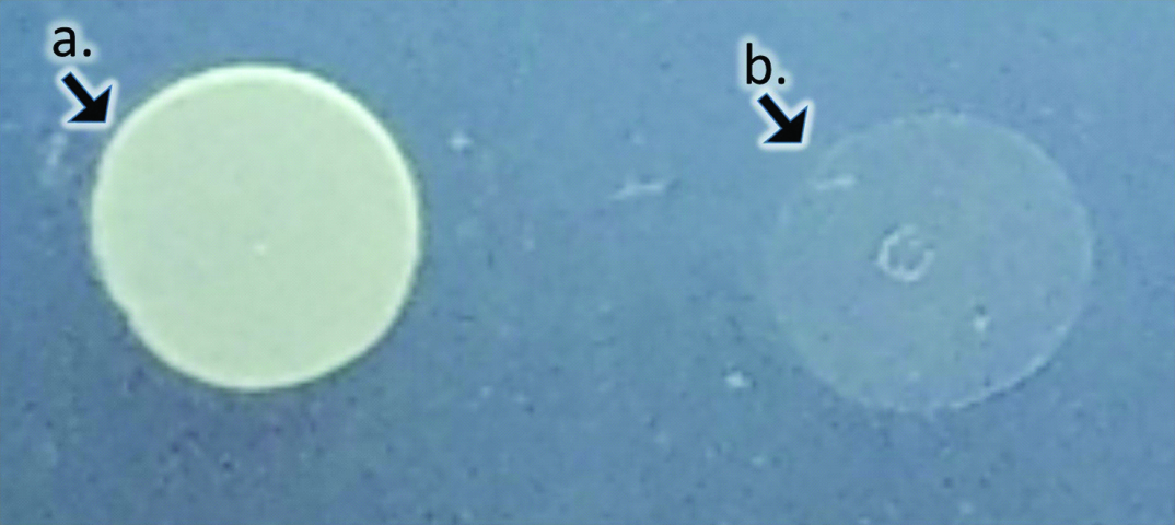
Interpretation for auxanographic carbohydrate assimilation technique: a) Well circumscribed opaque yeast growth indicative of assimilation of the test carbohydrate; b) Translucent to opalescent growth with the outline of initial inoculum alone indicative of lack of assimilation.
Results
The test results following incubation for selected carbohydrates along with control is shown in [Table/Fig-2]. The auxanographic carbohydrate assimilation profiles for; C.albicans, C.tropicalis, C.krusei, C.parapsilosis, C.glabrata, C.kefyr is presented in [Table/Fig-3]. Out of all the carbohydrates assimilated, Candida glabrata assimilated glucose and trehalose; C.krusei assimilated glucose only. Lactose is assimilated only by Candida kefyr. Cellobiose assimilation is positive for C. tropicalis which differentiates it from C. albicans and C. parapsilosis. Dulcitol is not assimilated by these Candida species.
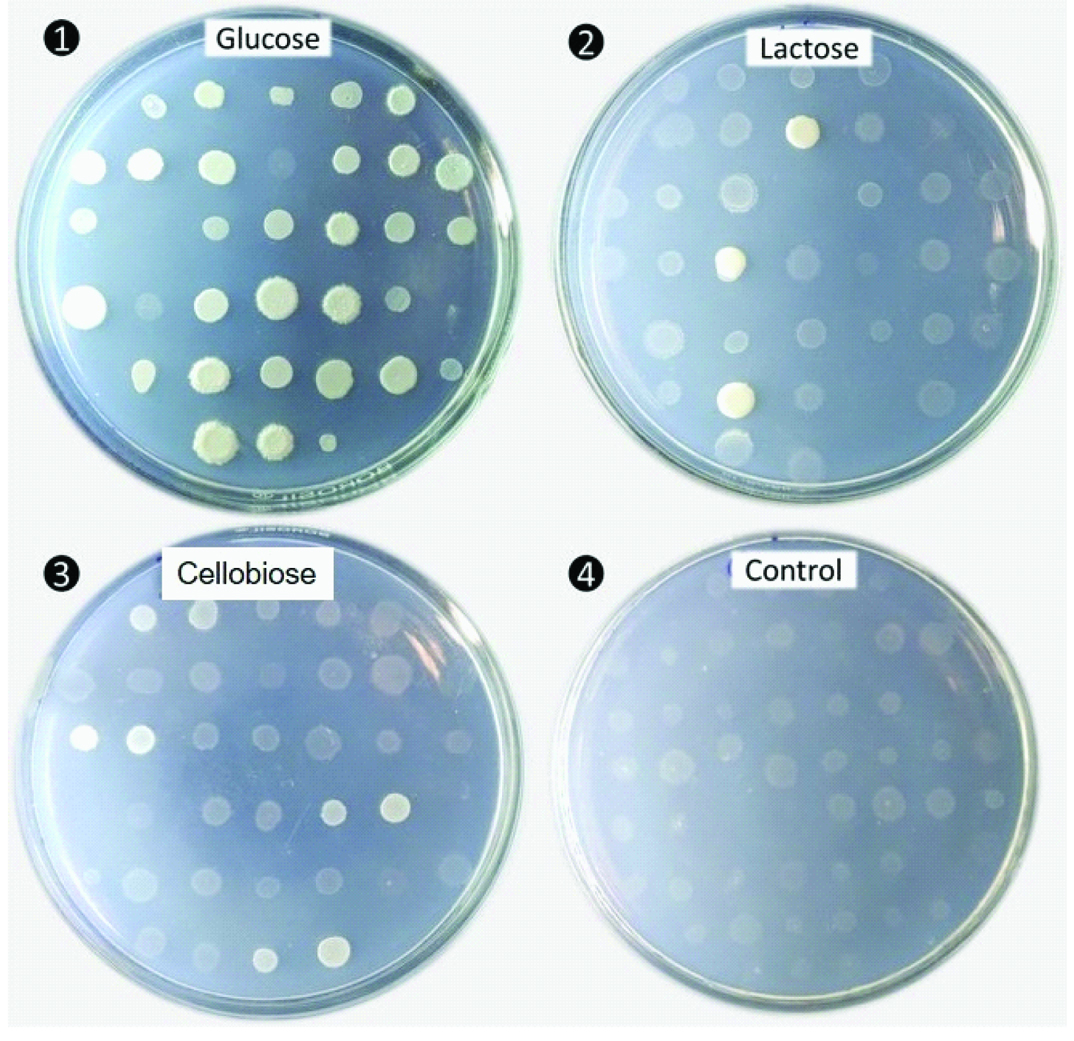
Auxanographic carbohydrate assimilation plate following incubation at 300C for 48 hours for selected carbohydrates (1) Glucose (2) Lactose (3) Cellubiose along with (4) Control.
Note-The control plate with translucent or opalescent outline indicative of a lack of assimilation. A large number of yeasts spotted thus can be screened for their assimilative properties for a specific carbohydrate.
Results of carbohydrate assimilation test for various Candida species. Glucose (Glu), Sucrose (Suc), Maltose (Mal), Lactose (Lac), Cellubiose (Cel), Raffinose (Raff), Trehalose (Tre), Xylose (Xyl), Galactose (Gal) and Dulcitol (Dul)
| Test Isolates(Wild / ATCC Strains) | Test carbohydrates |
|---|
| Glu | Lac | Suc | Mal | Tre | Xyl | Raff | Cel | Gal | Dul |
|---|
| Candida albicans | + | - | + | + | + | + | - | - | + | - |
| Candida tropicalis | + | - | + | + | + | + | - | + | + | - |
| Candida krusei | + | - | - | - | - | - | - | - | - | - |
| Candida parapsilosis | + | - | + | + | + | + | - | - | + | - |
| Candida glabrata | + | - | - | - | + | - | - | - | - | - |
| Candida kefyr | + | + | + | - | - | + | + | + | + | - |
Discussion
The technique described here has an advantage over some of the other methods in that each sugar is placed in a separate, individual plate so that the diffusion of adjacent sugars into the surrounding medium is prevented. Such diffusion sometimes occurs when performing the test in petri dishes, be it in wells, disks, or just by dropping the sugar onto the surface of the agar. The use of a sugar-free control plate eliminates false-positive interpretations that may result when traces of nutrient are carried over in the inoculum.
The methods used were modifications of the basic auxanographic technique as described by Adams and Cooper [5] and other investigators [7-9] and the accuracy was evaluated for the standard and clinical yeast isolates using the Haley Technique. In this method, un-starved yeast as spot inoculum has been used where as in the basic auxanographic technique as described by Adams and Cooper, the unstarved yeast for the heavy inoculum were added in the pour plates. Martin MV et al., observed that the un-starved yeasts and yeasts starved in various media for 24 hours and 48 hours gave the same readings for assimilation tests [6]. Further, the researchers also found that the temperature control of the molten agar is critical for the appropriate results [6]. Although, diverse species of yeasts of industrial and clinical importance exist, the correct identification of yeasts has been a prime requisite for researchers. In the food industry, it is vital to differentiate the small number of spoilage yeasts from the numerous environmental isolates. Patient management for yeast mycoses requires correct identification to assign appropriate treatment specific to pathogenic species. Their taxonomical discrimination is dependent on the types and numbers of biochemical substrates employed [8]. The use of chromogenic media to differentiate clinical isolates of yeast species was used [7,10]. However, chromogenic media have certain drawback in differentiating the emerging Candida species – C.famata, C.lipolytica [11,12] and C.auris [13,14]. Chromogenic agars are useful to identify mixed cultures with C.albicans but when non - albicans are present, these should be subcultured on SDA and identified accordingly using either biochemical or molecular tools [14].
More recently, yeast identification and taxonomy have been transformed by DNA-based methods [9]. In this study, the genus Candida, of industrial and emerging clinical importance [15] has been employed for demonstrating the utility of auxanographic carbohydrate assimilation method for its ease in large scale yeast identification. A wide armamentarium of yeast identification techniques ranging from; morphological, biochemical, physiological, proteomic (MALDI-TOF) and genotypic methods are currently available. Among this, the conventionally used methods like biochemical profiling have been exploited for automated machine based identification using API, VITEK etc. However, utility of these methods for extensive yeast identification has been beset with limitations attributed to discrepant identity [16]. Despite these advancements in technology, the auxanographic carbohydrate assimilation remains as the main stay for definitive yeast identification in resource limited settings. The carbohydrate assimilation tests find utility for yeast diversity studies exploring novel ecological niches. In such instances, the metabolic, proteomic and genotypic profiles for yeast identification has limited scope for characterization of a large spectrum of emerging and exotic yeasts with pathogenic potential [11,17]. This partly owes to lack of well curated pre-existing database aiding their conclusive identification. In such instances, the techniques described here facilitates testing of an extended range of carbohydrates in a cost effective manner with atleast a week long shelf life aiding with sufficient discrimination thresholds and enabling fundamental yeast biological studies.
Limitation
The current study validated this technique for the genus Candida only. Although, the methodology is adaptable to aid in the identification of other important yeast genera like Cryptococcus, Trichosporon, and Rhodotorula.
Conclusion
The present method represents procedural alterations that do not comprise, and apparently enhance, the accuracy of assimilation determinations while allowing large scale yeast identification by the auxanographic method to be more convenient and practical.
[1]. Freydiere AM, Guinet R, Boiron P, Yeast identification in the clinical microbiology laboratory: Phenotypical methodsMedical Mycology 2001 39(1):9-33. [Google Scholar]
[2]. Huppert M, Harper G, Sun SH, Delanerolle V, Rapid methods for identification of yeastsJournal Clin Microbiol 1975 2(1):21-34. [Google Scholar]
[3]. Oliveira GD, Ribeiro ET, Baroni FD, An evaluation of manual and mechanical methods to identify Candida spp. from human and animal sourcesRev Inst Med Trop Sao Paulo 2006 48(6):311-15. [Google Scholar]
[4]. Larone DH, Larone DH, Medically important fungi: A guide to identification 1987 New YorkElsevier [Google Scholar]
[5]. Cooper LH, Yeast identification methodsClinical Microbiology Newsletter 1979 1(4):01-04. [Google Scholar]
[6]. Martin MV, Schneidau JD, A simple and reliable assimilation test for the identification of Candida speciesAm J Clin Pathol 1970 53(6):875-79. [Google Scholar]
[7]. Pincus DH, Orenga S, Chatellier S, Yeast identification–the past, present, and future methodsMedical Mycology 2007 45(2):97-121. [Google Scholar]
[8]. Mickelsen PA, McCarthy LR, Propst MA, Further modifications of the auxanographic method for identification of yeastsJ Clin Microbiol 1977 5:297-301. [Google Scholar]
[9]. Land GA, Vinton EC, Adcock GB, Hopkins JM, Improved auxanographic method for yeast assimilations: A comparison with other approachesJ Clin Microbiol 1975 2:206-07. [Google Scholar]
[10]. Rajeevan S, Thomas M, Appalaraju B, Characterisation and Antifungal susceptibility pattern of Candida species isolated from various clinical samples at a tertiary care centre in South IndiaIndian J Microbiol Res 2016 3:53-57. [Google Scholar]
[11]. Naseema S, Uma P, Myneni RB, Yarlagadda P, Singamsetty S, A study of identification and antifungal susceptibility pattern of Candida species isolated from various clinical specimens in a tertiary care teaching hospital, Chinakakani, Guntur, Andhra Pradesh, South IndiaInt J Curr Microbiol App Sci 2016 5:71-91. [Google Scholar]
[12]. Nurat AA, Ola BG, Olushola SM, Mikhail TA, Ayodeji AS, Molecular and phenotypic identification of Candida isolates from pregnant women in Ogbomoso, Southwestern NigeriaInt J Reprod Contracept Obstet Gynecol 2016 5:317-22. [Google Scholar]
[13]. European Centre for Disease Prevention and Control. Candida auris in healthcare settings – Europe – 19 December 2016. Stockholm: ECDC; 2016 [Google Scholar]
[14]. Shetty N, Johnson E, Patel B, Lamagni T, Muller-Pebody M, Neely F, Guidance for the laboratory investigation, management and infection prevention and control for cases of Candida auris 2016 :1-6. [Google Scholar]
[15]. Tap RM, Betty H, Sue L, Ramli NY, Suppiah J, Hashim R, First isolation of Candida wangnamkhiaoensis from the blood of immunocompromised paediatric patientMycoses 2016 59(11):734-41. [Google Scholar]
[16]. Castanheira M, Woosley LN, Diekema DJ, Jones RN, Pfaller MA, Candida guilliermondii and other species of Candida misidentified as Candida famata: Assessment by the Vitek 2, DNA-Sequencing Analysis and MALDI-TOF MS in two global antifungal surveillance programsJournal Clin Microbiol 2012 :JCM-01686. [Google Scholar]
[17]. Kuan CS, Yew SM, Toh YF, Chan CL, Lim SK, Lee KW, Identification and characterization of a rare fungus, Quambalaria cyanescens isolated from the peritoneal fluid of a patient after nocturnal intermittent peritoneal dialysisPloS one 2015 10(12):e0145932 [Google Scholar]