Over past two decades there has been significant improvement in medical field in elucidating the underlying pathophysiology and genetics of Addison’s disease. Adrenal insufficiency (Addison’s disease) is a rare disease with an incidence of 0.8/100,000 cases. The diagnosis may be delayed if the clinical presentation mimics a gastrointestinal disorder or psychiatric illness. We report a case of Addison’s disease presenting as acute pain in abdomen mimicking clinical presentation of acute pancreatitis.
Case Report
A 50-year-old woman with no known premorbid illnesses was presented to the emergency room with sudden onset of severe upper abdominal pain, fatigue, nausea and vomiting for one day. There was no history of intake of NSAIDs, alcohol, spicy food, or history of peptic ulcer disease. Consent was taken from the patient.
Patient was conscious, co-operative but in agony due to the severe pain in abdomen. General examination was unremarkable and vital signs were stable. Abdominal examination revealed tenderness in the epigastric region. Systemic examination was otherwise unremarkable. The patient was admitted to the surgical ward and evaluated for suspected causes of acute upper abdominal pain.
Investigations: An emergency ultrasonography of the abdomen was performed that showed only mild hepatomegaly. Serum amylase and lipase levels were elevated (Serum Amylase-69 IU/l Serum Lipase- 318 IU/l). Other salient biochemical investigations revealed random blood sugar of 100 mg/dl, serum sodium 103 mEq/l\l, serum potassium 5.3 mEq/l, serum urea of 54 mg/dl, serum creatinine of 2.2 mg/dl, serum calcium 10 mg/dl. Complete haemogram obtained with peripheral smear were within normal limits.
A provisional diagnosis of acute pancreatitis with acute kidney injury was made and the patient was managed conservatively with no feeds administered orally. Intravenous fluids, proton pump inhibitors and anti spasmodic medications were administered along with parenteral tramadol to alleviate acute abdominal pain despite patient having abnormal renal function test. However, despite improvement in the patient’s amylase and lipase levels and creatinine levels within the subsequent three to four days, the patient complained to have persisting abdominal pain. Repeat biochemical investigations three days after admission revealed serum sodium of 110 mEq/l, serum potassium of 5.1 mEq/l, serum creatinine-1.5 mg/dl.
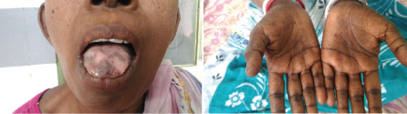
An internal medicine opinion was sought in view of persisting hyponatremia despite optimal hydration with parenteral fluids. Repeat systemic examination was consistent with previous findings and revealed only mild epigastric tenderness. However, a meticulous general examination revealed blackish hyperpigmentation of the palms, soles, and mucus membranes [Table/Fig-1]. On reviewing the history, the patient had claimed to have noticed these skin changes since the last one year. She also gave history of salt craving, malaise, fatigue, anorexia, mild bouts of self-limiting pain in abdomen and occasional vomiting after food intake that she ignored. Examination of vital signs revealed postural hypotension with a drop in the systolic blood pressure by 20 mmHg from the baseline in recumbent position.
Blackish hyperpigmentation of mucus membranes of tongue and palms.
In view of the persisting acute severe epigastric pain, progressive hyperpigmentation, postural hypotension and persisting hyponatremia with hyperkalemia despite adequate hydration, a diagnosis of hypocortisolism was considered.
The serum cortisol levels estimated at 8 am was 29.5 ng/ml (normal range – 49.9-249.9 ng/ml) and serum Adrenocorticotrophic (ACTH) levels were 1250 pg/ml (reference normal range: 0-46 pg/ml). A diagnosis of primary adrenal failure was made and screened for secondary causes. Radiograph of the chest was normal and showed no evidence of tuberculosis or sarcoidosis. ELISA test for HIV was negative. She was not consuming any medications like rifampicin, ketoconazole, fluconazole, phenytoin that can inhibit steroid hormone synthesis. CT scan of the abdomen done showed no features of adrenal metastasis. MRI brain to rule out pituitary tumour was done and it was normal. Screening for coexisting auto immune disorders like hypothyroidism was made and Thyroid Stimulating Hormone (TSH) was within normal limits. Final diagnosis of primary Addison’s disease of autoimmune aetiology was made; parenteral hydrocortisone and oral fludrocortisone were initiated resulting in a remarkable recovery of her pain in abdomen. Repeat values of sodium normalized (serum sodium 130 mEq/l). After switching over to oral prednisolone, the patient was discharged after appropriate counselling to continue the medications without fail. Patient was doing well on her last follow up one month after starting steroids.
Discussion
Thomas Addison, in 1855 described adrenal failure but still the disease remains under diagnosed with high rates of morbidity and mortality [1]. The clinical symptoms are usually non specific but disease may present as fatigue, weakness, anorexia, and weight loss, nausea and vague abdominal pain, postural dizziness, and musculoskeletal pains. Only 10% of patients present without hyperpigmentation which may delay diagnosis. Postural hypotension is very common with Addison’s disease [2].
Addison’s disease is a rare disease with an estimated prevalence four to 11 per 100,000 population. Autoimmune aetiology has an incidence of one per 100,000 [3]. Autoimmune Addison’s is isolated in 40% with the rest associated with autoimmune polyglandular syndrome [3]. Tuberculosis remains the most common cause in developing countries in comparison to autoimmune disorder being the leading cause in developed countries [3]. Other causes include congenital adrenal hyperplasia, congenital lipoid adrenal hyperplasia, X-linked adrenoleukodystrophy, familial glucocorticoid deficiency.
Various syndromes associated with Addison’s disease include Triple A syndrome, Smith-Lemli-Opitz syndrome, Kearns-Sayre syndrome. Rare causes of primary adrenal insufficiency include infection, haemorrhage, metastasis and infiltrations of the adrenal gland. Iatrogenic causes of primary adrenal insufficiency include bilateral adrenalectomy as a management of Cushing’s disease or after bilateral nephrectomy and administration of drugs like mitotane, etomidate, suramin, ketoconazole [4].
Primary adrenal insufficiency occurs when major portion of the adrenal cortex has been destroyed. Loss of both glucocorticoid and mineralocorticoid secretions causes primary adrenal insufficiency. Whereas, secondary adrenal insufficiency leads to loss of only glucocorticoid secretion because the adrenal itself is intact and still can be regulated by renin angiotensin system.
Aldosterone is the primary endogenous mineralocorticoid produced from the zona-glomerulosa of the adrenal cortex. This hormone is a part of the renin-angiotensin system. Aldosterone stimulates sodium-potassium ATPase pump activation on the distal renal tubules and collecting ducts leading to increased uptake of sodium and water from the tubular lumen with simultaneous excretion of potassium. So, in Addison’s disease, the deficiency of aldosterone leads to hyponatremia, dehydration, and hyperkalemia. Loss of sodium and water in turn leads to hypotension. Hyponatremia being a very common presentation with patients having craving for salt, it reflects mineralocorticoid deficiency and increased vasopressin secretion caused by cortisol deficiency, whereas, deficiency of glucocorticoids leads to ineffective gluconeogenesis and hypoglycemia. Hyperkalemia is present in 40% of the patients at initial diagnosis [4].
Addison’s disease presents with gastrointestinal complaints in form of diarrhea in 20% of cases. Adrenal insufficiency usually presents as acute abdominal pain especially during adrenal crisis [5]. Studies have shown that acute gastrointestinal manifestations usually preceded Addison’s crisis [6].
Relative Adrenal Insufficiency (RAI) is an important problem in patients with severe sepsis and septic shock, acute hepatic dysfunction, after cardiopulmonary arrest and several other diseases. As per De waele et al., the incidence of relative adrenal insufficiency is less in early stage of pancreatitis when compared to pancreatic necrosis which shows higher SOFA scores as well 28-day mortality [6].
In our case, patient presents with acute painful abdomen and was diagnosed of acute pancreatitis with elevated amylase and lipase levels which resolved in four days but abdominal pain persisted along with hyponatremia, weakness and postural hypotension together with history of hyperpigmentation for one year. Since all other conditions were ruled out, final diagnosis of autoimmune Addison’s disease which presented as acute pancreatitis was made.
Conclusion
The present case report underscores the importance of considering Addison’s disease amongst the causes of acute pain abdomen and unexplained hyponatremia. The initial mild acute pancreatitis might have precipitated and unmasked underlying adrenal insufficiency. The case also signifies the importance of reviewing medical history and performing repeated clinical examination in patients whenever the diagnosis is not achieved or is doubted. Specialists and residents in surgical specialties must be cautioned to foresee medical causes of acute pain abdomen whenever surgical causes are ruled out.
[1]. Alebiosu CO, Odessan O, Addisonès disease: A case reportAnnals of African Medicine 2003 2(2):85-87. [Google Scholar]
[2]. Sarkar SB, Sarkar S, Ghosh S, Bandyopadhyay S, Addison’s diseaseContemporary Clinical Dentistry 2012 3(4):484 [Google Scholar]
[3]. Srinivasulu P, Sree G, Bingi P, A case of Addison’s disease presenting as acute abdomen (common presentation of an uncommon disease)Indian Journal of Applied Research 2015 5(5):19-21. [Google Scholar]
[4]. Ten S, New M, Maclaren N, Addison’s disease 2001The Journal of Clinical Endocrinology and Metabolism 2001 86(7):2909-22. [Google Scholar]
[5]. Konda CS, Subramanian G, Gopalaratnam B, Adrenal insufficiency mimicking gastrointestinal disorder: A case reportInt J Sci Stud 2015 3(8):204-06. [Google Scholar]
[6]. De Waele J, Hoste E, Baert D, Heyndrickx K, Rijkckaert D, Thibo P, Relative adrenal insufficiency in patients with severe acute pancreatitisCritical Care 2007 11(2):1 [Google Scholar]