To solve problems related to human identification, the study of anthropometric characteristics is of fundamental importance. As bones of the body are last to perish after death, next to enamel of teeth, skeletal remains have been used for determining the sex of an individual [1]. The primary components of any skeletal analysis in forensic sciences are age and sex determination. As most bones used for sex determination are recovered in incomplete state, it is often necessary to use bones that are recovered intact e.g. the maxillary sinus. The MS remains intact even when the skull and other bones may be badly disfigured in victims who are incinerated, thus it can be used for identification [2].
The first paranasal sinus to develop is MS is located in the left and right maxillary bones and consists of two pyramidal shaped air filled cavities lined with mucosa. The MS tend to appear at the end of the second embryonic month and complete by the age of 18 to 20 years [2,3]. The shape and size of the MS varies amongst individuals, between genders, and in various populations. The MS stabilise after second decade of life and thus reliable measurements can be achieved by radiographic images [4].
Considering the complex structure of MS, diagnostic methods like Magnetic Resonance Imaging (MRI) and Computed Tomography (CT) are used as gold standard to evaluate the true anatomy of sinuses [5]. However, their use is limited by high cost, dose, and restricted accessibility. With the introduction of CBCT, these drawbacks have been overcome. CBCT can visualize and provide precise information about teeth and surrounding complex anatomical structures, as it is characterized by rapid volumetric image acquisition with high resolution and low dose radiation level. These advantages of CBCT make it a reliable tool for sex determination in forensic medicine [5].
To the best of our knowledge, most of the studies on sexual dimorphism of MS have been performed using CT scan images, few Indian studies using CBCT imaging modality has been reported till date [6]. Thus, the present study was designed to evaluate the size and volume of MS to determine gender by CBCT.
Materials and Methods
The present study was a descriptive study and the duration of the study was one year (April 2015 to March 2016). Hundred patients attending the Department of Oral Medicine and Radiology at The Oxford Dental College and Research Centre, Bengaluru, Karnataka, India were prospectively included in the study randomly, out of which 50 patients were males and 50 were females.
Patients aged between 20 to 50 years with complete dentition were selected as study sample and patients associated with periapical infections, periodontal pathologies and sinus pathologies were excluded from the study.
Study procedure was explained and an informed consent was taken from the selected patients. To perform the present study, ethical clearance was obtained from the ethical committee of Oxford Dental College and Research Centre, Bengaluru. Bilateral maxillary sinus images (left and right) were acquired for 100 patients (50 females and 50 males) using CBCT and evaluated for following parameters: width, length, height, area, perimeter and volume [Table/Fig-1] and for CBCT exposure, CBCT scanner (Kodak 9300 3D imaging system) was used with the following scanning parameters - 90kvp, 6mA and 11x13 cm field of view. The images were then reconstructed with the help of CS 3D imaging software.
Different parameters of maxillary sinus.
| S. No. | Parameter | Units | Reference points |
|---|
| 1 | Width | mm | Outermost point of the lateral wall to the medial wall. |
| 2 | Length | mm | Longest anterior to posterior measurement of the cavity. |
| 3 | Height | mm | Superior wall to lowermost point of the inferior wall of the sinus. |
| 4 | Area | cm2 | Area = Length × width |
| 5 | Perimeter | cm | Perimeter = 2 x length + 2 x width |
| 6 | Volume | cm3 | Volume = length x width x height x1/2 |
All data were subjected to descriptive and discriminative functional analysis and measurements were evaluated by using two fold magnification and modifiable screen brightness with a standardized slice thickness of 300 μm.
Each linear measurement was recorded with the help of CS 3D software for three times in three different measurement sessions at an interval of one week to minimize the memory bias. Mean value of each linear measurement was then calculated. Width and length distances were measured in the axial section [Table/Fig-2,3], whereas, the height distances were measured on coronal cross-section [Table/Fig-4]. Area, perimeter and volume were recorded manually [Table/Fig-1] for each MS (left and right).
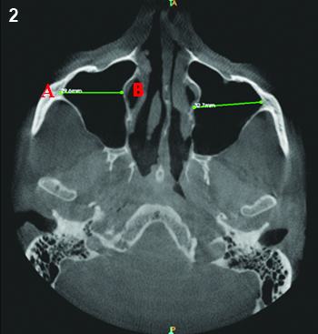
Measurement of maxillary sinus width in axial section
POINT A = Most lateral point on lateral wall of maxillary sinus
POINT B = Most medial point on the medial wall of maxillary sinus
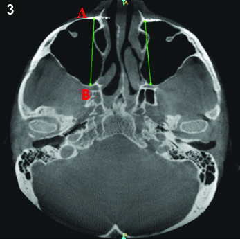
Measurement of maxillary sinus length in axial section.
POINT A = Most anterior point of the medial wall of the maxillary sinus
POINT B = Most posterior point of the medial wall of the maxillary sinus
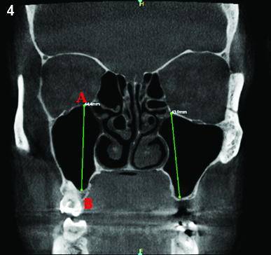
Mesurement of maxillary sinus height in coronal section.
POINT A = Most highest point of the sinus roof
POINT B = Most lowest point of the sinus floor
Statistical Analysis
Statistical analysis was done by calculating the mean and standard deviation of both maxillary sinuses measurements which were calculated and compared. All the measured parameters data was then subjected to discriminative statistical analysis and analysed using unpaired t-test.
Results
A sample size of 50 male and 50 female patients was considered for the study with their ages ranging between 20-50 years [Table/Fig-5].
Sample distribution according to gender and age group.
| Age Groups | Males | Females |
|---|
| n | % | n | % |
|---|
| 20-25 yrs | 14 | 28% | 22 | 44% |
| 26-30 yrs | 10 | 20% | 8 | 16% |
| 31-35 yrs | 4 | 8% | 10 | 20% |
| 36-40 yrs | 6 | 12% | 4 | 8% |
| 40-50 yrs | 16 | 32% | 6 | 12% |
| Total | 50 | 100% | 50 | 100% |
The right MS parameters (width, length, height, area, perimeter, volume) were measured and compared between males and females. The difference in mean value of the different parameters between males and females was not found to be statistically significant (p>0.05) [Table/Fig-6].
Comparison of different parameters of right MS between males and females.
| Parameter | Gender | n | Mean | Std. Dev | SE of Mean | MeanDifference | t | p-value |
|---|
| Right Maxillary Sinus Width (cm) | Male | 50 | 2.42 | 0.41 | 0.06 | -0.151 | -1.834 | 0.070 |
| Female | 50 | 2.57 | 0.41 | 0.06 |
| Right Maxillary Sinus Length (cm) | Male | 50 | 3.81 | 0.33 | 0.05 | 0.025 | 0.432 | 0.667 |
| Female | 50 | 3.78 | 0.23 | 0.03 |
| Right Maxillary Sinus Height (cm) | Male | 50 | 3.55 | 0.53 | 0.08 | 0.151 | 1.548 | 0.125 |
| Female | 50 | 3.40 | 0.44 | 0.06 |
| Right Maxillary Sinus Area | Male | 50 | 9.27 | 2.14 | 0.30 | -0.517 | -1.223 | 0.224 |
| Female | 50 | 9.79 | 2.08 | 0.29 |
| Right Maxillary Sinus Perimeter | Male | 50 | 12.45 | 1.30 | 0.18 | -0.253 | -1.003 | 0.318 |
| Female | 50 | 12.70 | 1.22 | 0.17 |
| Right Maxillary Sinus Volume | Male | 50 | 16.74 | 5.28 | 0.75 | -0.151 | -0.147 | 0.883 |
| Female | 50 | 16.89 | 4.97 | 0.70 |
The difference in mean value of the different parameters with respect to right MS between males and females was not found to be statistically significant (p>0.05).
The left MS parameters were measured and compared between males and females. The difference in mean left MS width (cm) between males and females was found to be statistically significant (p<0.05). The difference in mean values of the other parameters between males and females was not found to be statistically significant (p>0.05), [Table/Fig-7].
Comparison of different parameters left MS between males and females.
| Parameter | Gender | N | Mean | Std. Dev | SE of Mean | Mean Difference | t | p-value |
|---|
| Left Maxillary Sinus Width (cm) | Male | 50 | 2.40 | 0.43 | 0.06 | -0.183 | -2.071 | 0.041* |
| Female | 50 | 2.58 | 0.46 | 0.06 |
| Left Maxillary Sinus Length (cm) | Male | 50 | 3.78 | 0.33 | 0.05 | 0.067 | 1.067 | 0.288 |
| Female | 50 | 3.71 | 0.29 | 0.04 |
| Left Maxillary Sinus Height (cm) | Male | 50 | 3.56 | 0.57 | 0.08 | 0.179 | 1.707 | 0.091 |
| Female | 50 | 3.38 | 0.48 | 0.07 |
| Left Maxillary Sinus Area | Male | 50 | 9.13 | 2.12 | 0.30 | -0.491 | -1.196 | 0.234 |
| Female | 50 | 9.62 | 1.98 | 0.28 |
| Left Maxillary Sinus Perimeter | Male | 50 | 12.35 | 1.33 | 0.19 | -0.232 | -0.897 | 0.372 |
| Female | 50 | 12.58 | 1.25 | 0.18 |
| Left Maxillary Sinus Volume | Male | 50 | 16.58 | 5.69 | 0.80 | -0.010 | -0.009 | 0.993 |
| Female | 50 | 16.59 | 5.09 | 0.72 |
*denotes statistically significant difference
The difference in mean left maxillary sinus width (cm) between males and females was found to be statistically significant (p<0.05).
Gender determination using MS parameters was evaluated using discriminant analysis. Only the left MS width (cm) was found to be a significant factor in determining gender (p<0.05), [Table/Fig-7].
Classification function coefficients and accuracy level for each parameter in determining gender was done, the left maxillary sinus width was found to be the best discriminant parameter that could be used to study sexual dimorphism with an overall accuracy of 60% [Table/Fig-8]. The final result of the analysis shows that 68% of males and 74% of females were sexed correctly. The given parameters when used to identify the gender of a sample will be able to provide results with an accuracy of 71% [Table/Fig-9].
Classification function coefficients and accuracy level for each parameter in determining gender.
| Parameter | Male | Female | % Correctly Classified |
|---|
| Constant | Coefficient | Constant | Coefficient |
|---|
| Right Maxillary Sinus Width (cm) | -17.84 | 14.18 | -20.05 | 15.07 | 56% |
| Right Maxillary Sinus Length (cm) | -88.74 | 46.25 | -87.60 | 45.94 | 49% |
| Right Maxillary Sinus Height (cm) | -27.73 | 14.94 | -25.03 | 14.31 | 58% |
| Right Maxillary Sinus Area (cm2) | -10.33 | 2.08 | -11.43 | 2.19 | 54% |
| Right Maxillary Sinus Perimeter (cm) | -49.38 | 7.82 | -51.38 | 7.98 | 53% |
| Right Maxillary Sinus Volume (cm3) | -6.02 | 0.64 | -6.12 | 0.64 | 49% |
| Left Maxillary Sinus Width (cm) | -15.37 | 12.25 | -17.71 | 13.19 | 60% |
| Left Maxillary Sinus Length (cm) | -72.75 | 38.14 | -70.21 | 37.46 | 48% |
| Left Maxillary Sinus Height (cm) | -23.59 | 12.88 | -21.34 | 12.23 | 58% |
| Left Maxillary Sinus Area (cm2) | -10.60 | 2.17 | -11.69 | 2.29 | 59% |
| Left Maxillary Sinus Perimeter (cm) | -46.27 | 7.38 | -48.00 | 7.52 | 58% |
| Left Maxillary Sinus Volume (cm3) | -5.42 | 0.57 | -5.42 | 0.57 | 53% |
Accuracy of the final model in determining gender.
| Actual Gender | Predicted Gender | % Correctly Classified |
|---|
| Male | Female |
|---|
| Male | 34 | 16 | 71% |
| Female | 13 | 37 |
| Total | 47 | 53 |
Discussion
Identification of gender from remains of human skeletons is an important forensic procedure. It has been reported that the gender can be determined with an accuracy of 100% if entire skeleton is available. A total of 98% accuracy can be achieved from both the pelvis and the skull [7].
The current study was designed to determine the reliability and accuracy of MS dimensions measurement as a method for gender identification using CBCT on 100 patients (50 males and 50 females). After birth, the MS continues to pneumatize into the developing alveolar ridge as the permanent teeth erupt. At the age of 20, with the completion of the eruption of the third molars, the pneumatization of the sinus ends. It has been reported that genetic diseases, post infections and environmental factors can affect the sizes of MS. Considering this factor the patients with disease conditions were excluded from the study.
Comparison between male and female groups showed statistically insignificant differences on both the right and left sides with respect to the MS length, height, area, volume and perimeter [Table/Fig-6,7]. However, the female group showed statistically significant higher values for left side MS width with a p-value of 0.041 [Table/Fig-7]. In our study accuracy level for each parameter in determining gender was done, the left MS width was found to be the best discriminant parameter that could be used to study sexual dimorphism with an overall accuracy of 60%.
A study by Tambawala SS et al., for comparison between male and female groups demonstrated that the female group had statistically significant lower values for both the left and right MS in context to the length, height and width dimensions. In contrast to this study our results showed no statistical significant difference between genders in relation to right and left MS length and height dimensions and right MS width dimensions except for left MS width which was higher in females [6].
A discriminating analysis performed by Azhar A et al., on MS measurements showed that the left MS width was the best discriminate parameter (with an overall accuracy of 61.3%) [3]. The results obtained in our study were in agreement with Azhar A et al., study as left MS width was the best discriminative parameter with an overall accuracy of 60%.
Teke HY et al., estimated the mean value for the maximum width of MS and obtained almost similar results as that of our study in relation to the width of MS [8].
A study by Sharma SK et al., for measurements of MS volume and dimensions by CT scan for gender determination was done. The difference was statistically significant for sinus anterio-posterior dimension (length) and volume. The MS length was the best discriminant parameter with an overall accuracy of 69.81% [4]. Whereas, in our study the right MS length mean values in males and females showed statistically insignificant difference.
Amin MF and Hassan EI concluded that cephalo-caudal (height) measurement of the MS as most reliable predictor of sex with correct predictive accuracy of 70.8% in males and 62.5% in females respectively [9].
Studies were also conducted by Vidya CS et al., and Prabhat M et al., on similar population for gender identification by using different MS parameters (height, length, width and volume). Significant statistical difference (p<0.01) was found in the right MS volume between males and females [10,11], whereas, in our study the difference in MS volume was not statistically significant.
Many of the previous studies used MS width, length, height and volume to determine gender but in our study we have included area and perimeter in addition to the above mentioned parameters to find out if these can determine gender. However, in our study area and perimeter showed statistically insignificant values.
In the present study, the final result of discriminative analysis shows that 68% of males and 74% of females were sexed correctly. The given parameters when used to identify the gender of a sample will be able to provide results with an accuracy of 71%.
Prabhath M et al., and Ekizoglu O et al., reported the overall accuracy rates of MS to 77.15% and 83.3% respectively in predicting gender, which was higher when compared with our study prediction accuracy (71%) [11,12].
A study by Sharma SK et al., conducted a study on similar population as in our study and showed that 68.9% of females and 65.16% of males were sexed correctly and the overall percentage for sexing MS correctly was 67.03%. The prediction accuracy in our study is comparatively greater than this study [4].
A study by Teke HY et al., showed that the accuracy of MS measurements i.e., the ability to identify gender was 69.4% in females and 69.2% in males with a mean of 69.3% [8]. The results obtained in this study were almost similar to our study’s prediction accuracy (71%).
Saccucci M et al., conducted a study on gender determination using MS by means of CBCT. No statistical difference was found in patient’s MS volumes between genders and they concluded that it is not possible to use the MS to discern sexual difference in corpse identification [5]. Similarly our results also didn’t reveal much significant difference between males and females except for left MS width.
Most authors have reported significant differences in the measurements of MS between males and females [3,6,8,9,11]. Whereas, Saccucci M et al., reported no such differences [5].
The role of the MS in ethnic classification was established by Fernandes CL. They had found that European crania had significantly larger antral volumes than the Zulu crania. Males were found to have larger volumes than females. On the other side, Zulu male sinuses were narrower than Zulu female sinuses and race was found to be highly significant, with European sinuses being wider than Zulu sinuses. The length and height of Europeans were higher than that of the present findings of our study, while the width of was almost equal to that recorded in the present study [13].
Thus, the variations in some of the results of maxillary air sinus dimensions and volume in these studies are probably due to combination of many factors like different ethnic and racial groups with differences in body stature, skeletal size, height and physique; genetic and environmental factors; anatomical variations of sinus; difference in osteoclastic and osteoblastic activity and pneumatization process of sinus [4]. The difference in the results generated for different populations also suggested that this study can be done for different populations all over the world to generate such identification equations for sex determination. This also emphasizes the need to conduct this analysis on a larger study sample and in different population.
To the best of our knowledge, most of the studies on sexual dimorphism of MS have been done using CT images which carries high radiation exposure to the patients and also relatively high cost. With the advent of CBCT, studies can be planned on larger samples with low radiation exposure and relatively low cost. By conducting such studies a base line value can be generated for use in forensic applications.
Limitation
The limitation of this study included the small sample size due to the stringent inclusive and exclusive criteria of the study. Also, no statistical formula was used to determine the sample size of the study. Further studies are needed to conduct these analyses on a larger population.
One more limitation of the present study was that the samples were randomly selected. Further studies can be conducted by statistically determining the sample size.
Conclusion
The results of the present work showed that the MS exhibits anatomic variability between genders, as female group showed statistically significant higher values for left side MS width. But the other parameters showed no significant difference between genders. Therefore, the width of left MS can be used as an aid in forensic anthropology for gender determination. In our study CBCT images could provide adequate measurements for MS with low radiation exposure, thereby proposing the use of CBCT for evaluating MS dimensions in the field of forensic science.
We suggest that measurements of the MS together with other bones can be used for gender determination when the whole skeleton is not available.
The difference in mean value of the different parameters with respect to right MS between males and females was not found to be statistically significant (p>0.05).
*denotes statistically significant difference
The difference in mean left maxillary sinus width (cm) between males and females was found to be statistically significant (p<0.05).