As a way of improving these histological staining techniques, several stains have been modified and combined with various other stains to improve their effectiveness [2]. Among these techniques, Modified Gallego’s stain is one that has been derived from the modification of Lille’s stain which uses basic reagents like haematoxylin, carbol fuschin and aniline blue [3]. This stain can be used as one of the differential stains for the hard tissues of teeth and also for the pathological lesions which are characterised microscopically by fibrous stroma containing bone and cementum like material [4]. The present study was done to differentiate hard tissues of teeth and to identify the presence of hard tissue like component in various pathological lesions using Modified Gallego’s stain under light microscopy.
Materials and Methods
This prospective study was conducted at Sri Sai College of Dental Surgery, Vikarabad, India. The study included freshly extracted normal human teeth. Decayed/ root canal treated/fluoride affected teeth were not included. Control group included 10 teeth out of which five ground and five decalcified sections were prepared. Decalcification procedure was done using 8% formic acid+ 8% HCL (hybrid technique) for four days.
Study group included 10 oral calcifying lesions which were taken from the archives. The lesions included were calcifying epithelial odontogenic tumour containing Liesegang rings, odontome containing dentinoid and cementoid like material, odontogenic fibro-myxoma, osteoma and osteomyelitis containing bony tissue. Two slides of each lesion were prepared. These were stained with Modified Gallego’s stain and were viewed under light microscopy. The various colours taken by different hard tissues were observed and interpreted.
Sections were stained with Modified Gallego’s by using the following technique: The sections were initially deparaffinised, stained in haematoxylin for 8-12 minutes and rinsed in distilled water. Then, they were sensitised in mordant for two minutes and rinsed in distilled water (Mordant was prepared by mixing 200 ml of distilled water in 1.5 ml of concentrated Nitric acid with 1 ml of 40 per cent formaldehyde and 1.5 ml of Iron chloride). Later they were stained with 3 ml of carbol fuschin diluted in 50 ml of 0.2 percent acetic acid, rinsed in distilled water and washed in mordant for 1-2 minutes. Finally, these sections were stained with 0.01 per cent aniline blue in saturated picric acid solution for 30 seconds, dehydrated, cleared with xylene and mounted in DPX mounting media. The stained sections were viewed using routine light microscopy and were interpreted under 4X, 10X and 40X.
Results
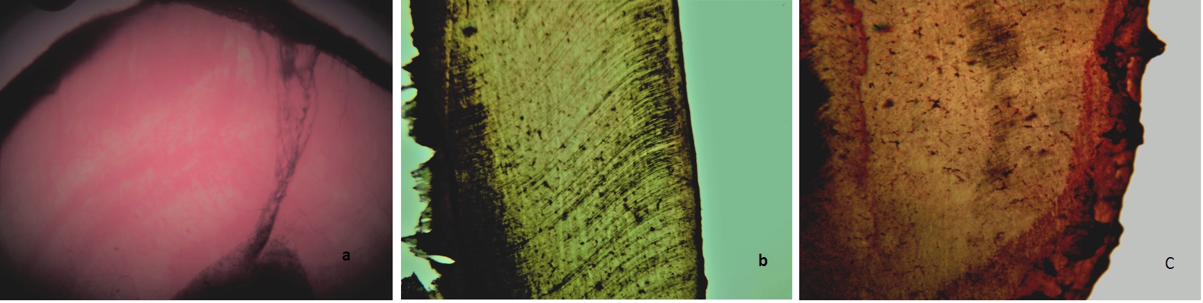
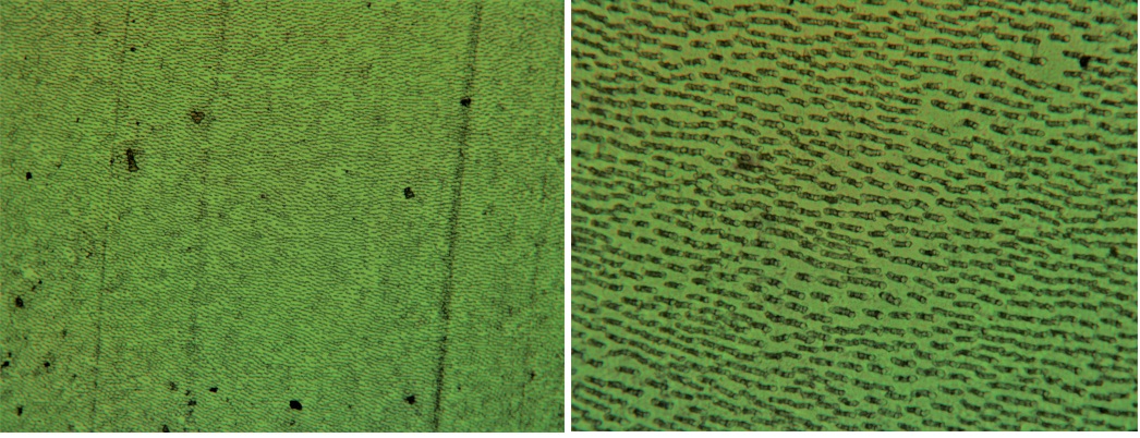
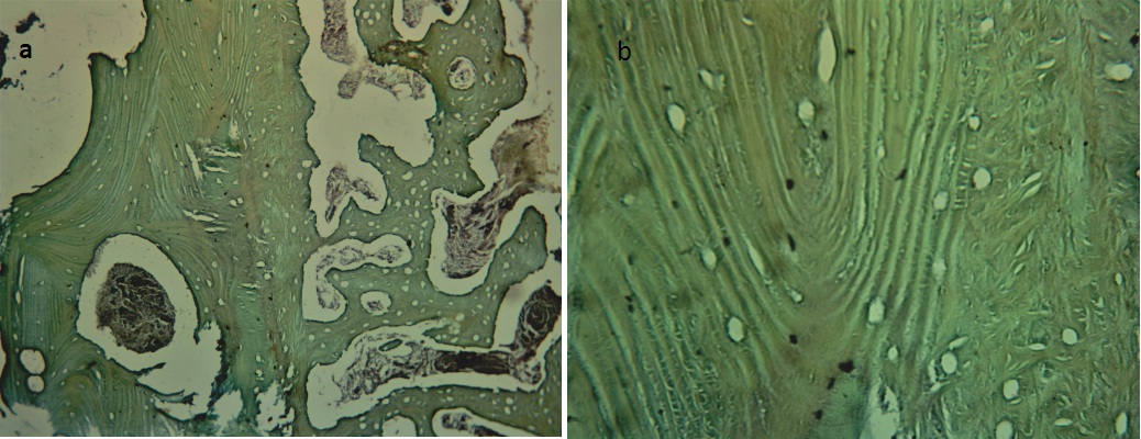
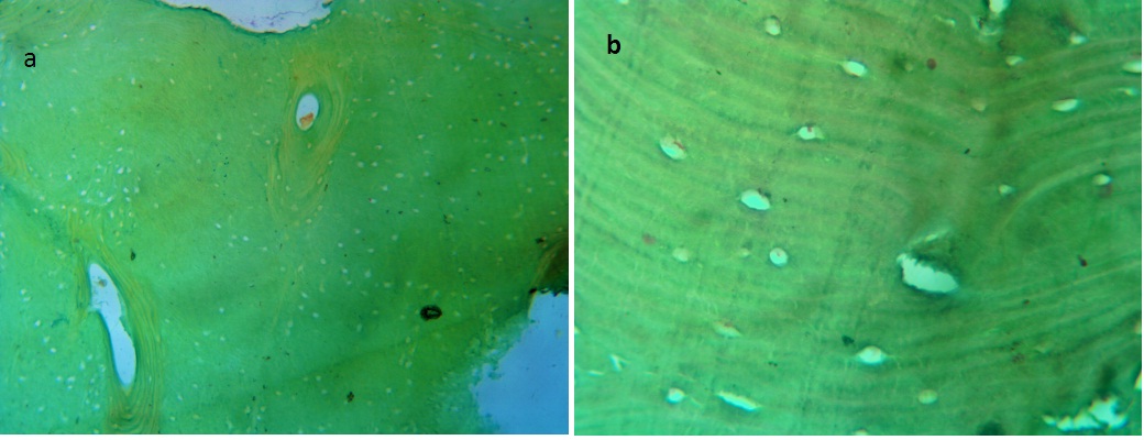
In the ground sections stained with Modified Gallego’s stain, it was observed that various hard tissues had taken different colours like enamel-pink, dentine-green and cementum-dark red [Table/Fig-1]. In the stained decalcified sections, dentin was stained with green colour [Table/Fig-2].
Photomicrograph demonstrating the ground section of normal tooth stained by Modified Gallego’s stain – 10X a) Enamel – pink; b) dentin- green; c) cementum – dark red.
Photomicrograph of Gallego’s stained decalcified section of tooth demonstrating dentin in green a) 4X; b)10X.
The results of staining various pathological lesions used in this study have been depicted in [Table/Fig-3,4,5,6,7 and 8].
Interpretation of staining of pathological lesions.
| Tissue Stained | Colour | Interpretation |
|---|
| Odontome | GreenRed | Dentin like materialCementum like material |
| Calcifying EpithelialOdontogenic Tumour | Green | Liesegang rings |
| OdontogenicFibro-Myxoma | Green | Bone |
| Osteoma | Green | Bone |
| Osteomyelitis | Green | Bone |
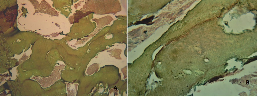
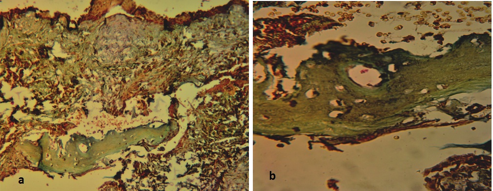
Photomicrograph of odontome showing dentin like material (green) and cementum like material (red) (Modified Gallego’s stain: a) 10X; b) 40X respectively).
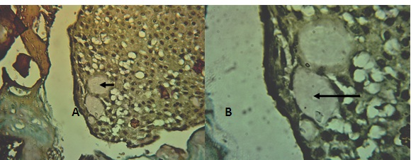
Photomicrograph of Calcifyinng epithelial odontogenic tumour demonstrating Liesegang rings {Modified Gallego’s stain: a) 10X; b) 40X)}.
Photomicrograph of Odontogenic fibromyxoma demonstrating normal bone (green) {Modified Gallego’s stain: a) 4X; b) 10X)}.
Photomicrograph of Osteomyelitis demonstrating bone (green) {Modified Gallego’s stain: a) 10X; b) 40X)}.
Photomicrograph of Osteoma demonstrating bone in green {Modified Gallego’s stain: a) 10X; b) 40X}.
Discussion
Tooth is usually composed of 3 different mineralised tissues–enamel, dentin and cementum [6]. Though the amount of calcification varies between these hard tissues, they are stained uniformly with H&E and are indistinguishable from each other [7]. When there are any calcified structures present in the oral pathological lesions, their identification often poses a problem and creates difficulty in diagnosis. The present study was done to differentiate between these hard tissues and to identify the calcified components present in the pathological lesions.
Various special stains like Von kossa, Alizarin red for the bone, Picro-thionin for dentin, Toulidine blue and Alcian blue for cementum are available but the use of single histo-chemical stain that differentiates between the hard tissues of tooth are rare [3].
Modified Gallego’s stain is one such stain which not only stains the decalcified sections but also differentially stains the hard tissues of tooth and the calcified structures present in the pathological lesions thus helps in obtaining a clear histological picture [8].
In the present study, the pathological lesions containing the hard tissue like components were taken. Odontoma refers to hamartomatous malformation rather than a neoplasm. This lesion occurs by the proliferation of both epithelial and mesenchymal components which results in the formation of hard tissues like enamel, dentin and cementum in an abnormal pattern [9]. When stained with Modified Gallego’s stain, these components were stained with different colours, dentin stained green and cementum red.
Calcifying odontogenic epithelial tumour, also known as pindborg tumour is classified as uncommon, benign odontogenic neoplasm of epithelial origin. Histologically, this tumour is characterized by the presence of polyhedral epithelial cells arranged in cords and sheets, amyloid like material and calcifications in the form of Liesegang rings [9]. These calcifications stained green with this stain which signify that these could be either dentin like or bone like material.
Osteoma is an uncommon oral lesion which usually occurs in young individuals. It is a slow-growing, benign neoplasm characterized by the proliferation of either cancellous or compact bone [9]. Histologically, it is composed of coarse, cancellous or dense compact bone. Osteomyelitis is defined as the inflammation of bone and bone marrow. This inflammatory process occurs in the cortical or medullary spaces of bone which extends away from the initial site of involvement. Microscopically, it demonstrates the presence of dense, irregular bony trabeculae with empty lacunae and little interstitial marrow tissue [10]. Along with these bony lesions, the normal bony component of odontogenic fibromyxoma was taken and stained to observe the difference between the pathological bone and normal bone, but no difference was observed between them.
A study was done by Gallego in 1954 in which he stained the ground, decalcified and frozen sections of normal tooth and bone. He observed that the hard tissues, dentin and bone were stained with green and cementum with red. Later in 2014, a similar study was done by Sandhya et al., in which few oral calcifying lesions were stained along with ground, decalcified sections of tooth and observed similar results. These findings suggest that Modified Gallego’s stain could be considered as one of the practical tool in diagnosis of these lesions [3].
Considering the above mentioned studies, calcifications in the various pathological lesions of the oral cavity were studied by taking decalcified and ground sections as control. These sections were stained and observed under light microscopy. The findings of the present study display similarity with the literature. Thus, the stain is capable of determining the hard tissue nature which was confirmed by selecting certain pathological lesions with calcified components.
Limitations
This staining procedure is technique sensitive and the intensity of the colour in the stained sections decreases as the time duration increases and becomes difficult to differentiate between the colours.
Conclusion
As Modified Gallego’s stain not only stains the hard tissue components of decalcified and ground sections but also helps in identifying the nature of calcified structures of various pathological lesions, this stain can be used as one of the differential stains and as an adjunct to routine H&E.