Rehabilitation of a Patient with Facial and Palatal Defect - A Case Report
Litty Francis1
1 Former Senior Resident, Department of Prosthodontics, Government Dental College, Thiruvananthapuram, Kerala, India.
NAME, ADDRESS, E-MAIL ID OF THE CORRESPONDING AUTHOR: Dr. Litty Francis, A-7 Quarters, Medical College, P.O, Thiruvananmthapuram-695011, Kerala, India.
E-mail: litty.franciz@gmail.com
Defects involving the face and maxilla present a challenge to the prosthodontists as these have a direct effect on aesthetics, function as well as the psychology of the patient. An array of problems awaits the clinician from restoring the previous contour of the oral cavity, facial form, etc. to the mental state of the patients. This article deals with the rehabilitation of a hemimaxillectomy patient with a facial defect, using an interim hollow bulb obturator and a silicone facial prosthesis, which helped to improve the general well being of the patient. Rehabilitative procedures provide patients considerable care so that they can continue their life with confidence.
Facial silicone prosthesis, Hemimaxillectomy, Hollow bulb obturator
Case Report
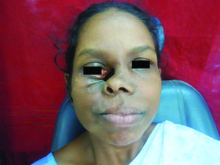
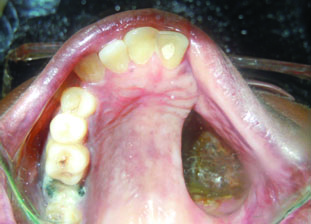
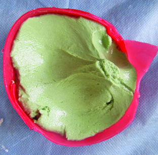
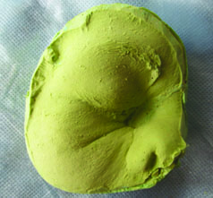
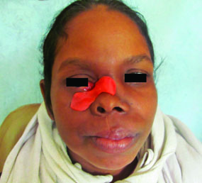
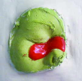
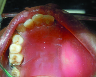
A 35-year-old female, who had undergone hemimaxillectomy for the management of Adenoid Cystic Carcinoma (ACC), was referred to the Department of Prosthodontics for the rehabilitation of facial and oral defects [Table/Fig-1,2]. On examination, the middle and posterior portions of the right maxilla were completely resected and there was a large defect involving the medial aspect of the right eye and nose. The intraoral defect and the facial defect were connected. The following steps were followed for the fabrication of facial prosthesis. The defect was packed with a piece of gauze and borders of the defect were boxed with wax sheets. The nostrils were blocked using cotton and a moulage impression was made with irreversible hydrocolloid impression material (ZELGAN) [Table/Fig-3]. The impression was boxed and poured in Type III dental stone [Table/Fig-4]. A wax pattern for the defect was sculpted and checked between the patient and cast for the accuracy of fit, orientation and aesthetics [Table/Fig-5]. The pattern was then invested [Table/Fig-6], flasked and dewaxed. Room temperature vulcanizing silicone material (MP Sai BioMed) was packed into the prepared mould and processed according to the manufacturer’s directions. Intrinsic colours were added to the silicone material before packing to match patient’s skin colour. The processed prosthesis was finished and tried on the patient for proper fit, contour and colour matching. The prosthesis had adequate retention due to extension into undercuts. The superior aspect of the prosthesis could not be merged as the corner of the eye contacted the prosthesis. This area was masked with the help of a spectacle of appropriate size.
Pre rehabilitation extraoral photograph.
Pre rehabilitation intraoral photograph.
Facial moulage impression.
Stone cast of the facial defect. (Images left to right)
Wax try-in of facial prosthesis.
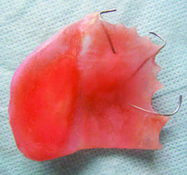
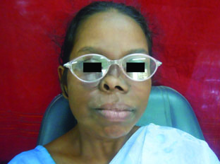
For the fabrication of hollow bulb, a preliminary cast of the maxillary arch with the defect, was made in Type II plaster from an alginate impression. The custom resin tray was fabricated and final impression was made with polyvinyl siloxane (ELITE HD Dentsply) impression material to record the entire defect and tooth structures. It was poured in Type III dental stone to obtain the master cast. The wax pattern was fabricated using modelling wax (Rolex, Dental Product, Delhi) with necessary clasps for retention. The pattern was then invested, dewaxed and the obturator was fabricated in heat cure acrylic resin [Table/Fig-7]. The defect portion was covered using autopolymerizing acrylic resin after filling the bulb with common salt. Once polymerization was completed, a few small holes were drilled in the bulb, through which the water was allowed to enter and dissolve the salt, making it hollow and weightless. After final trimming and polishing, it was inserted in patient’s mouth. Instructions were given on insertion, removal, and maintenance of the prosthesis. Swallowing evaluation was done and there was no leakage and regurgitation and thus feeding tube was removed. The was patient recalled was done after one week and was found to be comfortable with the prosthesis [Table/Fig-8,9].
Post rehabilitation extraoral photograph.
Post rehabilitation intraoral photograph. (Images left to right)
Discussion
Radical surgery for the management of facial tumours usually involves partial or total removal of orbital, nasal, antral contents and jaws creating facial and oral anatomic defects [1]. The morbidity associated includes oro-antral/nasal communications, inability to take food through the mouth, speech defects, depression, etc. The facial defects can be managed either by surgical reconstruction or prosthetic rehabilitation. Vascularized Free Tissue Transfer (VFTT), also known as free flap transfer, is now considered the gold standard for maxillofacial reconstruction and non-vascularized tissue transfer is no longer the accepted first-line treatment [2]. Facial prosthesis cannot replace reconstructive surgery, but in certain circumstances, it may be an alternative. Maxillofacial prosthetic management has clearcut advantages as it requires no extra surgery and the outcome can be more aesthetically satisfying.
The fundamental objective of prosthetic obturation is the closure of the surgical defect and isolation of mouth from the antrum and nose [3]. The prosthesis should restore facial contour, improve mastication and speech, provide lip support, and reduce drooling [4]. The obturator bulb creates a pressure resistant seal against mucosal tissues, thereby restoring speech and swallowing [5]. The prognosis of maxillofacial prosthesis depends on the postsurgical bony anatomy, number of abutment teeth present, location and size of the defect, post radiation mucosal changes, and neuromuscular coordination of the patient [6]. The sequence of treatment for a maxillectomy patient includes immediate surgical obturator at the time of surgery or soon thereafter, an interim obturator after initial healing (approximately three months), and a definitive obturator prepared after the tissues has stabilized [7].
Most of the hemimaxillectomy defects can be managed with a conventional simple obturator prosthesis using various clasps for retention. But in some cases, the defects may extend to contiguous structures like orbit, antrum, etc., and rehabilitation of such cases needs an extraoral prosthesis. A prosthetic replacement of an exterior part is termed as epithesis [8]. Extraoral prosthesis help to restore normal appearance, mitigates patient’s concerns and improves day to day life [9]. The hollow obturator can be an open or closed type. A closed obturator prevents the entry of fluid and reduces air space in the defect [10,11]. The weight of the prosthesis can be reduced up to 33% by fabricating a hollow obturator.
The field of maxillofacial prosthesis is rapidly advancing. The introduction of osseointegrated implants, 3D imaging techniques [12], advances in rapid prototyping technology [13], computerized colour formulation, new improved materials has all broadened the scope of maxillofacial prosthetic rehabilitation.
In this case report, rehabilitation of a patient with the facial and intraoral defect is discussed. The prosthesis had two parts: an interim hollow bulb obturator and a facial prosthetic part. The silicone facial prosthesis along with the spectacles gave the patient enough self confidence and the obturator helped the patient to discontinue the feeding tube and was relieved of the problem of regurgitation and nasal tone of speech. The social life improved tremendously and her attitude towards life also changed.
Conclusion
Maxillofacial defects can prevent a patient from returning to normal daily activities. Apart from providing treatment for the defect, the psychological aspect of the patients should also be considered. The trauma which they endure because of the social enigma has a profound effect. Many patients with these defects have been rehabilitated successfully with prosthesis. Knowledge of material science is the key to providing the best care for patients. The rehabilitation procedure needs careful planning of the surgical technique and prosthetic fabrication.
[1]. Spiro RH, Strong EW, Shah JP, Maxillectomy and its classificationHead Neck 1997 19(4):309-14. [Google Scholar]
[2]. O’Fearraigh P, Review of methods used in the reconstruction and rehabilitation of the maxillofacial regionJ Ir Dent Assoc 2010 56(1):32-37. [Google Scholar]
[3]. Turkaslan S, Baykul T, Aydin MA, Ozarslan MM, Articulation performance of patients wearing obturators with different buccal extension designsEur J Dent 2009 3(3):185-90. [Google Scholar]
[4]. Ortegon SM, Martin JW, Lewin JS, A hollow delayed surgical obturator for a bilateral subtotal maxillectomy patient: A clinical reportJ Prosthet Dent 2008 99(1):14-18. [Google Scholar]
[5]. Okay DJ, Genden E, Buchbinder D, Urken M, Prosthodontic guidelines for surgical reconstruction of the maxilla: A classification system of defectsJ Prosthet Dent 2001 86(4):352-63. [Google Scholar]
[6]. Cheng AC, Somerville DA, Wee AG, Altered prosthodontic treatment approach for bilateral complete maxillectomy: A clinical reportJ Prosthet Dent 2004 92(2):120-24. [Google Scholar]
[7]. Rilo B, Dasilva JL, Ferros I, Mora MJ, Santana U, A hollow-bulb interim obturator for maxillary resection. A case reportJ Oral Rehabil 2005 32(3):234-36. [Google Scholar]
[8]. Van Doorne JM, Extraoral prosthetics: past and presentJ Invest Surg 1994 7(4):267-74. [Google Scholar]
[9]. Butler DF, Gion GG, Rapini RP, Silicone auricular prosthesisJ Am Acad Dermatol 2000 43(4):687-90. [Google Scholar]
[10]. Oh WS, Roumanas ED, Optimization of maxillary obturator thickness using a double-processing techniqueJ Prosthodont 2008 17(1):60-63. [Google Scholar]
[11]. Kocacikli M, Yalug S, Yazicioglu H, Yilmaz C, Fabricating a hollow obturator with visible light-cured resin systemJ Prosthodont 2008 17(7):596-98. [Google Scholar]
[12]. Elbashti M, Hattori M, Sumita Y, Aswehlee A, Yoshi S, Taniguchi H, Creating a digitized database of maxillofacial prostheses (obturators): A pilot studyThe Journal of Advanced Prosthodontics 2016 8(3):219-23. [Google Scholar]
[13]. Jiang FF, Hou Y, Lu L, Ding XX, Li W, Yan AH, Functional evaluation of a CAD/CAM prosthesis for immediate defect repair after total maxillectomy: a case series of 18 patients with maxillary sinus cancerJ Esthet Restor Dent 2015 27(1):S80-89. [Google Scholar]