Unilateral Absence of Ethmoid Sinus and Nasal Turbinates: A Rare Case Report
Vinod Felix1, Narendrakumar Veerasigamani2
1 Consultant, Department of ENT, SUT Pattom Hospital, Trivandrum, Kerala, India.
2 Consultant, Department of ENT, Indoamerican Hospital, Vaikom, Kerala, India.
NAME, ADDRESS, E-MAIL ID OF THE CORRESPONDING AUTHOR: Dr. Vinod Felix, Consultant, Department of ENT, SUT Pattom Hospital, Trivandrum-695004, Kerala, India.
E-mail: dr.vinodfelix@gmail.com
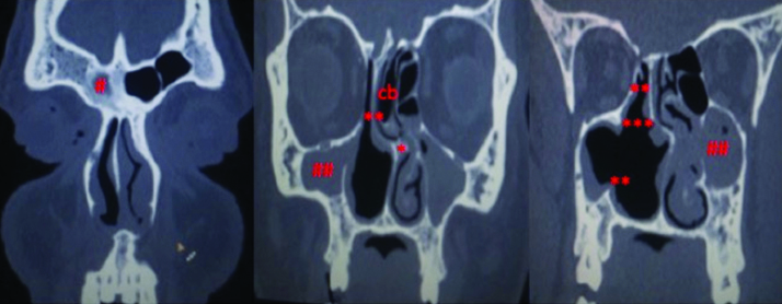
A variety of anatomical variation of paranasal sinus and nasal turbinates exist, as its development is a complex and long standing process. Computerized Tomography (CT) of the paranasal sinuses is a very valuable tool in diagnosing these variations. Preoperatively defining the anatomical variations of the intranasal structures is essential in performing the safe functional endoscopic sinus surgery and to avoid unnecessary complications. Several degrees and combinations of aplasias and hypoplasias have been reported. We report a case of 37-year-old male who presented with bilateral nasal block and rhinorrhea and his CT paranasal sinuses showed gross septal spur in left side, absence of right middle, inferior and superior turbinates, absent right ethmoid air cells, aplastic right frontal sinus, left concha bullosa with bilateral maxillary sinusitis.
Nasal cavity, Paranasal sinus, Sinusitis, Variations
Case Report
A 37-year-old male patient presented to our hospital with the complaints of bilateral rhinorrhea and nasal block for the past 6 months with no previous history of any nasal surgery, nasotracheal intubation or trauma. On anterior rhinoscopy, septum was deviated to left side with absent right inferior turbinate. Diagnostic nasal endoscopy and computerized tomography of the paranasal sinuses was done, which revealed presence of gross septal spur in left side, absence of right middle, inferior and superior turbinates, absent right ethmoid air cells, aplastic right frontal sinus, left concha bullosa with bilateral maxillary sinusitis [Table/Fig-1]. The patient was posted for functional endoscopic sinus surgery. We were aware of the congenital abnormality present in right side of paranasal sinuses, hence avoided seeking for the ethmoid sinus in that side and limited our surgery to maxillary sinus on that side. Postoperative period was uneventful with symptomatic relief of complaints.
Aplastic right frontal sinus (#), left concha bullosa(cb) with bilateral maxillary sinusitis (##), gross septal spur in left side (*), absence of right middle, inferior and superior turbinates(**), absent right ethmoid air cells (***).
Discussion
There are usually three nasal turbinates in each nasal cavity. The middle and superior turbinates are part of the ethmoid bone, while inferior turbinate is a separate bone. Rarely the fourth supreme turbinate may also present above the superior turbinate [1]. Paranasal sinuses and all turbinates arise from the cartilaginous nasal capsule [2]. Six to seven folds appear in the lateral wall of the nasal capsule during 9th and 10th week of gestation where they are separated from each other by corresponding grooves. These folds are fused into three to four crests in the following weeks [3]. Maxilloturbinal develops into separate inferior turbinate. First furrow is in between first and second ethmoturbinals. First ethmoturbinal regress without forming turbinates rather its descending part forms uncinate and ascending part forms Agger Nasi cell named as nasoturbinal. Second, third and fourth or fifth develops into middle, superior and supreme turbinates respectively [4]. During intrauterine life, lower three turbinates increases in length, while the supreme turbinate remains approximately at 5 mm length from 14th to 36th week and only 65% present in fetuses [2]. By 15th to 16th weeks gestation, all lower three turbinates are well formed. Many anatomical variations may occur during this development, but most of them remain asymptomatic [5]. During the 3rd and 4th fetal months, mucosa of the nasal cavities evaginates and develops into paranasal sinuses. Maxillary and ethmoid sinuses are rudimentary at birth whereas frontal and sphenoid sinuses are not developed. Ethmoid cells are more developed anteriorly at birth with pneumatization progressing in a posterior direction and its growth lasts until late puberty [6].
Middle turbinates are considered as a very useful landmark during endoscopic sinus surgery [1,5]. The turbinates are the physiologic structures, which participates in nasal cycle and are important for laminar airflow maintenance [5]. Secretions from the nose and paranasal sinuses are transported along the middle turbinate surfaces, and reaches nasopharynx. This process may get disrupted with the presence of middle turbinate variations resulting in secondary maxillary sinusitis [7].
Many turbinate variations are reported like paradoxical MT (middle turbinate), bifid MT/IT (Inferior turbinate), secondary MT, trifucate MT, accessory MT/IT, duplicate MT, MT hypogenesis, agenesis of MT/IT, Elongated ST (superior turbinate) and pneumatisation of the MT/IT/ ST. Among all, pneumatisation of the middle turbinate is the most common variation [1,8-10]. Total Absence of paranasal sinuses are also reported [6]. Ferrari M et al., reported a case of bilateral middle turbinate absence in a cadaveric study [11]. Aydil and Ozelik reported a case of unilateral agenesis of MT that was ipsilateral to septal spur and they suggested that presence of septal spur is substitute to the space normally occupied by MT [1]. But in our case septal spur was present on left side and agenesis of nasal turbinates was on right side along with absent right ethmoid cells, aplastic right frontal sinus, left concha bullosa and bilateral maxillary sinusitis which are very unique and these variations are due to temporary interruption of the prechordal plate during early embryogenesis. Most of the paranasal sinuses anatomical variations are asymptomatic and diagnosed as an incidental findings in routine radiological imaging.
Conclusion
Paranasal sinuses are more prone for anatomical variations. Among nasal turbinates especially, MT is the essential landmark in any nasal surgery. Computerized tomography plays a role in diagnosis and to identify paranasal sinuses variations. Accurate descriptions of anatomical variations are very important in achieving correct diagnosis and successful endoscopic sinus surgeries.
[1]. Aydil U, Ozelik T, Unilateral agenesis of middle nasal turbinateJ Laryngol Otol 2010 124(4):447-49. [Google Scholar]
[2]. Som PM, Naidich TP, Illustrated review of the embryology and development of the facial region, part 1: early face and lateral nasal cavitiesAJNR Am J Neuroradiol 2013 34(12):2233-40. [Google Scholar]
[3]. Gleeson M, Scott brown’s otorhinolaryngology Head and neck surgery 2008 7th editionGreat BritainEdward Arnold Publishing Ltd [Google Scholar]
[4]. Stammberger H, The messerlinger techniqueFunctional endoscopic sinus surgry 1991 PhiladelphiaB.C. Dekker [Google Scholar]
[5]. Gupta T, Aggarwal A, Sahni D, Morphometric evaluation of the middle turbinate in relation to endoscopic sinus surgeryClinical Rhinology: An International Journal 2012 5(3):103-06. [Google Scholar]
[6]. Korkmaz H, Korkmaz M, Total aplasia of the paranasal sinusesAllergy Rhinol 2013 4(2):e105-09. [Google Scholar]
[7]. Kim YW, Lee JH, Hong SL, Cho KS, Anomalous middle turbinate with choanal obstruction and maxillary sinusitis: A case reportJ Med Case Rep 2013 7:242 [Google Scholar]
[8]. Qudah MA, Extra middle turbinate lamellas: a suggested new classificationSurg Radiol Anat 2015 37(8):941-45. [Google Scholar]
[9]. Caylakli F, Yilmaz I, Hürcan C, Ozer C, Ozlüoğlu L, Unilateral inferior turbinate agenesis: a case reportEar Nose Throat J 2008 87(1):26-27. [Google Scholar]
[10]. Ozcan KM, Selcuk A, Ozcan I, Akdogan O, Dere H, Anatomical variations of nasal turbinatesJ Craniofac Surg 2008 19(6):1678-82. [Google Scholar]
[11]. Ferrari M, Tschabitscher M, Rezzani R, Fodella L, A case of middle turbinate absenceInternational Journal of Anatomical Variations 2015 8:12-14. [Google Scholar]