Any tooth which does not reach the occlusal plane even after two-thirds root formation can said to be impacted. As widely stated in the literature, mandibular third molar teeth are the most frequently impacted teeth. The position of these impacted third molars being most distally placed in the arch, their frequent association with a pericoronal flap makes this area less accessible to oral hygiene. One of the oldest and significant classification for impacted third molars is by Pell GJ and Gregory GT in 1933 [1]. In addition to this, the pressure exerted by the impacted third molars on the second molars makes the second mandibular teeth more prone to distal caries. Partially erupted mesioangular and horizontally impacted teeth accumulate plaque against the distal surface of the second molars thereby predisposing to distal cervical caries [2]. The gingival margin recedes thus exposing the cemento-enamel junction which aids in bacterial accumulation and thus encouraging root caries along distal surface in second mandibular molars. Detection of distal caries in a second molar is more difficult with a mesioangular third molar [3]. When the caries involves the radicular portion of the second molar, the restorative procedure becomes very difficult and such teeth often end up in extraction [4]. Caries involvement of the lower second molar and/or third molar is the second most common indication for removal of impacted third molars. The development of distal cervical caries in mandibular second molar is a protracted process which develops over the time and increases with continued exposure to the oral cavity [5]. One of reasons for development of distal caries in second molar and the impacted tooth itself is the delay in seeking dental care [6]. Even when the second molar is restored with the impacted third molar left untreated, recurrent caries would develop thus advancing the decay process and eventually leading to tooth loss [7]. An early removal of impacted third molar teeth, early restorative procedures involving second molars, maintenance of good oral hygiene would significantly reduce the morbidity associated with second mandibular molar teeth.
This study was aimed to assess the prevalence of distal caries in mandibular second molars due to impacted third molar teeth. The objectives of the study were: a) To predict the prevalence of distal caries in mandibular second molars due to impacted third molar teeth; b) To predict the most common type of third molar impaction associated with cervical caries of second molar teeth; c) To predict the most common age group with highest prevalence of distal caries involving second molars; d) To predict the gender variation in the prevalence of distal caries involving second molar teeth.
Materials and Methods
This is a retrospective study which included a total of 6000 OPGs of patients reporting to College of Dentistry, King Khalid University, Abha, Kingdom of Saudi Arabia for dental care. The data was retrieved from the computer software used in the institution to take OPGs and was subsequently decoded and entered into excel spread sheet. Descriptive analysis of the data was done and results were displayed as frequency table and graphs.
Each OPG was analyzed by a minimum of two investigators. OPGs with impacted third molar teeth were identified. Of these OPGs with impacted third molar teeth, prevalence of distal cervical caries in mandibular second molars was assessed. An excel spread sheet was designed to enter the data. Prevalence of distal cervical caries in mandibular second molars was analyzed in relation to age group, gender, specific type of impacted third molar causing distal caries in second molar was identified and recorded. Impacted mandibular third molar were classified as per the standard Pell GJ and Gregory GT classification as vertical, mesioangular, distoangular and horizontal [1].
Inclusion criteria included all Saudi nationals seeking dental care between 2009 to 2014 in the age group 21 to 45 years with normal eruption pattern of second molar teeth were included in the study.
Patients who had already extracted third molars or had associated pathologies like cysts, tumours were excluded from the study. Patients with developmental disorders like microdontia, presence of fourth molars, impacted second molars, supernumerary teeth, odontomes and with implants were also excluded from the study group.
Statistical Analysis
The data was cross checked for any discrepancies and then subsequently analyzed. Results were displayed as prevalence of distal cervical caries in second molar teeth due to impacted third molars in relation to age, gender and type of impaction.
Results
A total of 6000 OPGs were assessed which included patients over a period of five years (2009 to 2014). A total of 979 patients had impacted third molars (16.31%). [Table/Fig-1] shows distribution of distal cervical caries in second molar teeth according to age group, gender and type of impaction of third molar.
Distribution of distal cervical caries in second molar teeth.
| Gender | Male | Female | | |
|---|
| N | % | N | % | | | | |
|---|
| 213 | 56.4 | 164 | 43.6 | | | | |
| Age group | 21-28 Years | 29-36 Years | 37-45 Years | |
| N | % | N | % | N | % | | |
| 225 | 59.6 | 133 | 35.2 | 19 | 5 | | |
| Type of Impaction | Horizontal | Distoangular | Mesioangular | Vertical |
| N | % | N | % | N | % | N | % |
| 92 | 24.4 | 13 | 3.4 | 228 | 60.4 | 44 | 11.6 |
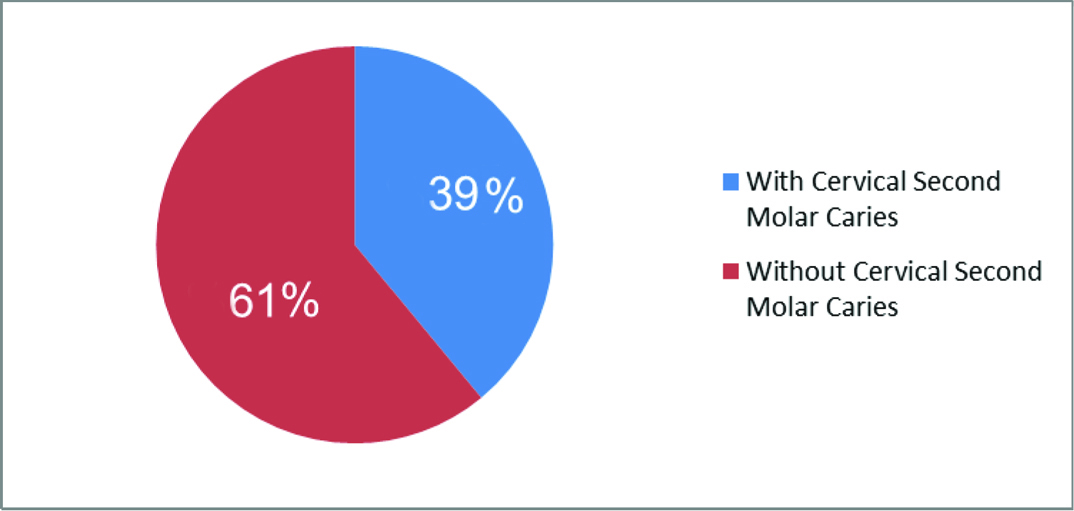
A total of 377 (39%) of the 979 patients with impacted third molars had distal cervical caries in second molar [Table/Fig-2].
Prevalence of distal caries in mandibular second molar due to impacted third molar teeth.
A total of 228 of the 377 patients had mesioangular impaction causing distal caries in second molars. This was closely followed by horizontal impaction (92 patients) causing distal caries in second molars. Distoangular impaction caused least caries in the second molars (13 patients having distal caries due to distoangular impaction). Age group 21-28 years had the highest prevalence of distal caries in second molar teeth due to impacted third molars (225 patients of the 377 patients with impacted teeth). The prevalence of distal caries in second molars is relatively high in males (213 patients) as compared to females (43.6% in females and 56.4% in males) [Table/Fig-1].
Discussion
Impaction of mandibular third molar is a common finding [8]. Distal caries of mandibular second molar is a frequently noted complication of impacted third molars [9]. Caries in the adjacent second molars, external resorption of the roots of adjacent mandibular second molars were among the radiographically detectable pathologic conditions around impacted mandibular third molars in a study conducted among Jordanian population [10].
The depth of impacted third molar and the occlusal angulation between the impacted tooth and the occlusal surface of the second molar influences the distal caries in second molar [8,11,12]. A total of 5% mandibular third molars are extracted due to distal cervical caries in mandibular second molar teeth [13]. This can be correlated with the study conducted by Nunn ME et al., involving 416 subjects. They stated that the second molars adjacent to absent third molars were at the lowest risk for developing pathology; whereas, second molars adjacent to soft tissue impacted third molars were at greatest risk [14]. One problem frequently encountered while assessing the radiolucency on the distal surface of second molars is whether it is due to caries or root resorption [15,16]. Even with good resolution and cross examination by two investigators, the problem still exists.
Ozec I et al., conducted a study among Turkish population to assess the prevalence and factors affecting the formation of second molar distal caries [8]. In their series, the prevalence of second molar distal caries was 20% and were of the opinion that significant effects on caries formation was due to contact point on the second molar cemento-enamel junction and increasing age. Falci SG et al., conducted a study involving 246 high quality periapical radiographs to assess the association between the presence of a partially erupted mandibular third molar and the existence of caries in the distal part of the second molars [17]. The prevalence rate of caries on the distal surface of the second molar in their series was 13.4%. The prevalence rate of distal caries in second mandibular molars due to impacted third molars in this present study was 39%. The total number of patients with impacted teeth in this study was 979. Of which 377 patients had distal caries in the second molars making up a prevalence rate of 39% which is significantly high as compared to other studies. Raheem AA et al., conducted a study involving 148 panoramic radiographs to assess the influence of mandibular third molar position on distal caries in mandibular second molar [9]. The majority of distal caries in the second molar (38.9%) was caused by horizontally impacted third molars in their patient series. This is contrary to the findings of our study wherein the mesioangular impactions have caused majority of distal cervical caries in second molars (61%). As compared to age group, in the present study age group 21- 28 years had the highest prevalence of distal caries in second molars.
Silva HO et al., conducted a study to analyze the dental caries on distal surface of mandibular second molar [18]. Similar to this present study, males had higher prevalence of distal caries than females in their series. However, patients above age group of thirty five years had a higher prevalence of distal caries in their study as against 21-28 years age group with higher prevalence in our study. Contrary to our study wherein mesioangular impaction is the major cause of distal caries in second molars, study conducted by Silva HO et al., showed vertical impaction as the major cause of distal caries in second molar. Allen RT et al., in their study also concluded that mesioangular position of impacted third molar was the major reason for causing caries on the distal surface of the second molar [19]. Our study also contradicts the view of Bruce RA et al., who stated that the prevalence of caries on the distal surface is an indication for the third molar removal that significantly increases with age [20]. In the present study, the higher age groups had lower prevalence of distal caries. This can be attributed to the fact that patients have neglected care, and as the caries advanced with symptomatic pain and infection, the second molars would have been extracted with age. This has led to the finding that prevalence of distal caries is higher in the younger age group. Also worth mentioning is the fact that most of the patients in this region present with missing first and second molars or grossly decayed (root stumps) second molar teeth. A prospective study design with long term follow up will assess the extracted second molar teeth due to cervical caries. Proper counseling and creating awareness would significantly conserve the second molars. Early prophylactic removal of impacted third molars will conserve the second molars. If the tooth is mesioangularly inclined, then we would stress on almost immediate removal of impacted third molars.
Since impacted third molar teeth do not play a significant role in mastication, occlusal load distribution and in maintaining occlusion, we suggest early prophylactic removal of impacted third molars. The percentage of crown structure lost in second molars due to cervical caries, the amount of finances spent in rehabilitation and the loss of natural tooth by extraction of second molars in advanced stages of caries indicates prophylactic removal of third molars. This study further emphasizes on the integration between specialties of operative dentistry, endodontics, oral diagnosis and oral surgeons. A patient may be counseled at the primary visit in oral diagnosis about the significance of third molar removal. This would stop further decay process of the involved second molar. When a patient has already been referred to the operative dentist or endodontist for restoration of second molar teeth, these specialties must counsel and advice the patient for prophylactic removal of third molars. Most of the patients try avoiding surgical procedure in fear of the pain, swelling and related factors.
Though this study included screening 6000 OPG’s over a period of five years, the design is retrospective which is a limitation in this study. For further assessment of the role of impacted third molars causing distal cervical caries in second molar teeth, a prospective study design with a long term follow is advised. With advanced diagnostic aids in clinical diagnosis and availability of CBCT, better analysis can be performed.
Conclusion
A total of 39% of the patients with impacted mandibular third molars caused distal cervical caries in second molars with mesioangular impaction being the most prominent type causing caries. Age group 21-28 years and male gender had a significantly higher incidence of cervical caries.