Azygos Lobe - A Rare Anatomical Variant
Gourahari Pradhan1, Satyajeet Sahoo2, Siladitya Mohankudo3, Yera Dhanurdhar4, Suman Kumar Jagaty5
1 Senior Resident, Department of Pulmonary Medicine, All India Institute of Medical Sciences, Bhubaneswar, Odisha, India.
2 Senior Resident, Department of Pulmonary Medicine, All India Institute of Medical Sciences, Bhubaneswar, Odisha, India.
3 Senior Resident, Department of Pulmonary Medicine, All India Institute of Medical Sciences, Bhubaneswar, Odisha, India.
4 Senior Resident, Department of Pulmonary Medicine, All India Institute of Medical Sciences, Bhubaneswar, Odisha, India.
5 Senior Resident, Department of Pulmonary Medicine, All India Institute of Medical Sciences, Bhubaneswar, Odisha, India.
NAME, ADDRESS, E-MAIL ID OF THE CORRESPONDING AUTHOR: Dr. Gourahari Pradhan, House No.052, I Block, Cosmopolis Apartments, Aiginia, Bhubaneswar-751019, Odisha, India.
E-mail: drghpradhan@gmail.com
Accessory lobe, Bronchopulmonary segments, Embryogenesis, Tear drop sign
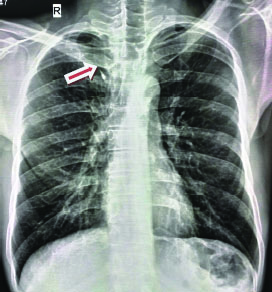
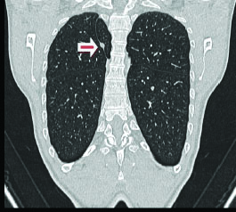
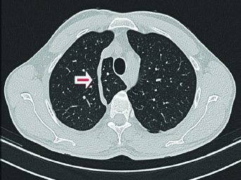
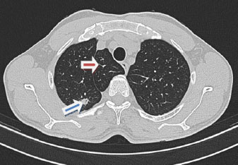
A 55-year-old male patient presented with intermittent sputum production for last two years. He was a clerk by profession and a non-smoker. He had a contact history of pulmonary tuberculosis. On investigation, tear drop sign was seen in chest radiograph [Table/Fig-1]. High resolution Computed Tomography (CT) scan of thorax confirmed presence of azygos lobe [Table/Fig-2,3] and revealed a small patch of consolidation in posterior segment of right upper lobe [Table/Fig-4]. Bronchoscopy was normal and bronchoalveolar lavage from right upper lobe was negative for tuberculosis and malignancy.
Chest X-ray with inverted comma sign / Tear drop sign in right upper lobe signifying azygos lobe (arrow).
Computed tomogram of thorax showing azygos lobe in coronal section (arrow).
Computed tomogram of thorax showing azygos lobe in axial section (arrow).
Computed tomogram of thorax showing patch of consolidation in right upper lobe (blue arrow) adjacent to azygos lobe (brown arrow).
In 1877, azygos lobe was first described by Heinrich Wrisberg. Azygos lobe has been reported in both the lungs [1]. Azygos lobe is a very rare but normal anatomic variant of right upper lobe seen in only 0.4% of population radiologically and in 1% specimen during anatomical dissection [1,2]. But it’s not a true accessory lobe as it does not have its own bronchus and does not correspond to a specific bronchopulmonary segment [2]. Azygos lobe is formed due to penetration of right posterior cardinal vein, one of the precursors of azygos vein into the apex of the lung instead of normal migration over it during embryogenesis [3]. Clinical importance of azygos lobe is that it may be confused with a bulla or abscess, a pulmonary nodule and a consolidated azygos lobe may mimick like a lung mass [2]. Lung tumours, pneumothorax, extralobar pulmonary sequestration has been reported to occur in azygos lobe [1]. Thoracic surgeons as well as physicians need to be aware of this rare anomaly.
[1]. Seiber W, Karcara N, Pant P, Pumonary azygos lobe - An anatomical variantKathmandu Univ Med J 2014 46(2):151-52. [Google Scholar]
[2]. Chabot-Naud A, Rakovich G, Chagnon K, Ouellette D, Beauchamp G, A curious lobeCan respir J 2011 18(2):79-80. [Google Scholar]
[3]. Mata J, Cáceres J, Alegret X, Coscojuela P, De Marcos JA, Imaging of the azygos lobe: normal anatomy and variationsAJR Am J Roentgenol 1991 156(5):931-37. [Google Scholar]