Neoumbilicoplasty in a Laparoscopic Port Site: Description of a New Technique and Review of Literature
Aravind Menon1, Alagesan Ganapathi2
1 Junior Consultant, Department of General and Laparoscopic Surgery, Shanthi Nursing Home, Surandai, Tirunelveli, Tamil Nadu, India.
2 Head and Senior Consultant, Department of General and Laparoscopic Surgery, Shanthi Nursing Home, Surandai, Tirunelveli, Tamil Nadu, India.
NAME, ADDRESS, E-MAIL ID OF THE CORRESPONDING AUTHOR: Dr. Aravind Menon, Junior Consultant, Department of General and Laparoscopic Surgery, Shanthi Nursing Home, Surandai, Tirunelveli-627859, Tamil Nadu, India.
E-mail: aravindmenonk@gmail.com
The umbilicus contributes significantly to the cosmetic appearance of the abdomen especially in women. Loss of umbilicus may result not only in cosmetic disfigurement but also in significant psychological effects. Omphalectomy may accompany certain surgical procedures like ventral hernia repairs and abdominoplasty. For such patients, many techniques have been described by various authors in literature for creation of a neoumbilicus for good cosmetic appearance. In this report, we describe how a laparoscopic port site was utilized to create a neoumbilicus in a patient who required omphalectomy as a part of large umbilical hernia repair. Satisfactory postoperative result promted us to report this novel technique of neoumbilicus creation. This can be utilized in patients who have been previously subjected to laparoscopic procedures. This is the first case description in literature where a laparoscopic port scar was utilized for neoumbilicoplasty.
Abdominoplasty, Flap, Laparoscopy
Case Report
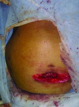
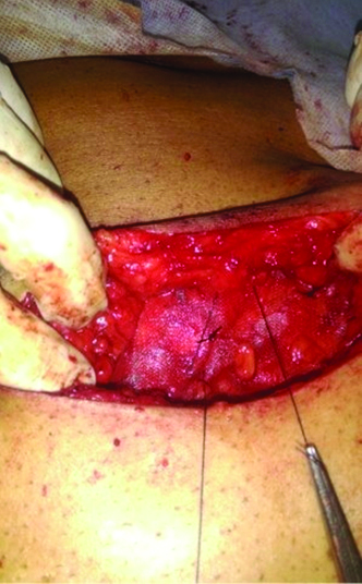
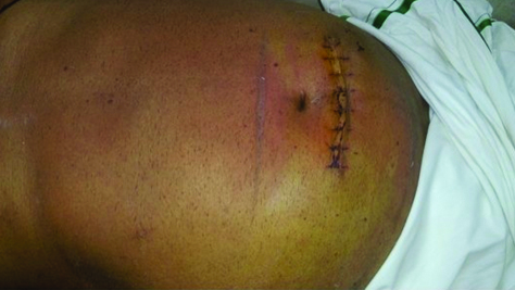
A 55-year-old male presented with a history of long standing umbilical hernia since 10 years with recent history of irreducibility. Patient had a history of laparoscopic cholecystectomy done for gallstones 12 years back. Clinical examination revealed a partially reducible 6x5 cm hernia involving the umbilicus with patchy discolouration of skin. An ultra sonogram done, confirmed the defect of 4x3 cm with omentum as content. Meshplasty was planned and a 10x7.5 cm polypropylene mesh was fixed on lay onto the rectus sheath after reduction of contents and repair of defect. Due to patchy discolouration of skin over the hernial site and fear of exposure of mesh, it was decided to do an omphalectomy [Table/Fig-1]. The previous laparoscopic supraumbilical 10 mm port site scar was converted into a neoumbilicus by using horizontal mattress nylon sutures taken from the subcuticle of the scar site and fixed onto the polypropelene mesh-anterior rectus sheath interface [Table/Fig-2,3]. Wound was closed with suction drain in situ. Postoperatively, the wound healed well with acceptable cosmesis and well formed neoumbilicus with adequate depth [Table/Fig-4,5].
Post mesh repair and omphalectomy.
Fixation of port site scar to the mesh-rectus sheath interface.
Well formed neoumbilicus with adequate depth. (Images left to right)
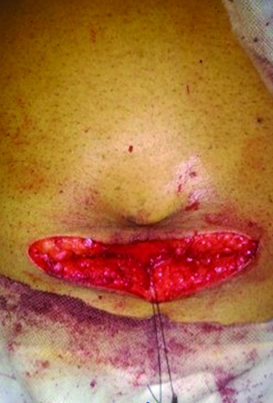
Appearance of neoumbilicus on postoperative day 2.
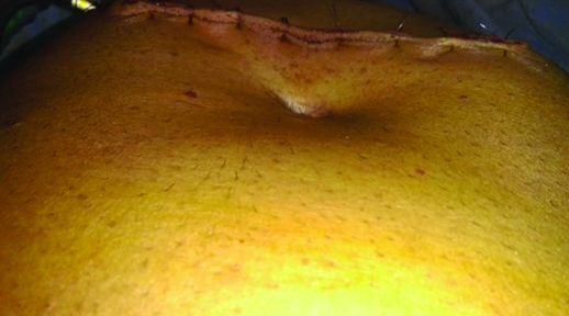
Appearance after two weeks with well formed neoumbilicus. (Images left to right)
Discussion
The umbilicus is an important aesthetic component of the abdomen and contributes significantly to the cosmetic appearance. But in certain surgeries like umbilical hernia repair with precarious viability of skin [1], urachal cancer [2] and neonatal omphalocele [3], it becomes inevitable to sacrifice the umbilicus as a part of the surgical procedure. In such situations, the onus is on the operating surgeon to create a cosmetically acceptable umbilicus in the anterior abdominal wall. Such a reconstructed neoumbilicus must have natural contour, prominent depth, minimal additional scars and prominent superior hooding [4]. In order to satisfy these conditions and reconstruct a neoumbilicus, authors have described a variety of techniques in literature. These can be broadly divided into techniques employing local flaps, special methods of suturing and grafting techniques.
Of these, local flap based reconstruction is perhaps the most common method employed. Donnabella A described the largest successful series of 162 patients who underwent surgery for morbid obesity where two rectangular flaps were fixed to rectus sheath and used to create a neoumbilicus [5]. Dessy LA et al., described double opposed Y flap technique in 10 patients and followed them for one year to report satisfactory cosmetic results [6]. Four flap techniques have been described by Lee Y et al., and Kim Y et al., individually [4,7]. While the technique elaborated by Lee Y et al., utilized four flaps created in the shape of an ‘X’, Kim Y et al., designed four transposition flaps and reconstructed neoumbilicus out of it. Senturk S et al., published a series of six patients in whom a new method of neoumbilicoplasty called dome procedure was described [8]. This was basically a V-Y advancement flap and gave gratifying cosmetic results postoperatively. Double half-cone umbilicoplasty technique was described by El-Dessouki L et al., which gave pleasing results in 6 to 18 months follow up [1]. Other flap techniques include inverted C-V flap [9], modified unfolded cylinder technique [10] and triangular flaps [11]. Other than flap based techniques, a suturing technique has been described by Bartsich SA and Schwartz MH which made use of purse string sutures [12]. The third type of reconstructive technique uses autografts like conchal cartilage which have employed the basic principles from microtia reconstruction [13]. [Table/Fig-6] shows the various techniques of neoumbilicoplasty described in literature with their outcomes [1,4–14].
Various techniques of neoumbilicoplasty described in literature and their outcome [1,4–14].
| Author | n(No of patients) | Technique described | Postoperative results | Follow up |
|---|
| Kim Y et al., [7] | n=1 | Four transposition flaps | Good, no complication | 11 months |
| Lee Y et al., [4] | n=2 | Four flap technique | Good aesthetic appearance | 8 years |
| Senturk S et al., [8] | n=6 | Dome V-Y advancement flap procedure | High patient satisfaction | - |
| Donnabella A, [5] | n=162 | Two parallel rectangular flaps | All anatomical components of umbilicus reproduced. No complications | - |
| Ozbek S et al., [10] | n=1 | Modified unfolded cylinder technique | Sufficient depth achieved | 2 years |
| Dessy LA et al., [6] | n=10 | Double opposing Y technique | Nine cases complete healing, one case partial dehiscence with delayed healing | 1-4 years |
| Hong YG, Cho JJ, [9] | n=1 | Inverted C-V flap | Good, no complications | - |
| Pfulg M et al., [11] | n=2 | Triangular skin flap folded onto itself | Natural appearance | 2 years |
| El-Dessouki N et al., [1] | n=10 | Double half cone flap | Good, one patient developed scar hypertrophy | 1.5 years |
| Bartsich SA, Schwartz MH, [12] | n=3 | Purse string method | Good, no added scarring | 8 months |
| Matsuo K et al., [13] | n=1 | Conchal cartilage composite graft | Satisfactory result | 12 months |
| Lee S et al., [14] | n=6 | Advancement flap umbilicoplasty | Good, one patient had wound dehiscence | - |
| In the present study | n=1 | Mattress suturing of laparoscopic port site | Adequate depth achieved | 2 weeks |
Conclusion
The technique described is a type of suturing technique which can be utilized in patients who have been previously operated by laparoscopy. Since the scar already exists, conversion into neoumbilicus gives the added advantage of disappearance of the laparoscopic port scar. The results were gratifying in the short period follow up with adequate depth and contour of umbilicus having been achieved. It is a simple technique, less time consuming and easy to replicate. However, it can be performed only in a specific subset of patients with laparoscopic port scar. This is probably the first case to be reported in literature where a laparoscopic port scar was utilized for neoumbilicoplasty.
[1]. El-Dessouki N, Shehata S, Torki A, Hashish A, Double half-cone flap umbilicoplasty: A new technique for the proboscoid umbilical hernia in childrenHernia 2004 8(3):182-85. [Google Scholar]
[2]. Park ES, Kim MS, Kim YB, Immediate umbilical reconstruction for urachal cancerJ Korean Soc Aesthetic Plast Surg 2003 9:59-62. [Google Scholar]
[3]. Frigo E, Rettinger-Schimmerl S, Rokitansky A, Umbilicoplasty in neonates with primary omphalocele closurePediatric Surgery International 1999 15(7):523-24. [Google Scholar]
[4]. Lee Y, Kwon C, Rhee S, Cho S, Eo S, Four flaps technique for neoumbilicoplastyArch Plast Surg 2015 42(3):351 [Google Scholar]
[5]. Donnabella A, Anatomical reconstruction of the umbilicusRevista Brasileira de Cirurgia Plástica 2013 28(1):119-23. [Google Scholar]
[6]. Dessy LA, Fallico N, Trignano E, Tarallo M, Mazzocchi M, The double opposing "Y" technique for umbilical reconstruction after omphalectomyAnn Ital Chir 2011 82(6):505-10. [Google Scholar]
[7]. Kim Y, Park E, Yi H, Park J, A novel technique for umbilical reconstruction using four transposition flapsArchives of Aesthetic Plastic Surgery 2016 22(2):96 [Google Scholar]
[8]. şentürk S, Özkan A, Gemici K, Efe D, The dome procedure: a new technique for the reconstruction of the umbilicusHernia 2015 20(4):505-08. [Google Scholar]
[9]. Hong YG, Cho JJ, Reconstruction of scarred umbilicus using an inverted C-V flap: a case reportJ Korean Soc Plast Reconstr Surg 2007 34(5):653-655. [Google Scholar]
[10]. Özbek S, Özcan M, Umbilicus reconstruction with modified ‘unfolded cylinder’ techniqueBritish Journal of Plastic Surgery 2005 58(4):500-503. [Google Scholar]
[11]. Pfulg M, Van de Sijpe K, Blondeel P, A simple new technique for neo-umbilicoplastyBritish Journal of Plastic Surgery 2005 58(5):688-691. [Google Scholar]
[12]. Bartsich SA, Schwartz MH, Purse-string method for immediate umbilical reconstructionPlastic and Reconstructive Surgery 2003 112:1652-55. [Google Scholar]
[13]. Matsuo K, Kondoh S, Hirose T, A simple technique for reconstruction of the umbilicus, using a conchal cartilage composite graftPlastic and Reconstructive Surgery 1990 86(1):149-51. [Google Scholar]
[14]. Lee S, DuBois J, Greenholz S, Huffman S, Advancement flap umbilicoplasty after abdominal wall closure: Postoperative results compared with normal umbilical anatomyJournal of Pediatric Surgery 2001 36(8):1168-70. [Google Scholar]