Fine Needle Aspiration Cytology (FNAC) is a preliminary diagnostic procedure for wide spectrum of non neoplastic and neoplastic lesions. It is safe faster and cost effective procedure. It is quick, easy to perform and has high grade specificity and sensitivity. It is used as a primary investigation procedure in all the suspected neoplastic conditions before any surgery [1].
Routinely various stains like Haematoxylin and Eosin (H&E), Romanowsky and Pap have been used for staining the FNAC smears. Romanowsky stains are routinely used for staining the blood films and air dried cytological smears [2]. The different types of Romanowsky stains available are MGG, Wright Giemsa stain, Leishman stain and Diff Quick. These stains allow better estimation of cell size, nuclear size, cell cytoplasm and identify ground substances by metachromasia [3,4]. MGG is routinely used in cytology for air dried smears in many laboratories. This stain demonstrates the cytoplasmic features but not the nuclear chromatin well and the procedure is time consuming and stain has tendency to precipitate [5]. Leishman stain is a good nuclear stain which is used widely in haematology [6].
LG cocktail is a new staining technique with very few studies in the literature describing its applicability in cytology. Few authors have opinioned that LG cocktail stain provides an excellent cytoplasmic and nuclear staining comparable to Pap stain and superior to MGG in both staining quality and diagnostic ability in exfoliative cytology and also has a potential application in the screening programmes [6]. Hence, the study was done to evaluate the quality of staining of LG cocktail in air dried cytology smears and compare the quality of staining of LG cocktail with MGG.
Materials and Methods
This prospective study was done at a tertiary care centre. The number of cases studied was 100 for duration of two months. Before starting the study ethical clearance was taken from the Institutional Ethical Committee. It included all the patients who were sent to cytology department for FNAC. Informed consent was taken from patient for the FNAC procedure. Cases with inadequate material were excluded from the study. Extra slides were made from the FNAC material. These smears were stained with MGG (Himedia, India) and LG cocktail - Combination of Leishman stain (Chromo span, India) and Giemsa stain (Nice, India) respectively.
A total of 200 slides were screened by two pathologists. The slides were stained with MGG by using conventional method. The LG cocktail was prepared by the using the following protocol [2]:
The unit volume of Giemsa was mixed with equal amount of distilled water to prepare Giemsa working solution (1:1 dilution);
An equal volume of Leishman’s stain was filtered and mixed with an equal volume of the above Giemsa working solution (1:1);
LG cocktail staining procedure [2]: The air dried smears were flooded with LG cocktail and left for one minute. An equal volume of buffer/distilled water was added and left for six minutes. The slides were washed in tap water, dried, cleared and mounted.
Both the slides of all the cases were labelled and numbering was done continuously to prevent bias. These blinded slides were analyzed by the two pathologists. The slides were viewed and compared by giving scores as: score 1= Satisfactory, score 2= Good, score 3= Excellent, based on five parameters i.e., overall staining, clarity of staining, cytoplasmic staining, nuclear staining and background material staining.
The maximum score for a single case was calculated taking into account all parameters and it was 15. The overall maximum possible score in the study was calculated by multiplying the number of cases by 15 for each of the two stains. QI of the stain was obtained by ratio of actual score obtained/maximum score possible.
Results
Out of the 100 cases 53 were males and 47 were females. All the 200 slides from 100 cases were viewed and scoring was given. FNAC of lymph node lesions (38) were the commonest followed by thyroid (30), breast (18), subcutaneous tissue (7), salivary gland (3) and others (4). The scores of LG cocktail stain was more in all the five parameters when compared to MGG stain [Table/Fig-1].
Comparison of scores between MGG and LG cocktail.
| Sl.No | Parameters | MGG | LG Cocktail |
|---|
| 1 | Overall staining | | |
| Satisfactory | 23x1=23(No. of cases x score) | 10x1=10 |
| Good | 67x2=134 | 45x2=90 |
| Excellent | 10x3=30 | 45x3=135 |
| Score | 187 | 235 |
| 2 | Clarity of staining | | |
| Satisfactory | 31x1=31 | 10x1=10 |
| Good | 59x2=118 | 34x2=68 |
| Excellent | 10x3=30 | 56x3=168 |
| Score | 179 | 246 |
| 3 | Cytoplasmic staining | | |
| Satisfactory | 43x1=43 | 10x1=10 |
| Good | 46x2=92 | 30x2=60 |
| Excellent | 11x3=33 | 60x3=180 |
| Score | 168 | 250 |
| 4 | Nuclear staining | | |
| Satisfactory | 36x1=36 | 10x1=10 |
| Good | 53x2=106 | 40x2=80 |
| Excellent | 11x3=33 | 50x3=150 |
| Score | 175 | 240 |
| 5 | Background material staining | | |
| Satisfactory | 37x1=37 | 10x1=10 |
| Good | 47x2=94 | 50x2=100 |
| Excellent | 16x3=48 | 40x3=120 |
| Score | 179 | 230 |
| Actual score obtained | 888 | 1201 |
| Maximum possible score | 1500 | 1500 |
| Quality Index | 0.59 | 0.80 |
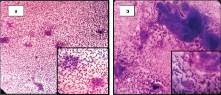
The overall staining, cytoplasmic staining and nuclear morphology staining was good with LG cocktail when compared to MGG stain [Table/Fig-2a,b]. The QI of LG cocktail was 0.8 and that of MGG was 0.59.
a) LG stain of thyroid FNAC showing a clear background staining and overall good staining characteristic (10X) and inset shows a well stained cytoplasm and nucleus of the Hurthle cells (40X); b) MGG stain of thyroid FNAC showing a dirty background (10X) and inset shows a Hurthle cells and thyroid follicular cells which are not clear (40X).
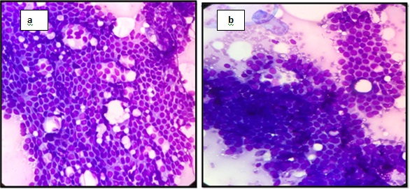
The differentiation between myoepithelial and ductal epithelial cells in the breast were well made out in the LG stain [Table/Fig-3a,b] which helped to arrive at the diagnosis.
a) L-G stain of breast FNAC showing better differentiation of ductal epithelial and myoepithelial cells (40X) when compared to MGG stain; b) 40X.
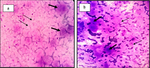
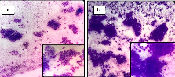
The background material like colloid (thick and thin colloid) in thyroid [Table/Fig-4a,b] and intercellular secretions in salivary gland acini [Table/Fig-5a,b] were better demonstrated in the LG cocktail when compared to the MGG and this helps to differentiate from the background over staining which can be confused for background material. The cytoplasmic and nuclear features of the lymphoid cells along with lymphoglandular bodies were well made out in the LG cocktail stain when compared to MGG [Table/Fig-6a,b].
a) L-G stain of thyroid FNAC showing better appreciation of thick colloid (thick arrow) and thin colloid (thin arrow) (40X) when compared to MGG stain; b) 400X.
a) LG stain of salivary gland FNAC showing good intercellular secretions in the salivary gland acini (40X) when compared to MGG stain; b) 40X.
a) LG stain of the lymph node showing better nuclear and cytoplasmic details of the lymphoid cells (40X) when compared to MGG; b) 40X.
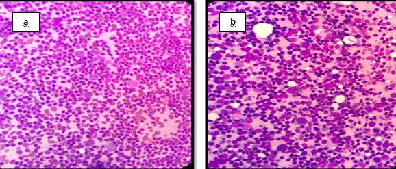
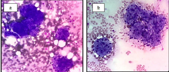
The nuclear features of malignant cells like nuclear chromatin and nucleolus were better appreciated in the LG cocktail stain when compared to MGG [Table/Fig-7a,b].
a) LG stain of breast FNAC showing malignant cells in groups (10X). Inset shows good nuclear details of the malignant cells (40X); b) MGG stain of breast FNAC showing malignant cells in groups (10X) and inset shows cellular details of individual cells are which are not made out properly (40X).
Discussion
FNAC is minimally invasive and the preferred technique for the diagnosis of wide spectrum of benign and malignant lesions. There are many factors which affect the interpretation of the FNAC smears. Among which the method of sampling and quality of staining are important. Sampling method is mainly dependent on the experience of pathologist and quality of staining depends on the type of stain and method followed for staining [1]. For staining the alcohol fixed FNAC smears commonly used stain is H&E whereas, for staining the air dried smears Romanowsky stain i.e., MGG stain is used [4].
Romanowsky stains are stains with differential staining capabilities of a combination of dyes. The major problem with these stains is instability. Later Giemsa, Leishman and MGG stains were developed over the ensuing years [4].
Leishman stain is used widely for staining the peripheral smears. It is also used intraoperatively for imprint cytology in ovarian neoplasms [7], in fluid cytology for cell count and cell type [8]. Leishman stain is a good nuclear stain, when used alone it stains the nucleus and extracellular ground substance intensely but understains the individual cells, 3-dimensional clusters and cytoplasmic granules. It is used very less in staining the FNAC cytology smears due to these limitations [2].
Giemsa stain is a good cytoplasmic stain but provides lighter staining of the cell nucleus and cytoplasmic granules. Hence, combination of these two stains forms the LG cocktail which is relatively a new staining technique which provides good nuclear morphology, crisp nuclear and cytoplasmic contrast, staining of cytoplasmic granules and metachromasia to the background material [9].
In a study done by Garbyal RS et al., cytoplasmic staining was found to be good with LG cocktail and excellent with MGG. But in the present study, it was observed that LG cocktail stain was better than MGG for cytoplasmic staining [2]. Garbyal RS et al., and Belgaumi UI et al., conducted a study which showed that nuclear staining, morphology and background staining were excellent with LG cocktail [2,5]. In the present study too, LG cocktail stain had same features when compared to MGG stain. Cytoplasmic granules were stained better in LG cocktail.
In any malignant case the diagnosis depends on the nuclear features. The nuclear features like variation and enlargement of the nucleus are exaggerated in air dried smears. If the background stain is too intense the visualization of the nuclear features in cell clusters is prevented. In such cases LG cocktail is a good staining option [1].
The overall staining and clarity of staining was good with LG cocktail when compared to MGG stain which is in accordance to the study done by Garbyal RS et al., [2]. In a pilot study done by Gajendra S et al., using LG cocktail for staining of peripheral blood and bone marrow smear examination, better nuclear and cytoplasmic staining was appreciated in LG cocktail as compared to Leishman stain or Giemsa stain when used alone. It also helped in morphological diagnosis and sub classification of mainly leukaemia cases. In anaemia cases polychromasia and RBC inclusions were better demonstrated. Ring forms of the plasmodium also showed better staining with LG cocktail [9].
The study done by Belgaumi UI et al., showed that LG cocktail provides comparable cytoplasmic staining and better nuclear staining to Pap in exfoliative cytology and also has potential application in the screening of oral cancer [5].
MGG stain is used commonly for air dried smears to look at the cytological features of the nucleus. It is an excellent stain, but the disadvantages are that it consumes more time of about 45 minutes, is of high cost, it is a laborious multistep procedure, needs prior fixation, working solution is to be prepared every day and has tendency to precipitate due to which there is high background staining [10].
However, LG cocktail staining requires no fixation, procedure will be complete within 10 minutes and is less expensive. Therefore, in addition to excellent staining characteristics other features that go in favour of LG cocktail are the ease of staining technique, time required for staining and the cost factor. This staining can be preferred when the case load is more and also in cancer screening programmes.
Limitation
The limitation of this study is the sample size and duration. The other stains like H&E and Pap stain were not taken for comparing the cytological parameters.
Conclusion
In this study comparing the LG cocktail and MGG staining of air dried fine needle aspiration smears, LG stain was found to have good overall staining characteristic with a QI of 0.8 as compared to 0.59 of MGG stain. Hence, this cocktail can be used for staining routinely of air dried smears to provide good staining quality which adds overall efficacy of the report generated. It also proves to be economical by saving time and manpower.