Introduction
With the use of various surgical techniques, types of implants, the preoperative assessment of cochlear dimensions is becoming increasingly relevant prior to cochlear implantation. High resolution CISS protocol MRI gives a better assessment of membranous cochlea, cochlear nerve, and membranous labyrinth. Curved Multiplanar Reconstruction (MPR) algorithm provides better images that can be used for measuring dimensions of membranous cochlea.
Aim
To ascertain the value of curved multiplanar reconstruction algorithm in high resolution 3-Dimensional T2 Weighted Gradient Echo Constructive Interference Steady State (3D T2W GRE CISS) imaging for accurate morphometry of membranous cochlea.
Materials and Methods
Fourteen children underwent MRI for inner ear assessment. High resolution 3D T2W GRE CISS sequence was used to obtain images of cochlea. Curved MPR reconstruction algorithm was used to virtually uncoil the membranous cochlea on the volume images and cochlear measurements were done.
Results
Virtually uncoiled images of membranous cochlea of appropriate resolution were obtained from the volume data obtained from the high resolution 3D T2W GRE CISS images, after using curved MPR reconstruction algorithm mean membranous cochlear length in the children was 27.52 mm. Maximum apical turn diameter of membranous cochlea was 1.13 mm, mid turn diameter was 1.38 mm, basal turn diameter was 1.81 mm.
Conclusion
Curved MPR reconstruction algorithm applied to CISS protocol images facilitates in getting appropriate quality images of membranous cochlea for accurate measurements.
Introduction
An important consideration in the management of children presenting with congenital sensorineural hearing loss is cochlear morphology. Assessment of cochlear morphology is an essential tool prior to cochlear implantation [1]. High resolution Computed Tomography (CT) and MRI of the inner ears is routinely performed before the child is considered for cochlear implantation in order to rule out morphological abnormalities of the cochlea [2].
Membranous cochlear length and diameter measurements will further facilitate the surgeon to decide on the type of implant and the length of electrode required and depth of insertion for a particular child to get optimal results [3].
Various methods have been employed to measure cochlear dimensions in imaging. However, due to the complexity of the shape of the membranous cochlea, each of these methods of measurement has their own drawbacks [3–5].
Curved multiplanar reconstruction is an algorithm applied to get virtually straightened images of curvilinear structures from 3D volumetric data obtained by CT or MRI. This helps in better appreciation of such structures which are otherwise difficult to trace due to their differing orientations in different planes. This algorithm has been used to demonstrate structures like nerve roots to look for compressions in spinal imaging, main pancreatic duct, urinary tract etc. [6–8]. However, no studies have been done applying this algorithm to uncoil membranous cochlea, which is likely to give more accurate length measurements of the structure.
Aim
To ascertain the value of curved multiplanar reconstruction algorithm in high resolution 3D T2W GRE CISS imaging for accurate morphometry of membranous cochlea.
Materials and Methods
An observational study was conducted in a tertiary care hospital, between January to June 2016, involving children presenting with congenital sensorineural hearing loss and referred for MR imaging. High resolution 3D T2W GRE CISS imaging of the inner ears was performed in addition to comprehensive brain screening sequences followed in the department using a MRI scanner (Philips, Achieva, 1.5T).
Eighteen such children underwent MRI during period of study. Out of these 18 children, 2 children were excluded as they had obvious cochlear anomalies (common cavity, incomplete partition). Another 2 cases were not considered from study for poor quality images obtained due to technical and patient related factors. The MRI studies of the remaining 14 children, who had no gross cochlear anomalies and who were considered prospective candidates for cochlear implants, were evaluated in detail. Thereby, a total of 28 cochlea were assessed, which is the effective sample size in this study. This sample size was considered sufficient, based on precision based sample size calculation formula, to meet the objectives of this study.
MRI Sequence Protocol
High Resolution 3D T2W GRE CISS sequence
Aquisition -Axial plane
Radiofrequency coil – Head coil
Time of Repetition (TR) – 1100 ms
Time to Echo (TE) - 264 ms
Slice thickness- 1 mm
Base resolution – 320
Phase resolution – 101%
Phase encoding direction R>>L
Voxel size – 0.6x0.6x1 mm
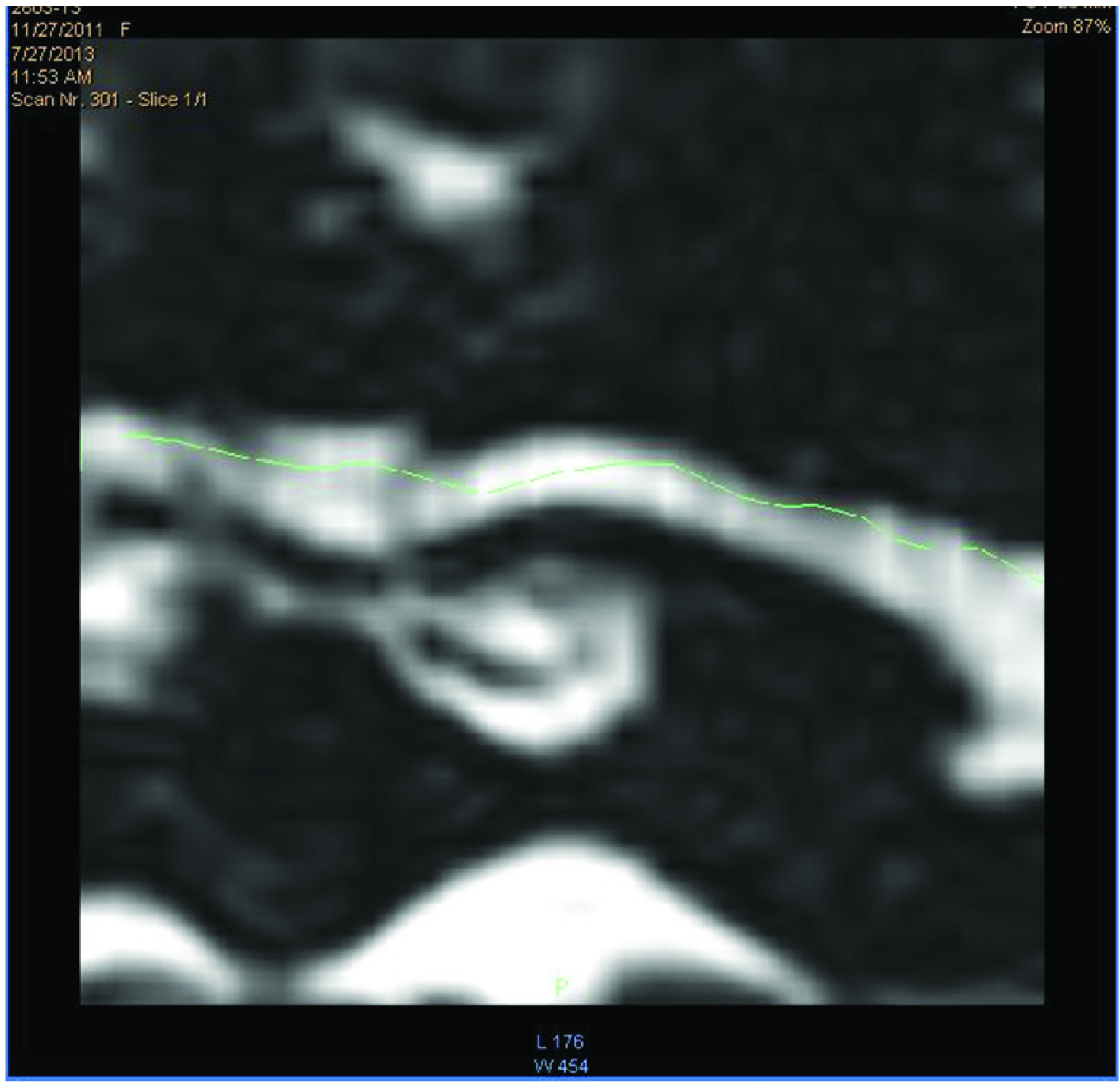
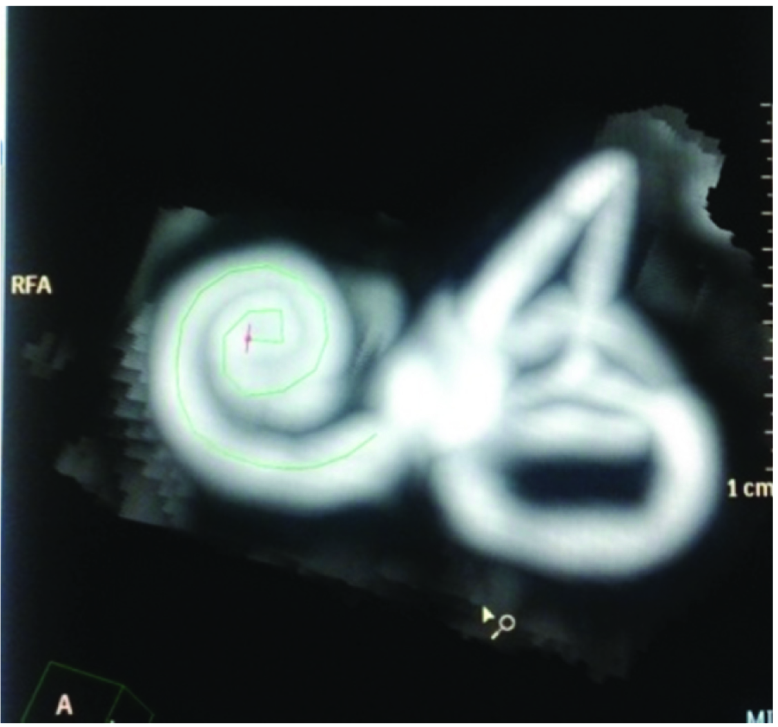
Curved MPR reconstruction algorithm was used to virtually uncoil the membranous cochlea from the volume data obtained from the high resolution 3D T2W GRE CISS images as shown in [Table/Fig-1,2].
High resolution 3-Dimensional T2 Weighted Gradient Echo Constructive Interference Steady State: Curved MPR reconstruction image of the membranous cochlea.
High resolution 3-Dimensional T2 Weighted Gradient Echo Constructive Interference Steady State sequence of MRI of inner ear: Volume rendered MIP image of the membranous cochlea – Axis of projected uncoiled membranous cochlea seen overlapped on the Volume rendered MIP image.
Results
Virtually uncoiled images of membranous cochlea of appropriate quality were obtained after applying curved multiplanar reconstruction algorithm to the volume data images acquired through high resolution 3D T2W GRE CISS protocol. Images were considered of appropriate quality if all the coils of the membranous cochlea could be identified as distinct segments when compared with the source images, as also the apex of cochlea and the junction with vestibule.
The membranous cochlear length was measured using digitized ruler in millimetres, along the axis of the virtually uncoiled cochlea from its apex to the point of its opening into the vestibule, which is identified as an area of abrupt change in calibre. The maximum diameters of each of the turns of the membranous cochlea were measured in the source images of the same sequence.
Study had 14 children with age range between 2 to 9 years. Eight of them were males and six were of female gender. A total of 28 cochlea were studied and measurements were taken. There were no morphological abnormalities detected in any of the cases included in the study.
Mean membranous cochlear length in the children was 27.52 mm. The longest cochlea measured 29.4 mm in a three-year-old, consistent with the observation that, cochlea are fully developed at birth. The shortest measured length was 25.36 mm.
Maximum apical turn diameter of membranous cochlea observed was 1.13 mm, mid turn diameter was 1.38 mm, basal turn diameter was 1.81 mm. Measurements of individual cochlea are depicted in [Table/Fig-3].
Cochlear dimensions observed.
| Age (yrs) | Gender | Side | Cochlear length in millimetres | Diameter of apical turn in millimetres | Mid turn diameter in millimetres | Diameter of basal turn in millimetres |
|---|
| 1. | 5 | M | RtLt | 28.3329.20 | 1.161.19 | 1.241.56 | 1.862.25 |
| 2. | 2 | F | RtLt | 26.4525.97 | 1.121.07 | 1.251.35 | 1.671.89 |
| 3. | 5 | F | RtLt | 25.3626.78 | 1.191.17 | 1.381.46 | 2.341.96 |
| 4. | 9 | F | RtLt | 28.3627.74 | 1.201.17 | 1.761.62 | 2.342.12 |
| 5. | 3 | F | RtLt | 26.6825.83 | 1.171.21 | 1.561.51 | 2.272.10 |
| 6. | 3 | M | RtLt | 25.6327.12 | 1.211.23 | 1.451.41 | 1.871.95 |
| 7. | 3 | M | RtLt | 29.4428.92 | 1.091.15 | 1.381.36 | 2.041.83 |
| 8. | 8 | M | RtLt | 28.4927.96 | 1.071.12 | 1.231.19 | 1.641.59 |
| 9. | 3 | M | RtLt | 27.6226.95 | 0.941.12 | 1.121.25 | 1.671.87 |
| 10. | 4 | F | RtLt | 27.4826.28 | 1.071.15 | 1.361.32 | 2.342.12 |
| 11. | 3 | F | RtLt | 28.6827.94 | 1.181.21 | 1.291.28 | 1.91.87 |
| 12. | 5 | M | RtLt | 26.8026.23 | 1.020.92 | 1.501.53 | 1.781.85 |
| 13. | 3 | M | RtLt | 28.3628.12 | 1.121.13 | 1.341.23 | 2.041.95 |
| 14. | 7 | M | RtLt | 28.7229.12 | 1.131.18 | 1.271.43 | 1.981.86 |
Discussion
Cochlear morphology assessment is vital prior to performing any surgical intervention to place cochlear implant in a suitable candidate. Most of such candidates are children with congenital sensorineural hearing loss [1]. Various types of cochlear implants with varying lengths of electrodes are available in the market and selection of such implant along with depth of insertion of the electrode should be tailored to each individual based on the cochlear morphological assessment.
Imaging of cochlea not only helps in identifying prospective candidates for implants but also gives a visual assessment of the likely difficulties that can be encountered on the operating table.
High resolution CT imaging of cochlea gives a better insight into bony framework of the inner ear. Anomalies related to cochlear maldevelopment may be picked up easily. Bony cochlear dimensions can be measured on CT accurately with volume rendering algorithms [9]. However, CT is not suitable to evaluate membranous cochlea, membranous labyrinth, cochlear nerve and possible obstructions to implantation [10,11].
To overcome the drawbacks of CT, MRI was proposed to evaluate inner ear abnormalities in prospective candidates for cochlear implantation. A 3D-CISS MRI for inner ear assessment was first used by Casselman JW et al., [12]. Membranous cochlea, cochlear nerve were better assessed on these images.
In addition to identification of developmental anomalies of the cochlea, measurement of the normal appearing cochlea is being seen as a prerequisite, in view of the wide variety of implants and surgical techniques employed for cochlear implantation. Length of electrodes and depth of insertion are some parameters which can be matched for every individual prior to the intervention by doing morphometry of the membranous cochlea [3].
Measurements have been done on the volume images of cochlea obtained by high resolution CISS imaging protocols on MRI [5]. However, it has been very cumbersome to measure due to the curve of the cochlea and spiral shape. Multiple small segments were marked on volume images and the values were added, leading to high rates of manual error and abnormal under/over-assessment of dimensions. Moreover, due to 3D spiral shape of cochlea, assessment of length was arbitrary as only 2D measurements were possible.
In order to overcome this it is proposed to utilise curved MPR reconstruction algorithm to virtually uncoil the 3D structure of membranous cochlea obtained from same CISS protocol images, after which length measurements are possible with more precision.
The mean length of cochlea obtained in our study (27.52 mm) is similar to results obtained in study by Sobrinho P et al., (17-26 mm) and Grover M et al., (29.8), who found that cochlear lengths in Asian population are smaller [5,13].
Curved MPR algorithm is readily available in all MRI machines that can be utilised for inner ear assessment, irrespective of manufacturer. It is an under-utilised application with a large potential. No additional imaging needs to be done to use this protocol. It can be easily applied to all high resolution 3D imaging sequences. This implies no additional financial burden on the consumer.
Limitation
There were some difficulties encountered initially in this study in forming good curved MPR reconstructions. With repetition and more practice, acceptable quality images can be reconstructed which will be beneficial in more accurate length measurements.
The diameters of cochlear turns were however, measured on source images in this study as it was difficult to identify each turn on uncoiled cochlea. With more practical application and expertise, it is possible to mark the turns on uncoiled cochlea and all measurements can be performed on single image.
This study was primarily restricted to the objective of introducing the application of curved MPR algorithm to virtually uncoil membranous cochlea as an aid for measurements, which has not been documented till date, in this field. Hence, no comparative group or study was considered necessary. Inspite of this the measurements obtained in this study were comparable to other studies as described earlier [5,13].
Conclusion
Cochlear morphometry is an important prerequisite prior to cochlear implantation. Curved MPR reconstruction algorithm applied to CISS protocol images facilitates in getting appropriate quality images of membranous cochlea. It is proposed to include cochlear morphometry as a part of reporting of MRI of inner ears in prospective candidates for cochlear implantation and curved MPR images will facilitate accurate measurements.
[1]. Gleeson TG, Bresnihan LM, Gaffney R, Brennan P, Viani L, High resolution computed tomography and magnetic resonance imaging in the pre-operative assessment of cochlear implant patientsJ Laryngol Otol 2003 117:692-95. [Google Scholar]
[2]. Silberman B, Garabedian É, Denoyelle F, Moatti L, Roger G, Role of modern imaging technology in the implementation of pediatric cochlear implantsAnn Otol Rhinol Laryngol 1995 104:42-46. [Google Scholar]
[3]. Connor SE, Bell DJ, O’Gorman R, Fitzgerald-O’Connor A, CT and MR imaging cochlear distance measurements may predict cochlear implant length required for a 360 degrees insertionAJNR Am J Neuroradiol 2009 30(7):1425-30. [Google Scholar]
[4]. Pelliccia P, Venail F, Bonafé A, Makeieff M, Iannetti G, Bartolomeo M, Cochlea size variability and implications in clinical practiceActa Otorhinolaryngologica Italica 2014 34(1):42-49. [Google Scholar]
[5]. Sobrinho P, Lazarini F, Roberto P, Hea Jung Y, de Abreu Júnior L, de Sá Meira L, A method for measuring the length of the cochlea through magnetic resonance imagingRevista Brasileira de Otorrinolaringologia 2009 75(2):261-67. [Google Scholar]
[6]. Inukai C, Inukai T, Matsuo N, Shimizu I, Goto H, Takagi T, Usefulness of curved coronal MPR imaging for the diagnosis of cervical radiculopathyNo Shinkei Geka 2010 38(3):251-57. [Google Scholar]
[7]. Maher MM, Kalra MK, Sahani DV, Perumpillichira JJ, Rizzo S, Saini S, Techniques, clinical applications and limitations of 3D reconstruction in CT of the AbdomenKorean Journal of Radiology 2004 5(1):55-67. [Google Scholar]
[8]. Silverman SG, Leyendecker JR, Amis ES Jr, What is the current role of CT urography and MR urography in the evaluation of the urinary tract?Radiology 2009 250:309-23. [Google Scholar]
[9]. Teissier N, Van Den Abbeele T, Sebag G, Elmaleh-Berges M, Computed tomography measurements of the normal and the pathologic cochlea in childrenPediatr Radiol 2010 40(3):275-83. [Google Scholar]
[10]. Nikolopoulos TP, O’Donoghue GM, Robinson KL, Holland IM, Ludman C, Gibbin KP, Preoperative radiologic evaluation in cochlear implantationAm J Otol 1997 18(6 suppl:S):73-74. [Google Scholar]
[11]. Guirado CR, Martinez P, Roig R, Mirosa F, Salmerón J, Florensa F, Three-dimensional MR of the inner ear with steady-state free precessionAJNR 1995 16:1909-13. [Google Scholar]
[12]. Casselman JW, Kuhweide R, Deimling M, Ampe W, Dehaene I, Meeus L, Constructive interference en steady state-3DFT MR imaging of the inner ear and cerebellopontine angleAJNR 1993 14:47-57. [Google Scholar]
[13]. Grover M, Mishra P, Gupta G, Jangid M, Cochlear duct length: are we giving it adequate Importance?Otolaryngology – Head and Neck Surgery 2013 149:P219 [Google Scholar]