Effectiveness of Video Demonstration over Conventional Methods in Teaching Osteology in Anatomy
Angela A Viswasom1, Abraham Jobby2
1 Professor and Head, Department of Anatomy, Travancore Medical College, Kollam, Kerala, India.
2 Professor and Head, Department of Forensic Medicine, Travancore Medical College, Kollam, Kerala, India.
NAME, ADDRESS, E-MAIL ID OF THE CORRESPONDING AUTHOR: Dr. Angela A Viswasom, Professor and Head, Department of Anatomy, Travancore Medical College, Thattamala P.O, Kollam - 691020, Kollam, Kerala, India.
E-mail: drangelakb@gmail.com
Introduction
Technology and its applications are the most happening things in the world. So, is it in the field of medical education. This study was an evaluation of whether the conventional methods can compete with the test of technology.
Aim
A comparative study of traditional method of teaching osteology in human anatomy with an innovative visual aided method.
Materials and Methods
The study was conducted on 94 students admitted to MBBS 2014 to 2015 batch of Travancore Medical College. The students were divided into two academically validated groups. They were taught using conventional and video demonstrational techniques in a systematic manner. Post evaluation tests were conducted.
Results
Analysis of the mark pattern revealed that the group taught using traditional method scored better when compared to the visual aided method. Feedback analysis showed that, the students were able to identify bony features better with clear visualisation and three dimensional view when taught using the video demonstration method. The students identified visual aided method as the more interesting one for learning which helped them in applying the knowledge gained. In most of the questions asked, the two methods of teaching were found to be comparable on the same scale.
Conclusion
As the study ends, we discover that, no new technique can be substituted for time tested techniques of teaching and learning. The ideal method would be incorporating newer multimedia techniques into traditional classes.
Learning objectives, Multimedia tools, Video assisted learning
Introduction
Over the years, methods of teaching Anatomy have gone through three stages, from simple observation to dissection of cadavers and now to Computer Assisted Learning (CAL) [1]. Visual aided methods, be it computer assisted learning or video films are effective way to overcome the negative effects of the increasing class size, expanding curriculum, time pressure on the students and shortage of trained faculty [2]. The new advances in technology have helped create teaching material which is more accessible and interactive. Though many commercial packages are available these are frequently not in a language and accent acceptable to the student and do not mostly cater to the Learning Objectives of the program. The application of new information technology has revolutionized the traditional methods of teaching Human Anatomy [3].
The traditional or conventional method of teaching Osteology is when a teacher demonstrates bony features in small groups with the student having firsthand experience with a bone in their hands. Films or videos have been used for many years as a method of supplementing the teaching of Anatomy. The use of multimedia makes the subject more appealing for the habitual computer user who realises the information from non-conventional sources [4]. Among the potential advantages of video and multimedia techniques is the step by step demonstration of process, attracting and holding class attention. The advantages of the use of the multimedia tools include more retention and long term learning [5].
Multimedia has been used to teach osteology to students using powerpoint presentations and commercially available CDs. Power point presentations have been identified to lack spontaneity, flexibility and non-linearity [6]. The use of commercial CD was avoided in this study because most of the available CD did not conform to the learning objectives envisaged for the study. Thus, an innovative visual aided method was developed which required total involvement by the teacher and could be adjusted as required by the progress of the class.
Aim
To assess and compare the impact, learning outcome and student satisfaction from use of interactive video demonstration techniques in teaching osteology with traditional methods.
Materials and Methods
The comparative study was done on 94 students admitted to First MBBS in the 2014 to 2015 batch at Travancore Medical College, Kollam district. After obtaining due permission from the Institutional Ethics committee, informed consent was taken from the students and the study was conducted. The students were divided into two groups A and B. Validation of the groups was done taking into consideration their second sessional marks.
The students were arranged in the ascending order of marks and alternate students were assigned to each group. The students were taught an overview of the bones of upper limb and lower limb using conventional method (teacher explains various bony features with help of diagram on black board assisted by demonstration of features to the students in small groups) and visual aided method (teacher explains various bony features with help of a magnified image projected on the screen). The projector used for the purpose was EPSPON Document camera ELPDC11. The bones were focussed using the document reader and the magnified image projected on a white board.
Group A was taught the bones of the upper limb using visual aided technique and the examination was conducted. The same group was again taught the bones of the lower limb using traditional method and comparison of the two methods obtained using the feedback form which was peer reviewed. On the same day Group B was taught the bones of the upper limb using traditional method and the exam was conducted. Following this Group B was taught the bones of the lower limb using visual aided method and the feedback obtained.
Valuation of the examination was done by the teacher who has taken the class using an answer key which has been prepared during question paper validation. The marks obtained by the two groups were tabulated, mean obtained and analysed statistically with Mann Whitney U Test. The p-value <0.05 is to be considered as significant. The feedback experience of the students were also tabulated and analysed.
Results
The mean marks of the students taught using traditional method was 5.43 while for the visual aided method was 4.59. The marks were subjected to tests of normality and the following data obtained.
From the normality tests, it is confirmed that, the two groups do not follow the normal distribution, so we choose non parametric method for analysing the data [Table/Fig-1].
Table showing distribution of data.
| Group | Kolmogorov-Smirnov | Shapiro-Wilk |
|---|
| Statistic | DF | Sig. | Statistic | DF | Sig. |
|---|
| Conventional Teaching | 0.161 | 49 | 0.003 | 0.951 | 49 | 0.039 |
| Video Demonstration | 0.133 | 46 | 0.041 | 0.952 | 46 | 0.057 |
DF: Degree of Freedom, Sig.: Significance
From the mean, it can be understood that, students scored higher marks in conventional method of teaching than video demonstration method.
The data when analysed with Mann Whitney U test showed a p value less than 0.05, which means that, there is a statistically significant difference in marks between the two groups [Table/Fig-2].
Table showing significance difference between conventional teaching method and video demonstration method.
| Group | Frequency | Mean | Standard Deviation | p-value(Mann-Whitney U Test) |
|---|
| Conventional Teaching | 49 | 5.43 | 2.041 | 0.047 |
| Video Demonstration | 46 | 4.59 | 1.796 |
The next part of the study was the analysis of the feedback form. Each question in the feedback form was analysed separately.
Analysis of Feedback
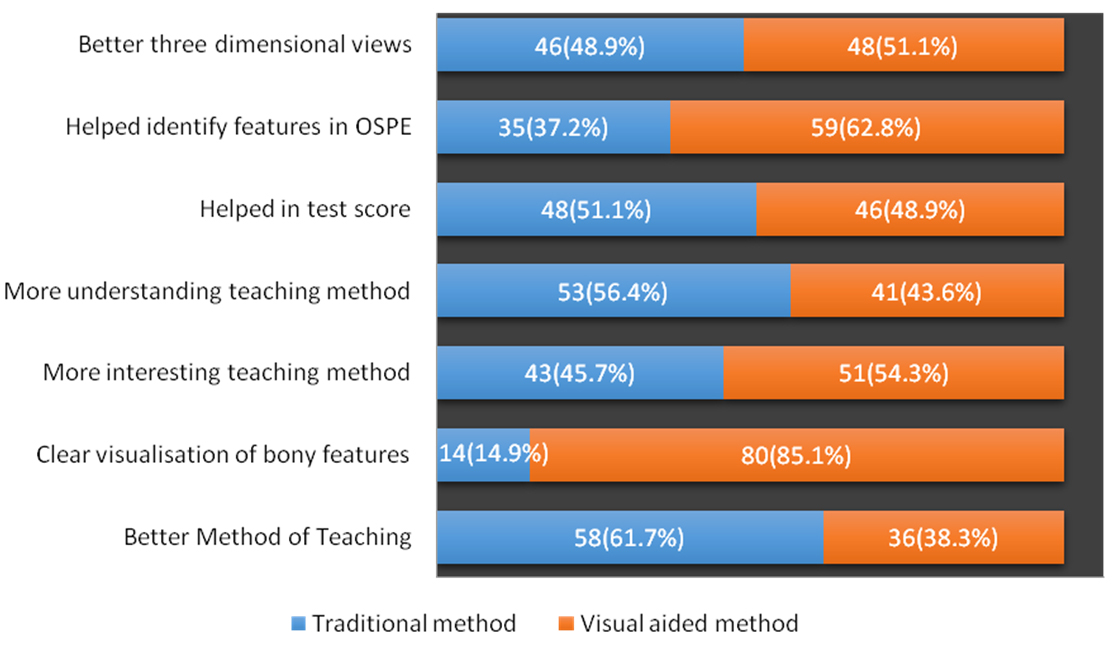
It is clearly seen that, the video demonstration enabled the students to visualize the bony features clearly; better than the conventional method. It also aided them in the Objective Structured Practical Examination (OSPE). The conventional method was identified as the better method of teaching which helped understanding [Table/Fig-3].
Showing the results of feedback comparing traditional and visual aided methods.
Discussion
The human anatomy is an ideal subject with which the use of new teaching tools will be beneficial to both students and instructors. Changing the Macroscopic Anatomy curriculum is a challenging task and it is necessary to evaluate educational methods to determine which are the most effective and efficient. The study was planned to identify innovative teaching techniques in osteology to large groups of students. It has been observed that the study of anatomy benefits from incorporation of new methodologies based on computer and information sciences [1]. The recent changes in the staff pattern and the unavailability of trained faculty due to the mushrooming of new medical colleges require newer techniques for effective teaching. Another limiting factor for small group teaching in osteology is the lack of availability of dried bones in adequate number to the students.
In the present study the students have scored statistically significantly (p-value 0.047) better marks in the traditional method than in the visual aided method. There is no positive improvement in the marks of students using the advanced technology. This is in conformity with previous reports that there is no clear superiority or beneficence of the use of multimedia techniques [2]. In a study conducted on 176 third year medical students, it was noted that, no additional learning benefits was seen in students taught with video assisted methods when compared to those with traditional methods. No clear superiority was noted for video assisted methods over traditional methods [7]. In a study comparing traditional cadaver dissection with computerized self directed course no clear advantage could be identified for the technological resources [8]. Earlier studies have clearly shown, the effectiveness of visual aided method in the academic performance with statistical significance. According to a similar study conducted in Delhi, the mean test marks were in the line of 51.35% using traditional method and 57.23% using visual aided method and the difference was statistically significant [9]. Similarly, a study conducted for evaluating interactive media in Saudia Arabia, the average total score by traditional method was 2.97 as compared to multimedia supported anatomy teaching which was 4.1 and the results were found to be statistically significant [10]. Clear increase in academic performance among students receiving visual procedures like animations during theoretical teaching has been observed [11].
Analysis of the feedback reveals that the students were able to visualise bony features better and this is in clear agreement with most of the previous studies [9]. The study also opined that practical classes were better when using multimedia methods [10]. As is observed in previous studies identification of features was easier with a better three dimensional view [9].
Analysing the result from a student’s point of view it can be understood that the student prefers the traditional way of teaching reinforced with visual aids. The reason for the preference could be that the student is used to learning with close interaction with the teacher as is the technique used in the traditional method of teaching osteology. Another factor here is that, the teacher is not well versed in handling classes with the new techniques and needs to be trained better in multimedia teaching. From the teachers point of view the visual aided method was less cumbersome and time was available for repetitive reinforcement.
To identify specific areas in the field of osteology which can be taught by video assisted learning, this study should be extended and conducted across a batch and newer teaching methods be incorporated into the first year teaching schedule.
From the above discussions, it can be clearly understood that the implementation of multimedia is not a substitute for dedicated faculty and positive interaction with the students. However, the use of Visual aided methods in teaching anatomy will become more effective in regions where visualisation otherwise is difficult like perineum. Even when implementing newer techniques, it should be done with proper planning and training of the faculty involved. Training of faculty can be done within the department by conducting microteaching sessions and peer evaluation.
Conclusion
Though the question remains unanswered i.e., which is a better teaching method: Traditional or visual aided, this study has thrown light on the fact that a blended system of teaching will be useful in addressing the issues highlighted at the beginning of the study. It should be considered that, the study should be done on various topics before a conclusion can be reached. With implementation of new MCI guidelines there will be shortage of both faculty and time for teaching. Innovative techniques, Teaching learning methods and tools should be envisaged to overcome the problems faced in the changing scenario. Implementation of visual aided method with proper planning and training of faculty would be useful to overcome the pitfalls of shortage of time and faculty.
DF: Degree of Freedom, Sig.: Significance
[1]. Trelease R.B, Anatomical Informatics: Millenial perspectives on a newer frontierAnat Rec 2002 269:224-35. [Google Scholar]
[2]. Smith TL, Ruocco A, Jansen BJ, Digital video in education. Proceedings of the ACM Computer Science Education Conference, New Orleans 1999 :122-126. [Google Scholar]
[3]. Paalman J, New frontiers in Anatomy educationAnat Rec (New Anat) 2000 261:47 [Google Scholar]
[4]. Elizondo-Omana RE, Morales-Gomez JA, Lopez Guzman S, Hernandez IL, Ibarra RP, Cavazos Vilchez F, Traditional teaching supported by computer assisted learning for macroscopic anatomyThe Anatomical Record (Part – B New Anat) 2004 278B:18-22. [Google Scholar]
[5]. Galvan SM, Visciglio S, Andreotti C, The effects of the use of image technologies in the learning of factic subjects upon students of vetinary anatomyRev Chil Anat 1999 17:1-15. [Google Scholar]
[6]. Estes A, Ressler S, Welch R, Hanus J, Seminar on communication skillsExceed teaching workshop 2009 http://www.asce.org/file/ppt/exced/USMA-09-Seminar-VI-chalkboard.ppt [Google Scholar]
[7]. Sultana CJ, Levt J, Rogers R, Video vs CD ROM for teaching pelvic anatomy to third year medical students a comparisonJ Reprod Mod 2001 46(7):675-77. [Google Scholar]
[8]. Bukowski EL, Assessment outcomes: Computerized instruction in a human gross anatomy courseJ Allied Health 2002 31:153-58. [Google Scholar]
[9]. Chopra J, Rani A, Rani A, Verma RK, Traditional versus computer assisted teaching of human osteology: a randomized control trial studyIndian Journal of Basic and Applied Medical Research 2012 5(2):370-74.www.ijbamr.com [Google Scholar]
[10]. Al-Hayani A, Abd El Aziz GS, Evaluation of using the interactive multimedia in teaching AnatomyBanha medical journal 1/2008 [Google Scholar]
[11]. Periera JA, Meri A, Masdeu C, Molina-Tomas MC, Martinez Carrio A, Using video clips to improve theoretical anatomy teachingEur J Anat 2004 8(3):143-46. [Google Scholar]