Case 1
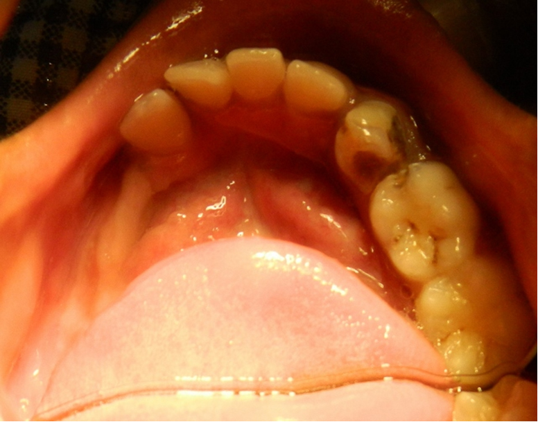
A boy of 10-year-old reported with chief complaint of missing teeth in his lower right back tooth region since one year. History revealed removal of deciduous molar tooth due to decay one year back. On clinical examination, it was noted that the child had decreased vertical dimension of occlusion on the right side due to the absence of the entire set of posterior mandibular teeth of the same side [Table/Fig-1].
Showing mandibular arch with Kennedy’s class 2 situation;
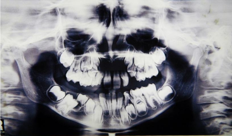
On radiographic examination, it was noted that the right first permanent molar was impacted due to the obstruction posed by the developing premolar. It was noticed that an epithelial-lined radiolucent well-defined developmental cavity surrounded the crown of the unerupted first permanent molar at the cementoenamel junction and could also be the possible aetiological factor to prevent its eruption [Table/Fig-2].
Showing preoperative orthopantamograph with impacted mandibular right permanent molar (46) obstructed by premolar in the pathway of eruption.
Therefore, a treatment plan was drafted to surgically remove the impacted premolar along with the epithelial lining and expose the first permanent molar and later subject it to histopathological examination. To improve the child’s appearance, enhance the clarity of speech, and maintain the health of the underlying alveolar ridge till the eruption of other permanent teeth and to improve the masticatory function; it was decided to fabricate a Cu-sil like denture for the lower arch and a Nance palatal space maintainer on the upper arch.
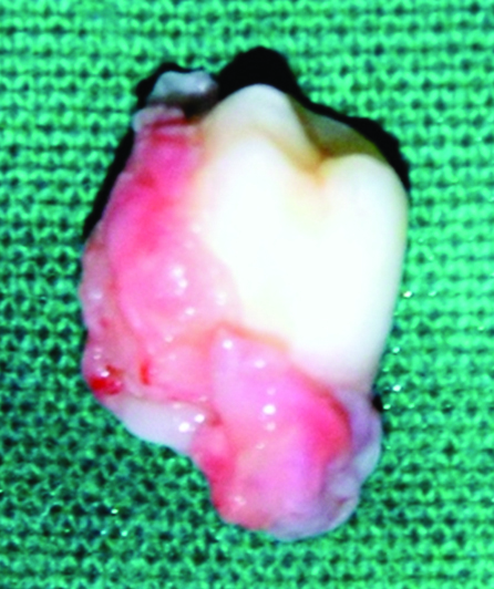
The patient was scheduled for surgical extraction of the premolar; which was removed under local anaesthesia. Post extraction sutures were placed and allowed to heal for one week. The extracted tooth along with the epithelial lining was sent for histopathological examination [Table/Fig-3].
Showing extracted mandibular permanent premolar (45) along with the epithelial lining attached to the tooth;
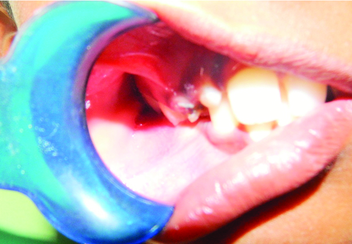
After satisfactory healing of the socket, the sutures were removed and the prosthetic phase was initiated. A few problems were anticipated in the prosthetic phase as the patient showed decreased vertical dimension of occlusion with the upper right primary second molar and first permanent molar occluding directly onto the lower alveolar ridge on the same side [Table/Fig-4]. On the left side the terminal plane relationship observed was mesial step molar relation and flush terminal plane with first permanent molars.
Showing decreased vertical dimension of occlusion with the upper right primary second molar and first permanent molar occluding directly onto the lower alveolar ridge.
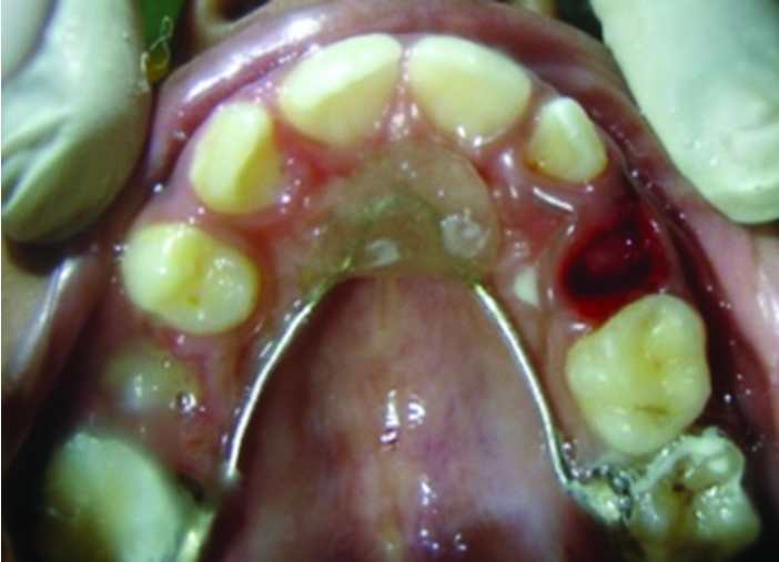
Primary impressions were made using irreversible hydrocolloid material and primary casts were obtained. The lower arch showed Kennedy’s Class II situation and a decision to make a Cu-sil like denture was made with the opinion of prosthodontist. The upper arch showed decayed mobile right primary canine and erupting left second premolar. The first premolar was prematurely erupted. It was decided to extract the right primary canine under local infiltration followed by the Nance palatal arch space maintainer [Table/Fig-5].
Showing Nance palatal arch space maintainer with extracted maxillary deciduous first molar (54) and erupting maxillary permanent second premolar (25);
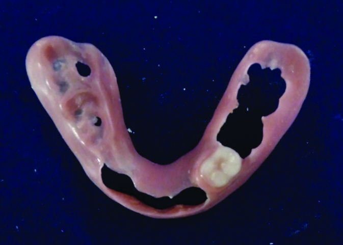
Considering the age of the child and the teeth, which were in active, erupting phase, it was decided to construct the temporary denture based on the primary cast itself without the need for border molding and secondary impressions. The aim of the Cu-sil like denture was to increase the vertical dimension, prevent the occurrence of any deleterious oral habits and also to guide the teeth in the proper path of eruption. After obtaining the lower cast a temporary denture base was constructed in self-cure acrylic resin and checked for the fit in the patient’s mouth. The fit was satisfactory; however, it was not possible to place acrylic teeth on the right side as there was insufficient space due to the decreased vertical dimension of occlusion. Hence, the denture base itself acted as a vertical stop with grooves to accommodate the opposing tooth. Nevertheless, there was a slight increase in vertical dimension which was favourable for the treatment. The extracted left first primary molar was replaced using an acrylic tooth in the Cu-sil like denture, which acted as a removable functional space maintainer [Table/Fig-6]. The dentures were then processed conventionally using heat cure acrylic resin with holes for the remaining natural teeth and flanges which fit accurately into the sulcus. The patient was recalled for routine post-insertion check ups to monitor the erupting permanent teeth.
Showing Cu-sil like denture with vertical stop with grooves to accommodate the opposing tooth on the right side.
Case 2
An 11-year-old boy reported to the clinic with a chief complaint of pain in his lower left back tooth region since six to seven months. On clinical examination a pulp polyp was noticed on left mandibular permanent first molar (36) along with missing right and left permanent mandibular canine, premolars, and right first molar (46,45,44,43,35,34,33). The mandibular arch showed Kennedy’s Class II modification I situation [Table/Fig-7].
Showing mandibular arch with Kennedy’s Class II modification I situation;
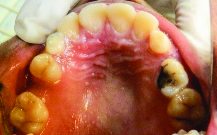
On taking a detailed case history the parent of the child revealed that the child had undergone multiple teeth extraction at the age of seven years. Ever since then the left lower posterior arch had been completely edentulous and the right lower posterior arch had only the presence of the first permanent molar. The lower central and lateral incisors were completely erupted along with the erupting lower left canine. The patients maxillary arch revealed mixed dentition with the presence of the maxillary deciduous canine on both sides (53,63) and second molar on the left side (65) [Table/Fig-8]. The maxillary right second premolar was missing/unerupted and left deciduous maxillary canine (63) and second molar (65) were grade III mobile. The primary right maxillary canine showed smooth surface caries on the labial surface.
Showing maxillary arch with mixed dentition with the presence of deciduous maxillary canine bilaterally (53,63) and left second molar (65);
The treatment plan included root canal therapy for tooth #36 which was diagnosed as chronic hyperplastic pulpitis followed by placement of glass fiber post with core build-up using composite. The deciduous left maxillary canine (63) and first molar (64) were indicated for extraction. The right maxillary deciduous canine (53) was indicated for aesthetic restoration.
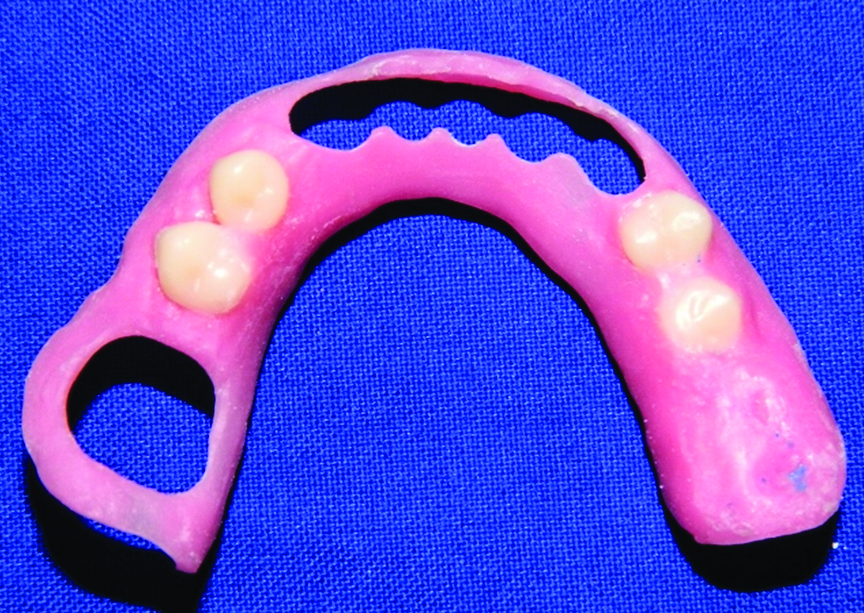
Root canal therapy was completed with respect to mandibular first permanent molar (36) after which the tooth was prepared to receive a stainless steel crown. Prior to the stainless steel crown cementation, a temporary crown was fabricated using tooth colored acrylic resin. This acrylic crown was placed with the aim of increasing the vertical dimension of occlusion by 2-3 mm. After cementation of temporary crown, maxillary and mandibular impressions were made using irreversible hydrocolloid material. The temporary denture base was constructed using modelling wax and the patient’s jaw relations were recorded and artificial acrylic semi-anatomic teeth were arranged for maximum intercuspation. Following which a Cu-sil like denture was fabricated using heat-cure acrylic [Table/Fig-9].
Showing Cu-sil denture for the mandibular arch with holes to accommodate teeth.
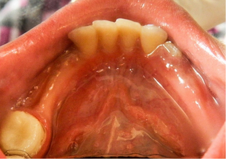
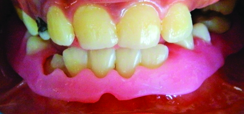
Since, the vertical dimension was increased by 3 mm, the patient was kept under observation during the use of this denture [Table/Fig-10].
Showing increased vertical dimension of occlusion with maximum intercusption;
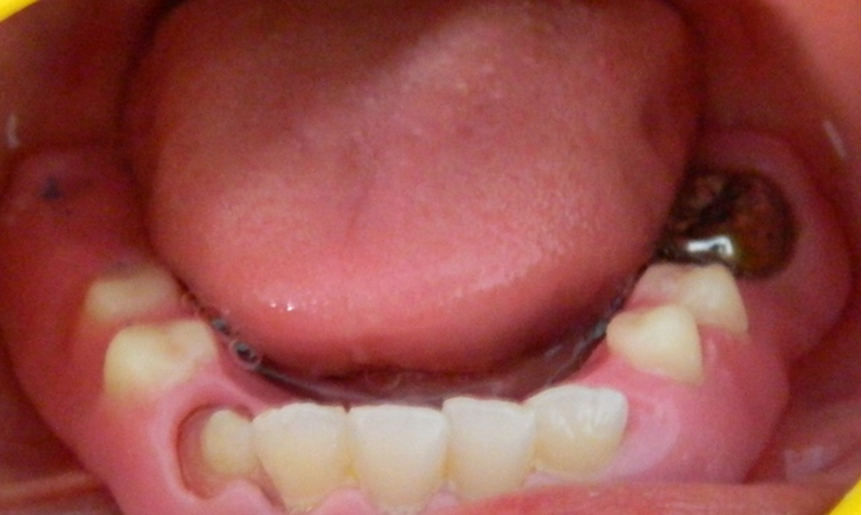
The patient reported with no discomfort or any temporomandibular joint pain. Hence, this denture was considered satisfactory. The temporary acrylic crown on tooth #36 was then replaced with a permanent stainless steel crown [Table/Fig-11]. The patient was recalled for routine post-insertion check ups to monitor the erupting permanent teeth.
Showing cemented stainless steel crown with respect to mandibular permanent first molar (36) with Cu-sil like denture.
Discussion
Children with unique cases where an extensive edentulous spaces created with premature loss of multiple primary teeth and failure of permanent teeth to erupt timely into the spaces; results in a sequelae of malocclusions like reduced vertical dimension of occlusion, defects in speech and phonetics, changes in facial appearance of the patient such as deepening of nasolabial groove, loss of labio-dental angle, narrowing of lips and negative psychological effects in their everyday activities like mastication and aesthetics. Situations when the permanent teeth are still erupting and/or do not satisfy the requirements of an abutment tooth for a fixed space maintainer; a transitional denture can be planned [1].
A relatively new type of transitional dentures is the Cu-sil denture [2,3]. Its trademarked modified denture used for replacement in elderly patients [3]. Cu-sil denture is one of the simplest forms of a removable partial denture. The literature review revealed very few case reports [1,4–8] and a review [9] on types of dentures [10] describing Cu-sil as one of the denture type. A transitional denture with holes to accommodate the existing natural teeth is called as Cu-sil denture [3]. Cu-sil denture is commonly fabricated in adult with very few permanent teeth who do not desire to extract the remaining permanent teeth [2,3]. It is made up of acrylic, featuring a soft elastomeric gasket used to clasp the neck of each natural tooth so that it seals and food and fluids will not enter below the denture base, also gives cushioning and splinting effect [3]. A resilient liner used to fill the gap between the denture base and the tooth so that it seals [2]. Various dentures have been introduced for different purposes [9,10].
In this case report, Cu-sil like denture was fabricated for two children with extensive edentulous arches with very few erupted permanent teeth, (most of the teeth were yet to erupt) which could not be utilized as an abutment teeth for fixed space maintainer. A transitional denture basically serves as a treatment option for patients presenting with very few erupting permanent teeth, in compromised condition and multiple edentulous spaces created by the premature loss of primary teeth. In our cases the natural teeth are accommodated within the denture through perforations made in the denture base. Because of the morphology and angulation of the natural teeth it will not maintain a seal. These children were also candidates for reduced vertical dimension of occlusion with anterior deep bite [11] like in our case. In such scenario Cu-sil like denture can be planned which requires less effort, chair side and laboratory time. It doesn’t require any special impression technique nor tooth preparation.
Advantages of Cu-sil Like Denture
Cu-sil denture cases require no adjustments upon insertion and no post-insertion adjustments too. Upon seating, it adapts comfortably to the tissues [3–10]. Vertical dimension of occlusion and original bite can be maintained till the permanent teeth erupt completely. Tissue response is not altered, therefore, the permanent teeth continue to erupt undisturbed. Denture stability and retention is achieved even when only one or two permanent teeth are present [11,12]. Proprioception is maintained, the potential psychological impact is avoided, and fewer trauma is realized as the patient can achieve clarity of speech, mastication and aesthetics [12]. Cu-sil like partial dentures eliminates the clasps and preserves the enamel of the young newly erupted permanent teeth. These dentures stabilize, cushion and splint teeth with an elastomeric gasket that provides retention and seals out food, therefore, maintains a good oral hygiene [13].
Disadvantages of Cu-sil Like Denture
The functional duration of soft liner used will be of short duration viz., three years. It needs frequent corrections. Entire gingival margin of remaining teeth, which is covered may lead to plaque accumulation. Cu-sil lower dentures are prone to fracture when grounded against the upper, natural teeth in elderly patients [13]. But these disadvantages can be overcome by regular dental visits and maintaining a proper oral hygiene practice [14].
Clinical Implications of Cu-sil Like Denture
Cu-sil like denture regulates and stabilizes erupting permanent teeth into the arch. It is also easy to replace lost primary teeth/unerupted permanent teeth on the Cu-sil like denture, hence, it also acts as a functional removable type of space maintainer. Whenever, the permanent tooth starts erupting in the future, existing denture can be modified to provide space for the erupting tooth. This treatment modality does not require any special armamentarium and materials. In our cases, the patients found it comfortable to wear the appliance along with ease to speak and masticate. Also, there was a remarkable improvement in facial appearance of the patient as the vertical dimension of occlusion was increased. Published literature on Cu-sil like denture used in elderly patients [1,4–8] but one report presented with six-year-old male child with ectodermal dysplasia have been used [4]. In our case, both patients were normal. So, use of Cu-sil like denture in paediatric age group is better clinical perspective as removable functional space maintainer using the same principles as that of Cu-sil denture like in our case supported by literature [4]. Cu-sil dentures are a viable option when a patient presents with less than optimal conditions to rehabilitate them with conventional partial denture [4].
Conclusion
Cu-sil like denture is a promising alternative for paediatric group of patients with unique edentulous conditions wherein multiple primary teeth are missing with very few permanent teeth erupted which cannot be used as abutment teeth for space maintainer. Cu-sil like denture not only acts as a removable functional space maintainer, but also promotes healthy stimulation of the mucosa to maintain alveolar bone to promote eruption of permanent teeth.
[1]. Khandelwal M, Punia V, Saving one is better than none-Technique for Cu-sil like dentures-A case reportAnnals and Essence of dentistry 2011 3(1):41-45. [Google Scholar]
[2]. Walter JD, A study of partial denture design produced by an alumni group of dentists in health service practiceEur J Prosthodont Rest Dent 1995 3:135-39. [Google Scholar]
[3]. Cu-Sil® Available from: http://cu-sil.com. (Latest Accessed on 30-10-2016) [Google Scholar]
[4]. Srividya S, Lakshmi S, Tayab T, Rai K, Ila S, Rehabilitation of patients with Cu-sil dentures: Trends in prosthodontics and dental implantology 2012 3(1):20-22. [Google Scholar]
[5]. Kansal G, Goyal S, Deepika Cu-sil denture: A novel conservative approachUnique journal of medical and dental sciences 2013 1(2):56-58. [Google Scholar]
[6]. Jain AR, Cu-sil denture for patients with few remaining teeth-A case reportInternational journal of research in dentistry 2014 4(4):98-103. [Google Scholar]
[7]. Jain JK, Prabhu CRA, Al Zahrane M, Al Esawy MS, Ajagannanavar SL, Pal KS, Cu-sil dentures – a novel approach to conserve few remaining teeth: Case reportsJ Int Oral Health 2015 7(8):138-40. [Google Scholar]
[8]. Vinayagavel K, Sabarigirinathan C, Manoj kumar PA, Jeyanthi kumara T, Kanmani M, Rajakumar M, Hybrid prosthesis- overdenture cum Cu-sil dentureIOSR Journal of Dental and Medical Sciences (IOSR-JDMS) 2015 14(6):49-52. [Google Scholar]
[9]. Nagapal A, Katna V, Gupta P, Kumar A, Rometra V, Kashyap KK, Specialized dentures: An individualistic approachAnnals of Prosthodontics and Restorative Dentistry 2015 1(1):9-15. [Google Scholar]
[10]. Types of dentures. Available from: https://healdove.com/oral-health/Types-of-Dentures. (Last Accessed on 30-10-2016.) [Google Scholar]
[11]. Levin AC, Shifan A, Lepley JB, Preservation of occlusal vertical dimension in overdenturesJ Am Dent Assoc 1995 5:838-39. [Google Scholar]
[12]. Winkler S, Essentials of complete denture prosthodontics 2009 2nd edU.S.A.Ishiyaku Euro America Inc:22-34.:384-402. [Google Scholar]
[13]. Disadvantages of Cu-sil partial dentures. Available from: http://crowchilddenture.com/News/ArtMID/9873/ArticleID/2460/Disadvantages-of-Cu-Sil-Partial-Dentures.aspx, (Latest Accessed on 30-10-2016.) [Google Scholar]
[14]. Zarb GA, Bolender CL, Prosthodontic treatment for edentulous patients 2004 12th editionMosby:3-5. [Google Scholar]