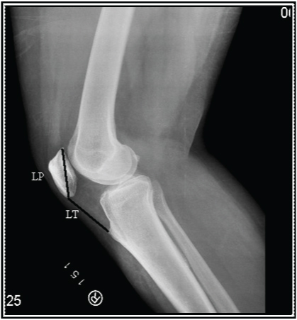
Patellar height is one of the important parameter in assessing patellar stability [1–4]. Its importance has been reflected through attempts by renowned scientists to measure the patellar height by such an index which is simple, accurate, practical and also reproducible. As a result, several radiological indices have been used to measure the height of the patella of which “IS Index” is the most popularly used method so far, as it fulfills all the above criteria [5]. Insall and Salvati in 1971 introduced this index from lateral view X-ray film of knee joint taken at 30° angle of flexion [4]. “IS index” is the ratio of LT and LP where LT indicates length of distal part of tendon of quadriceps femoris (i.e., ligamentum patellae) extending from distal pole of patella to tibial tuberosity and LP means diagonal length of Patella [6].
Though the “IS index” was initially based on radiological findings on lateral view of the knee joint in X-ray films, different studies have been conducted to measure the “IS index”, namely through cadaveric dissection, sagittal MRI and ultrasonography, keeping the principle of the index constant [7–9]. Though it is the most popular method of measurement it has many drawbacks including difficulty to identify the soft tissue [10]. So for accurate measurement it is wise to take the help of MRI which is expensive and cannot be applied to all the cases for routine measurement of patellar height particularly when it is very important in sports medicine.
In the present study, a clinical method, based on the criteria of the IS index has been adopted to compare, contrast and correlate the findings with a radiological method which is well validated, in respect to the patellar height among a population in North Bengal, India.
In this background, the present study is an important attempt to measure the patellar height by “IS index” by two separate methods-viz., radiological and clinical, comparing both the findings and to establish a cut off value of patellar height for clinical methods.
Materials and Methods
A cross-sectional institution based observational study was conducted among the adolescents (age above 10 years) and adults admitted in the orthopaedics indoor of North Bengal Medical College and Hospital, Darjeeling, West Bengal, India with any reason other than knee related disorders, during the year 2013–2014.
The patients were subdivided into two groups, one group with patients aged 10 to 20 years and another group with patients aged ≥20 years. The patients excluded were those having any acute condition, receiving intravenous fluid, those who cannot cooperate during clinical examination, patients with splint and compressive bandages.
Finally a total number of 93 subjects were included in this study. Permission was duly sought from the concerned departments as well as institutional authorities and the Ethical committee at North Bengal Medical College and Hospital before proceeding for the study.
The selected patients were informed about the nature of the study and the procedures involved and permission was obtained for both clinical examination and radiographic investigation. Consents were obtained from all the study subjects. The patient’s name, address, age, sex, date of admission, occupation, chief complaint, history of present illness, past illness, other associated illness, complaint of knee pain if any, were all noted as per the format through direct interview of the patients and assistance was taken from available hospital records.
Examination of both the lower limbs was done meticulously with special references to measurement of patellar height by “IS index”. Next, outline of patella, identifying its upper border, apex, and two lateral borders were marked. A small horizontal line on the patella was drawn in the midway between upper border and apex. Another short vertical line was drawn midway between its medial and lateral borders at the level of maximum breadth. At the point of intersection of the two above mentioned lines, was considered as the centre of patella. The most prominent point on tibial tuberosity was marked. The distance i.e., the maximum available diagonal Length of the Patella (LP) was measured by means of measuring tape. The distance from the lower pole of patella to tibial tuberosity was measured (LT). The ratio of LT/LP was calculated to derive “IS index”. Similarly X-ray of lateral view of knee joint was taken from which “IS index” was calculated [Table/Fig-1].
Lateral view radiograph of knee joint to measure IS index.
LP= Length of patella, LT= Length of ligamentum patellae

Statistical Analysis
SPSS version 16.0 was used for the statistical analysis (student t-test).
Results
In the present study 186 knee joints were subjected to measurement for patellar height by both clinical and radiological methods in respect to gender and two different age groups (above/equal to and below 20 years) [Table/Fig-2].
Age and sex distribution of the study population (n=93).
| Age group | Number of cases |
|---|
| Males | Females |
|---|
| < 20 years | 18 | 24 |
| ≥ 20 years | 24 | 27 |
It was found that, there was no statistically significant difference between the mean values obtained by clinical method of measurement, and the to conventional one (IS Index) for both the genders and age groups in both right and left sides [Table/Fig-3,4].
Measurement values as per “IS index” and clinical method in both the genders and age groups in right lower limb (n=93).
| ClinicalMethod | IS Index | Student t-test valueat df =1 | p-value |
|---|
| Male< 20 years | M-0.993SD-0.168 | M-0.997SD-0.119 | 0.024 | >0.05* |
| Female< 20 years | M-1.033SD-0.208 | M-1.039SD-0.148 | 0.360 | >0.05* |
| Male≥ 20 years | M-1.067SD-0.172 | M-0.989SD-0.072 | 2.091 | >0.05* |
| Female≥ 20 years | M-0.989SD-0.186 | M-1.011SD-0.173 | 0.391 | >0.05* |
df= Degree of freedom, M= Mean, SD= Standard deviation, * = Non significant
Student t-test applied
Measurement values as per “IS index” and clinical method in both the genders and age groups in left lower limb (n=93).
| ClinicalMethod | IS index | Student t-test value atdf =1 | p-value |
|---|
| Male< 20 years | M-1.011SD-0.214 | M-0.996SD-0.133 | 0.436 | >0.05* |
| Female< 20 years | M-1.023SD-0.198 | M-1.047SD-0.165 | 0.722 | >0.05* |
| Male≥ 20 years | M-1.066SD-0.175 | M-1.017SD-0.102 | 1.503 | >0.05* |
| Female≥ 20 years | M-1.013SD-0.196 | M-0.990SD-0.117 | 0.756 | >0.05* |
df=Degree of freedom, M= Mean, SD= Standard deviation, * = Non significant
Student t-test applied
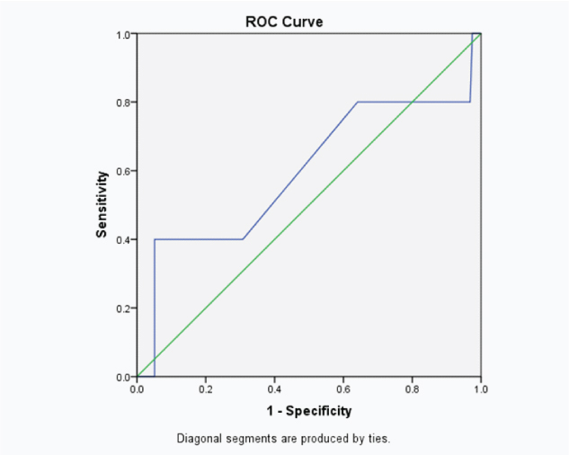
On further assessment, to explore a cut off value for clinical measurement it was seen that, a value of 0.98 cm gives sensitivity of test of 80% and the specificity of 36% with area under the ROC curve 0.596 which is not satisfactory [Table/Fig-5,6].
Different percentages of sensitivity and specificity at different cut off values of clinical measurement.
| Cut off Points | Sensitivity(True Positives) | 1 – Specificity(False Positives) |
|---|
| 0.9350 | 0.800 | 0.692 |
| 0.9850 | 0.800 | 0.641 |
| 1.0100 | 0.400 | 0.308 |
Area under the curve=0.596
The graph showing the relation between sensitivity and (1-specificity) at different cut off values of clinical measurement.

Discussion
There are so many methods of measurement of patellar height. They can be classified broadly into two groups- direct and indirect methods depending upon whether femur or tibia is taken as a reference point in relation to patella respectively [5]. Though femoro-patellar compartment is more important in cases of anterior knee pathologies due to alteration of patellar height but there are some disadvantages of using femur for measuring patellar height. It has to be measured from lateral radiograph in fixed flexion angle-900 and if patella alta is diagnosed, with 900 angle in flexion, the patella looks towards the ceiling not towards the torso [5]. The commonly used four methods namely (The Insall-Salvati, Modified Insall-Salvati, Blackburn-Peel and Caton-Deschamps) are indirect methods [6,11–13].
Among all the methods IS index is the most popular, accurate and simple due to the following factors. It can be obtained from lateral radiograph without any specific flexion angle. It is easy to remember the range of normal value. In the present study we have measured patellar height using conventional radiograph following the IS index.
There are some studies in China done by Leung YF et al., and India done by Upadhyay S et al., have shown that the IS index does not bear the same normal value for Western population and the population of China or India where squatting, cross-legging and kneeling are customs [14,15]. The value is slightly higher for the latter population. But our study failed to show such difference significantly though the value is slightly more following clinical method but this difference was not statistically significant.
Patella Alta is associated with several clinical conditions. Significant cause of recurrent patellar dislocation can be associated with Patella alta [13,14,16–24]. This dislocation due to patella alta had been explained in the light of delay in tendo-femoral contact and concomitant increase in the magnitude of patello-femoral contact with increased flexion of knee [25].
The diagnosis of patella alta should be considered in all cases of knee pain [18]. In a study, 45 patients of unilateral patello-femoral pain syndrome were examined and results suggested that high-riding of the patella due to long patellar tendon (patella alta) was the only definite finding in the affected knees. Kannus PA in 1992 concluded that idiopathic retropatellar pain is closely associated with Patella Alta after analyzing the result of this study [26].
Jerold EL and John AC in 1975 measured a series of roentgenograms of knees with chondromalacia patellae, knees with apophysitis of tibial tubercle (Osgood-Schlatter disease) [24]. The IS index in patients with chondromalacia patellae was 0.86 (patella infera). It may be the cause or effect. But other studies support the view that patella alta is a cause of symptomatic idiopathic chondromalacia patellae [22,27]. The IS index was 1.2 for those with apophysitis of tibial tubercle (Osgood-Schlatter disease) in the study done by Jerold EL and John AC [24]. Patella alta is classically described as the residuum of Osgood-Schlatter disease in other studies too [22,27]. But according to another study where patella infera is associated with Osgood-Schlatter disease may be the result of short tendon due to inflammation and fibrosis and patella infera is not the cause of the lesion [28].
Hence, it seems that patella alta should receive more importance in all the cases of knee pain [19]. Some of the cases were associated with Sinding-Larsen Johansson’s disease; ruptured ligamentum patellae, genu recurvatum pathology and trauma of extensor apparatus of knee, malposition, gonarthrosis or supratrochlear erosion of the femur [14,18].
Limitation
Firstly, it does not include the relevant pathological conditions like Osgood Schlatter’s disease, Larsen Johanssen’s disease or recurrent dislocation of patella. Secondly, it is important to do a large scale study for identifying the accurate cut off value for clinical measurement of patellar height.
Conclusion
There is no statistically significant difference of IS index between radiological and clinical methods. So, one can opt for clinical measurement techniques for measurement of patellar height as a primary procedure. If the measurement is above the cut off value (0.98 in the present study) the person should undergo more sensitive tests like MRI.
df= Degree of freedom, M= Mean, SD= Standard deviation, * = Non significant
Student t-test applied
df=Degree of freedom, M= Mean, SD= Standard deviation, * = Non significant
Student t-test applied
Area under the curve=0.596