Fat Free Pleomorphic Lipoma of Oral Cavity: A Rare Entity
Kannan Ranganathan1, Seema Alice Mathew2, Nellimad Sreedharan Sreena3, Nagarajan Lavanya4
1 Professor and Head, Department of Oral Pathology, Ragas Dental College and Hosptal, Chennai, Tamil Nadu, India.
2 Senior Lecturer, Department of Oral and Maxillofacial Surgery, Ragas Dental College and Hosptal, Chennai, Tamil Nadu, India.
3 Former Postgraduate Student, Department of Oral Pathology, Ragas Dental College and Hosptal, Chennai, Tamil Nadu, India.
4 Reader, Department of Oral Pathology, Ragas Dental College and Hosptal, Chennai, Tamil Nadu, India.
NAME, ADDRESS, E-MAIL ID OF THE CORRESPONDING AUTHOR: Dr. Nagarajan Lavanya, Reader, Department of Oral Pathology, Ragas Dental College and Hospital, 2/102, East Coast Road, Uthandi-600119, Chennai, Tamil Nadu, India.
E-mail: lavanyabds@yahoo.com
Pleomorphic lipoma is a rare, benign, soft tissue neoplasm that characteristically occurs as a subcutaneous mass in the posterior neck or upper back and rarely in the tonsillar fossa and oral cavity. Histologically, pleomorphic lipoma contains varying amounts of mature fat, areas of spindle and pleomorphic cells, floret giant cells and thick rope – like collagen in a myxoid stroma. Pleomorphic lipoma with scanty fatty elements is called the fat free variant of pleomorphic lipoma. The combination of meagre amount of fat and presence of pleomorphic elements gives a pseudosarcomatous picture under the microscope leading to misdiagnosis and over treatment. Here, we report a case of fat free pleomorphic lipoma, first of its kind in the oral cavity and discuss the diagnostic features and differential diagnosis.
Floret giant cells, Ropey collagen, Soft tissue neoplasm
Case Report
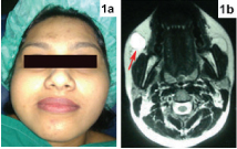
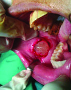
A 24-year-old female presented with a chief complaint of a slowly growing painless swelling of her right cheek for the past one year. Extraoral examination of the face revealed a diffuse swelling 2 cm lateral to the right corner of the mouth [Table/Fig-1a]. There was no punctum or sinus opening. Bimanual palpation revealed a freely movable well circumscribed subcutaneous lesion, with no adherence to the overlying skin or underlying buccal mucosa. Magnetic Resonance Imaging (MRI) scan revealed a well defined T2/STIR hyperintense, T1 isointense radiopaque heterogenous mass, lateral to the facial vein in the right cheek [Table/Fig-1b]. The lesion measured 18.7 mm x 18.4 mm x 14.7 mm in the subcutaneous plane of right cheek. The adjacent skin, surrounding fat plane, buccal mucosa and muscles appeared unremarkable. An excisional biopsy was performed under general anaesthesia through an intraoral approach. Blunt dissection was performed through the buccal mucosa and buccinator muscle. The lesion was removed in toto and was sent for histopathological examination [Table/Fig-2].
a) Diffuse swelling 2 cm lateral to the right corner of the mouth. b) A well-defined T2/STIR hyperintense, T1 isointense radiopaque lesion measuring 18.7x18.4x14.7 mm in the subcutaneous plane of right cheek (red arrow).
Intraoperative photograph showing in toto removal of the lesion.
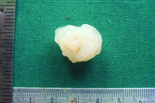
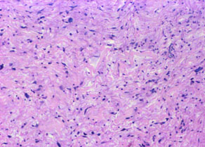
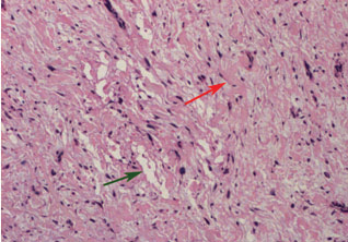
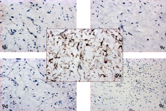
Grossly, the resected specimen was well circumscribed, measuring 2 cm x 1.5 cm x 1 cm, white in colour and had a jelly like consistency [Table/Fig-3]. The formalin-fixed, paraffin-embedded sections were then stained with haematoxylin and eosin. The sections showed well circumscribed mass, composed of scattered areas of pleomorphic spindled cells in a predominantly myxoid stroma. Amidst these areas of pleomorphic cells, numerous thick short fragments of “ropey” collagen were found. Negligible amount of fat like areas and few multinucleated giant cells were observed. A higher magnification revealed that the giant cells had a concentric floret like arrangement of hyperchromatic nucleus around an eosinophilic cytoplasm [Table/Fig-4,5,6,7 and 8]. Immunohistochemical study with CD34, CD68, Ki-67, S100, SMA were done. Positive immunostaining of the tumour cells was observed only with CD34 [Table/Fig-9]. Based on the clinical, histopathological and immunohistochemical findings, a diagnosis of fat free pleomorphic lipoma was given. The patient was recalled three and six months later for a re-evaluation, and patient was found to be disease free.
Specimen photograph showing translucent jelly like colour of the specimen.
Low power view of the lesion shows a mixture of pleomorphic spindled cells and thick ropey collgen (H&E; 10X).
Negligible amount of fat like areas (green arrow) seen amidst pleomorphic spindled cells. Rope like collagen seen here (red arrow) (H&E;10X).
High power view of the pleomorphic spindle cells with hyperchromatic bizarre nucleus (H&E; 20X). (Images left to right)
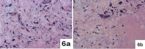
Coarse rope like collagen randomly distributed in diffuse myxoid stroma (H&E; 20X).
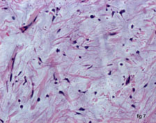
Floret like multinucleated giant cell with radially arranged hyperchromatic nuclei and central eosinophilic cytoplasm, (red arrow) (H&E; 20X).

a) Positive immunoreactivity with CD34 (IHC, 20X); negative immunostaining with; b) CD68 (IHC, 20X); c) Ki-67 (IHC, 20X); d) S-100 (IHC, 20X); e) SMA (IHC, 10X).
Discussion
Pleomorphic lipomas are uncommon soft tissue neoplasms of the subcutaneous tissues of the head and neck region and were first described by Shmookler and Enzinger in 1981 [1]. Only five cases have been reported in the oral cavity [2,3] and three in the perioral regions of tonsil, retropharyngeal region and hypopharynx [4–6]. Low fat/fat free variants are rarer; only nine cases have been reported in the literature. Of these reported cases of fat free variants of pleomorphic lipoma, three were located in head and neck, two in the shoulder, two in the axilla and one was a intradermal tumour of the nose. These cases had a male predominance and a mean age of occurence was 55–60 years [4,7,8]. The present case describes a fat free variant of pleomorphic lipoma in a 24-year-old female and first of its kind in the oral cavity.
Pleomorphic lipomas are variant of the spindle cell lipomas, earlier described as separate entities, spindle cell lipomas and pleomorphic lipomas are now considered to be the same family with overlapping clinical, histologic, immunohistochemical and cytogenetic features [2]. Histologically, on one end of the spectrum, classic spindle cell lipomas, show relatively equal amounts of spindle cells and mature fat tissue, while pleomorphic lipoma shows lots of pleomorphic elements with few spindle cells, multinucleated floret giant cells and varying amounts of fat, sometimes none. Both present with characteristic thick ropey collagen, mast cells and a myxoid/ mucoid matrix. The term spindle cell/pleomorphic lipoma has been suggested for cases showing overlapping feature [1,2]. Our case had very few spindled cells and scanty adipose elements and consisted predominantly of pleomorphic cells and multinucleated floret giant cells. With these features, a diagnosis of fat free variant of pleomorphic lipoma was made. The CD34 positivity of the spindled, pleomorphic cells confirmed the diagnosis.
Although a variant of lipoma, the diagnosis of pleomorphic lipoma is hugely based on the non lipomatous component of the lesion rather than the lipomatous component. Identificatiion of pleomorphic lipoma becomes straight forward when considerable amount of fat is present in the lesion along with its characteristic histological features of ropey collagen and floret giant cells. Diagnosis is usually difficult when the spindled component is more, exhibits rare atypical mitotic figures and amount of fat tissue is barely discernable. Another complicating factor is the CD34 positivity of most lesions in the differential diagnosis.
Histopathological differential diagnosis includes atypical lipomatous tumours/well differentiated liposarcoma, Myxo Fibro Sarcoma (MFS), neurofibroma, giant cell fibroblastoma and giant cell angiofibroma. Atypical lipomatous tumours/well differentiated liposarcomas present with atypical, pleomorphic spindle shaped cells and occasional multinucleated floret giant cells. The identification of ropey collagen in pleomorphic lipoma, cytogenic aberrations (MDM2 amplification) and CD34 negativity in atypical lipomatous tumours/well differentiated liposarcomas differentiates them [1,3,9].
MFS consists of pleomorphic spindle cells, greater mitotic activity and occasional multinucleated giant cells in a myxoid stroma. Characteristic elongated, curvilinear, thin walled blood vessels with peripheral condensation of the hyperchromatic spindle cells are seen. Also, certain MFS are recently reported to be CD34 positive [10]. Prominently vascular pleomorphic lipomas and its fat free variant should be recognized and distinguished from MFS, by presence of ropey collagen and the absence of characteristic curvilinear vasculature and lack of mitotic activity.
Neurofibroma, giant cell fibroblastoma and giant cell angiofibroma are spindled lesions which occasionally present with multinucleated (floret) type of giant cells and CD34 positivity of tumour cells. The presence of mast cells in neurofibroma and in pleomorphic lipomas is also a potential pitfall to diagnosis. S100 positivity and wavy nuclei of the neural cells help to identify neurofibroma [11]. Giant cell angiofibromas are recognized by the pseudovascular spaces filled with amorphous eosinophilic material and perivascular hyalinization [12]. Giant cell fibroblastoma occurs in infants and children and is identified by stellate spindled cells, giant cells that line the angiectoid spaces and perivascular extravasation of lymphocytes in an onion-skin manner [13,14].
Conclusion
Fat free pleomorphic lipoma is a rare lesion and this is the first reported case in the oral cavity. The absence of fat and presence of pleomorphic elements makes the histopathological diagnosis quite challenging. Although a variant of lipoma, the key to diagnosis lies in identifying the non lipomatous elements. The lesion requires a simple surgical excision and rarely recurs. Knowledge about the archetypal histopathologic features of the lesion and other closely resembling lesions, along with the immunophenotype is necessary to avoid a misdiagnosis or overdiagnosis of sarcoma and save the patient from a mutilating surgery.
[1]. Goldblum JR, Folpe AL, Weiss SW, Enzinger and Weiss Soft Tissue Tumours 2014 6th edChinaElsevier [Google Scholar]
[2]. Vecchio G, Amico P, Caltabiano R, Colella G, Lanzafame S, Magro G, Spindle cell/pleomorphic lipoma of oral cavityJ Craniofac Surg 2009 20:1992-94. [Google Scholar]
[3]. D’Antonio A, Locatelli G, Liguori G, Addesso M, Pleomorphic lipoma of the tongue as potential mimic of liposarcomaJ Cutan Aesthet Surg 2013 6:51-53. [Google Scholar]
[4]. Wang L, Liu Y, Zhang D, Zhang Y, Tang N, Wang E, A case of ‘fat-free’ pleomorphic lipoma occurring in the upper back and axilla simultaneouslyWorld J Surg Oncol 2013 11:145 [Google Scholar]
[5]. Lee HK, Hwang SB, Chung GH, Hong KH, Jang KY, Retropharyngeal spindle cell/pleomorphic lipomaKorean J Radiol 2013 14:493-96. [Google Scholar]
[6]. Arista-Nasr J, Tapia B, Rodriguez-Ribero D, Magallon-Sesma V, Hernandez J, A case of pleomorphic lipoma of the hypopharynxRev Invest Clin 1998 50:259-61. [Google Scholar]
[7]. Sachdeva MP, Goldblum JR, Rubin BP, Billings SD, Low-fat and fat-free pleomorphic lipomas: A diagnostic challengeAm J Dermatopathol 2009 31:423-26. [Google Scholar]
[8]. Lin X, Wang Y, Liu Y, Sun Y, Miao Y, Zhang Y, Pleomorphic lipoma lacking mature fat component in extensive myxoid stroma: A great diagnostic challengeDiagn Pathol 2012 7:155 [Google Scholar]
[9]. Minic AJ, Well-differentiated liposarcoma mimicking a pleomorphic lipoma-A case reportJ Craniomaxillofac Surg 1993 21:124-26. [Google Scholar]
[10]. Smith SC, Poznanski AA, Fullen DR, Ma L, McHugh JB, Lucas DR, CD34-positive superficial myxofibrosarcoma: A potential diagnostic pitfallJ Cutan Pathol 2013 40:639-45. [Google Scholar]
[11]. Stanger K, De Kerviler S, Vajtai I, Constantinescu M, The riddle of multinucleated “floret-like” giant cells and their detection in an extensive gluteal neurofibroma: A case reportJ Med Case Rep 2013 7:189 [Google Scholar]
[12]. de Andrade CR, Lopes MA, de Almeida OP, León JE, Mistro F, Kignel S, Giant cell angiofibroma of the oral cavity: A case report and review of the literatureMed Oral Patol Oral Cir Bucal 2008 13:E540-43. [Google Scholar]
[13]. Gómez-Mateo Mdel C, Monteagudo C, Nonepithelial skin tumours with multinucleated giant cellsSemin Diagn Pathol 2013 30:58-72. [Google Scholar]
[14]. Jha P, Moosavi C, Fanburg-Smith JC, Giant cell fibroblastoma: An update and addition of 86 new cases from the Armed Forces Institute of Pathology, in honor of Dr. Franz M. EnzingerAnn Diagn Pathol 2007 11:81-88. [Google Scholar]