Aesthetics is of paramount value for patients approaching any prosthodontic treatment. Among the varied dental prosthesis, fabrication of a natural looking complete denture is not an easy task [1]. The anterior teeth are significant in restoration of aesthetics, lip support and phonetics. The determination of size of maxillary anterior for an edentulous patient becomes difficult in the absence of any pre extraction records [2,3]. For any patient, it is prudent that the dental aesthetics be in harmony with the facial aesthetics. The aesthetic restoration of the edentulous patient has an important psychological effect. It improves the self esteem and self confidence of a patient and, therefore, is an important part of the oral rehabilitation treatment [4]. Umpteen literature regarding the selection of the size of the anterior teeth exist yet no solid single correlating element exists till date. Measurements like cephalic index, Berry’s index, Sear’s index, bizygomatic width index, ala of the nose, inter pupillary distance, inter canthal distance, inter commisural width are researched till date [5–8]. Iris of the human eye is now an evident component which is considered to be a unique biometric marker for personal identification. Digital acquirement of iris image is not uncommon today [9].
Thus, the purpose of this research was to identify if any correlation existed between the iris length and length of the maxillary central incisor to use as a guide for the selection of maxillary denture teeth. Achievement of a correlation element can aid the clinician in the selection of artificial tooth.
Materials and Methods
A pilot cross-sectional observational study was performed at the Department of Prosthodontics, SRM Dental College, Chennai, Tamilnadu, India to analyse the correlation between visible length of iris and the length of maxillary central incisor tooth. A 10% sample projected for the larger parent study of size 200 was opted. An ethical clearance was obtained from the Institutional Review Board. Informed consent was accomplished from the subjects interested to participate in the study.
Twenty Indian dental students in the age group of 18-26 with a gender ratio of 1:1 participated in the pilot study. Subjects having full complement of dentition with no restoration, malocclusion, dental caries, fracture, attrition, abrasion of tooth, orthodontic appliance were included. Subjects with hypermetropia, myopia, microcornea, macrocornea, congenital anomalies, orbital tumours, trauma, scars of iris were excluded. Dental irregularities like crowding, microdontia, macrodontia were also not included. Subjects using contact lenses were excluded.
Clinical technique followed
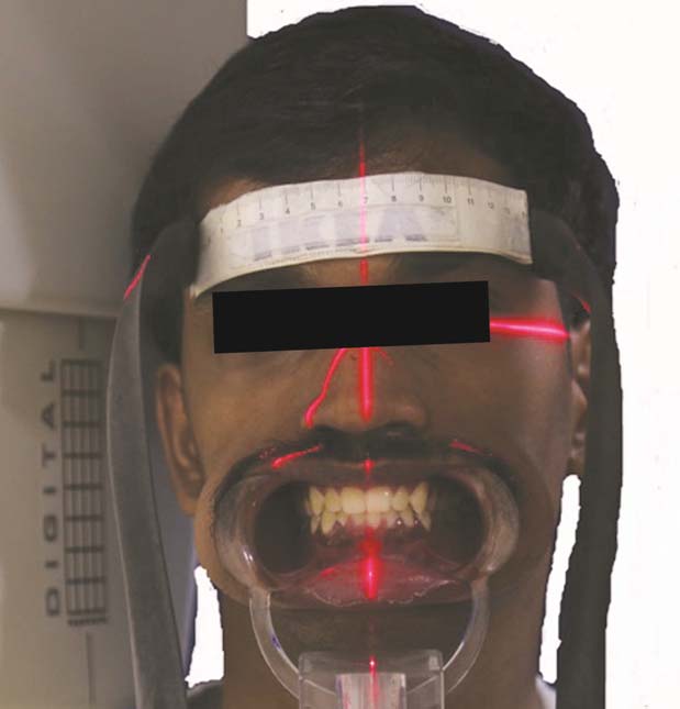
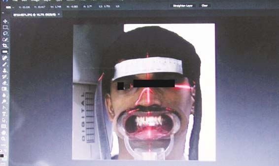
Facial photographs vividly revealing the anterior teeth and the eyes were made using a higher end digital camera (CANON EOS 600D). The camera was used with EF-S Mount 50 mm lens 1.8 with effective focal length of 61 AF points. The maximum resolution offered by this camera was 21.0 megapixels with CMOS sensor. The images were taken with the help of tripod positioned in front of the subject. The camera lens was kept five feet away from the subject and it was standardized by a marking on the floor. Subject was seated on a comfortable chair in an upright position and was instructed to look straight. The subjects head was supported by the chin rest of a panoramic radiographic machine (Planmeca Proline PM 2002 CC; Planmeca Oy, Helsinki, Finland). A flexible ruler was secured onto the subject’s forehead to quantify and eradicate any photographic error. The midline light of the machine was used to center the face. Cheek retractors were used for ease of accessing the maxillary anterior teeth that can be recorded in the photograph [Table/Fig-1]. The photographic images were made instantly with gaze lasting a few seconds by just one trained photographer in the same set up. Once the image was captured it was imported into the desktop, saved as JPEG files and analysed using the image analysing software (Adobe Photoshop Creative cloud). The dimensions of iris and tooth were taken only on right side. The measurements were made using the ruler tool in the software. For the iris visible length measurement, the pointer was positioned on the medial aperture height tangential to iris and the mouse button was clicked. The navigator on the corner of the screen gave the measurement in the scale. Same procedure was followed to measure the length of the tooth from the zenith point to the centre of the incisal edge [Table/Fig-2]. Each parameter was measured and recorded three times and the average value was calculated. Once the data was obtained, they were subjected to statistical analysis. SPSS version 16 statistical software was used. Pearson’s correlation coefficient was used to determine if association existed between length of the Iris and length of the maxillary central incisor.
Stabilization for facial photograph;
Variable dimension measurement of the right side of iris and the maxillary central incisor.
Results
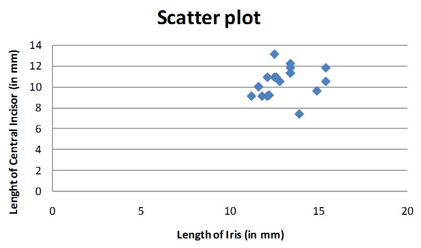
Data was analysed with Karl Pearson’s Coefficient of Correlation. The mean value of length of central incisor was 10.39 mm and the mean value of the length of iris was 12.9 mm. The Pearson correlation analysis revealed only a small correlation between iris and central incisor lengths as shown in [Table/Fig-3,4]. The scatter plot demonstrated non-linear relationship between the length of Iris and central incisor [Table/Fig-5].
| Mean | Std. Deviation | N |
|---|
| Length of Iris | 12.915 | 1.2313 | 20 |
| Length of central incisor | 10.390 | 1.3572 | 20 |
N - No of dental students
Pearson’s correlation coefficient.
| Mean | Length of Iris | Length of Central Incisor |
|---|
| Length of Iris | Pearson Correlation | 1 | 0.233 |
| Sig. (2-tailed) | | 0.322 |
| N | 20 | 20 |
| Length of central Incisor | Pearson Correlation | 0.233 | 1 |
| Sig. (2-tailed) | 0.322 | |
| N | 20 | 20 |
Scatter plot showing correlation between variables.
Discussion
Aesthetics is of paramount value for patients approaching any Prosthodontic treatment. With leaps and bounds of development in dental materials, science and technology, providing prosthesis with supreme aesthetics is now possible in clinical practice [1]. Among the various dental prostheses, fabrication of a natural looking complete denture is not an easy task. Very uncommonly completely edentulous patients report with any preoperative data for selection of artificial denture teeth. Maxillary anterior teeth which project with maximum visibility not only play a role in dental aesthetics but also impart great title in facial aesthetics [10]. Numerous studies have been done to identify specific facial landmarks that would assist in selection of anterior teeth [6,7,11]. Yet no definitive guidelines and landmarks exist in selection of artificial anterior teeth. Amidst the different landmarks adopted till date, the correlation of the measurement of eyes with the widths of maxillary anterior teeth is one corner not analysed yet.
In the recent years, iris of the eye recognition has emerged as an important and powerful biometric characteristic. With uniqueness of iris, it is considered as a stable internal organ that can be used as a person’s durable identity for years. Thus, a research was attempted to determine if a correlation existed between dimension of iris and width of maxillary central incisor.
The iris of eye is an internal organ protected by the cornea and controls the amount of light entering the eyes. It is formed at 10 months of age and remains unchanged throughout the lifetime. Even as age advances, iris doesn’t change in its biometric temper. Depending upon the intensity of light, the diameter of pupil constricts and dilates without altering the dimension of the iris [12].
The popularisation of digital photography in medical scientific research is well evident methodology employed for achieving measurements. In the present study, a higher end digital camera was used to capture the facial photograph for the assessment of oculometric and tooth dimension standards. The correlation with measurements was then estimated using digital analysis software program. The use of digital facial photography is a non-invasive method employed that can be costumed as a lower cost option. In clinical practice its easy availability, storage facility, possibility of repeated assessment, and direct analysis without the need of photographic printing escalates its extensive usage in the field of scientific research [13]. Nunes et al., has proven that digital eye measures are reliable and comparable in accuracy to manual measurements [14].
Previous researches done on identification of a correlation between tooth and anthropometric parameters employed direct measurements from dental casts, anatomical landmarks, with the help of a ruler or calliper like device. Such objective analysis renders a chance of error incorporation when manually estimated. Sellen et al., in his research demonstrated that a computer can be effectively used for the tooth and facial measurements [7]. Hence, a computer with standard analysis software for dimension evaluation was followed in the current study.
Enormous research has been done to establish an association between anthropometric values and maxillary anterior teeth in dentate patients to aid in selection of denture teeth in completely edentulous subjects. Various anthropometric measurements like cephalic index, Berry’s index, Sear’s index, bizygomatic width index, ala of the nose, inter pupillary distance, intercanthal distance, intercommissural width are researched till date. In the current study, a mean central incisor length of 10.39 mm was observed in Indian population. An 11.69 mm length from zenith to incisal edge of extracted maxillary central incisor was noted in white subjects in a study done by Magne P et al., whereas Gillen RJ et al., found the average length to be 10.4 mm as achieved in this research [15,16]. Visible iris length measurements found was 12.9 mm in correspondence to the results obtained by Miot HA in his study [13]. Inference demonstrated only minimal association between the two variables supporting the research done by Smith BJ et al., who studied the relationship between the width of the nose and the natural anterior teeth. He found no significant correlation between both the variables [17]. Al Wazzan KA et al., did a study to associate intercommissural width with intercanine distance, but could not achieve any positive outcome [8]. Deogade SC et al., performed a study on Indian population to identify an interrelationship between intercanthal distance, interalar and intercanine width. He concluded depicting no significant relationship to use these landmarks as preliminary mode of artificial teeth selection [18].
Nonetheless, there were a few constraints in this study, where facial photography was used for capturing the iris diameter instead of objective devices like Corneal Topographer, Slit Lamp and Reticle, loupe, which can provide explicit measurement of Horizontal Visible Iris Diameter (HVID). Direct measurement of the tooth using digital calliper can also be adopted for dental evaluation. More than one operator can be employed on a vast sample size (more than 100) to investigate the association between the variables. Thus further improvisations provide a platform for subsequent research associated to this study.
Direct tooth measurements from dental casts or digital calliper can be adopted. As only minimal correlation between two variables has been obtained further improvisations with horizontal visible iris diameter can be done.
The present study was conducted on the individuals of the age group 18 to 26 years and further studies can be conducted including few more age groups and a larger sample size.
Conclusion
The mean proportional measurements of the maxillary central incisor length from zenith to incisal edge and the length of visible iris from medial aperture height tangential to iris inferred only a small Pearson’s correlation coefficient statistically with r-value <0.3. However, with an achievement of average tooth dimension of 10.39 mm and iris measurement of 12.9 mm, an association factor has been realized. The visible iris length from medial aperture tangential to iris is 2.5 mm more than tooth length. Thus clinically, if a completely edentulous patient reports without any previous extraction data, the selection of maxillary central incisor denture tooth length can become extremely apparent by using the iris measurement and then deducting 2.5 mm to achieve the tooth length. This study provides a definitive ground for more future research using iris measurements.
N - No of dental students