Solving the Mystery of the Antero Lateral Ligament
P H Sonia Farhan1, Rathi Sudhakaran2, Jai Thilak3
1 Undergraduate Student, Amrita Institute of Medical Sciences, Amrita University, Kochi, Kerala, India.
2 Professor, Department of Anatomy, Amrita Institute of Medical Sciences, Amrita University, Kochi, Kerala, India.
3 Professor, Department of Orthopaedics, Amrita Institute of Medical Sciences, Amrita University, Kochi, Kerala, India.
NAME, ADDRESS, E-MAIL ID OF THE CORRESPONDING AUTHOR: Dr. Rathi Sudhakaran, Professor, Department of Anatomy, Amrita Institute of Medical Sciences, Amrita University, Kochi- 682041, Kerala, India.
E-mail: rathis@aims.amrita.edu
Introduction
The cruciate ligaments are essential for the antero-posterior stability of the knee joint. In Anterior Cruciate Ligament (ACL) rupture, though reconstructive surgery is a widely accepted and proven procedure, there is still an unacceptably high re-injury rate. The fact that the rotational instability persists even after the surgical reconstruction of ACL injury has evoked a new interest in the study of the soft tissue structures on the anterolateral aspect of the knee joint. The stability of the knee joint was found to improve dramatically if ACL reconstruction is accompanied with the reconstruction of the anterolateral soft structures of the knee.
Aim
To identify the attachment and observe the measurable parameters of Antero Lateral Ligament (ALL) and its relationship with the adjacent bony landmarks.
Materials and Methods
Twenty six cadaveric specimens of knee joints were collected from the Department of Anatomy, Amrita School of Medicine, Amrita Institute of Medical Sciences, Kochi and were dissected for the anterolateral ligament. Various parameters of ALL in extended knee- the length, width at midpoint and at the femoral-tibial attachments and thickness– were measured. The relationship of femoral attachment of ALL with the lateral femoral epicondyle as well as the tibial attachment with the Gerdy’s Tubercle (GT) and head of fibula were also noted.
Results
ALL was identified in all the 26 cadaveric knee specimens. It was 39.2±7.2 mm in length, 6.5±2.7 mm in width at femoral attachment and 7.4±3.4 mm at tibial attachment, while the thickness was 1.0±0.5 mm. At the femoral attachment it was 7.1±3.4 mm proximal to and 4.0±2.9 mm posterior to the lateral epicondyle while at the distal attachment it was 20.4±3.1 mm posterior to the GT and 21.33±4.6 mm anterior to the head of the fibula.
Conclusion
The ALL was found to be a distinct, supporting anatomical structure on the anterolateral aspect of the human knee. There is a high incidence of ALL lesions in ACL injuries which causes high-grade pivot-shift. The reconstruction of ALL along with that of ACL could lead to a decrease in the re-injury rates. The anatomical descriptions and the morphometry of ALL may be of great value to the orthopaedic surgeons in performing a more effective reconstructive surgery of ACL.
Anterior cruciate ligament, Gerdy’s tubercle, Segond fracture
Introduction
Though the major ligaments of the knee have been studied thoroughly, the soft tissue structures around it especially to the lateral aspect of the knee are not well defined. The high re-injury rate even after an ACL reconstructive surgery attracted the attention of anatomists, orthopaedic surgeons and radiologists to the stabilizing soft tissue structures on the antero-lateral aspect of the knee. In the recent past some studies done by Claes S et al., and Dodds AL et al., brought forward the presence of a ligamentous structure, the ALL on the anterolateral aspect of the knee [1,2]. The morphological parameters of ALL studied by Claes S et al., in 2013, described that in 97% of the cases the origin of ALL is from the lateral femoral condyle, just in front of the Fibular Collateral Ligament (FCL), descending till its insertion onto tibia midway between GT and head of fibula [1]. No standard anatomical term was used to refer to this soft tissue structure in the earlier times [3]. It was not even listed in the Terminologica Anatomica as a separate structure.
In 1879 Paul Segond mentioned the ALL for the first time in orthopaedic literature. According to him it was a pearly, resistant fibrous band attached to the anterolateral aspect of proximal tibia. Paul Segond described Segond fracture as commonly associated with ACL injury which he attributed to the ALL [4].
Anatomical details of this ligament were studied extensively by many. In 1948, Last RJ called it as ‘short lateral ligament’ [5]. Terry GC et al., referred to it as ‘capsulo osseous layer of the iliotibial tract’ [6,7], while La Prade RF et al., called it as ‘midthird lateral capsular ligament’ [8]. In 2001, Campos JC et al., used the term ‘lateral capsular ligament’ [9].
Vieira EL et al., described ALL as arising from the lateral femoral condyle and inserting onto the midportion of proximal tibia, halfway between GT and fibular head [10]. Some studies suggested an association of ALL with ACL injuries in the genesis of knee instability [10–12].
Studies show that mere reconstructive surgery of ACL does not give adequate stability to the knee joint. This information has developed a paradigm shift in the management of ACL injuries. Recent studies have proved that ALL injury correction along with ACL reconstruction provides more rotational stability to the knee joint [13]. The role of anatomists in providing background information on this ligament will be vital for the orthopaedic surgeons in performing functionally effective reconstructive surgeries involving both the ACL and ALL. This understanding has encouraged many to undertake detailed anatomical studies on ALL.
Present study on the anatomy of ALL was aimed at defining the attachments of ALL and its length, width and relationship with prominent bony landmarks and nearby soft tissue structures.
Materials and Methods
Twenty six embalmed human cadaveric knees were collected from the Department of Anatomy, Amrita School of Medicine, Amrita Institute of Medical Sciences, Kochi, Kerala, India, and were dissected for the anterolateral ligament. The skin on the lateral aspect of the extended knee was reflected. The Iliotibial Band (ITB), extensor apparatus, the short head of the biceps femoris and its tendon were cleared of subcutaneous fat tissue. The ITB was then cut just above the lateral femoral condyle and reflected distally upto its tibial insertion at the GT. The Lateral Collateral Ligament (LCL) was palpated and the lamina covering the LCL was then carefully incised posteriorly and parallel to the LCL.
A distinct fibrous structure running distally from lateral femoral condyle to the proximal tibia, posterior to GT, was identified. It was clearly delineated from the capsule following which all the visible fibres of the ligamentous structure were isolated and dissected out.
Special care was taken not to damage fibers intersecting with the proximal LCL. Furthermore, the lateral meniscus, the LCL and the popliteus tendon were carefully separated to study the relationship with the ALL.
A qualitative and a quantitative characterization of the ALL was done. A digital caliper with an accuracy of 0.01 mm (Mit500196-20; Mitutoyo, Japan) was used to measure various parameters of the ALL. The measurements observed were length of ALL, width at the femoral and tibial attachments and at the midpoint, distance between the center of tibial attachment of ALL to GT and to the tip of the fibular head, distance between femoral attachment of the ALL and the tip of the lateral epicondyle.
Results
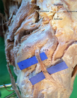
In all the 26 cadaveric knees dissected, a distinct ligamentous structure was identified at the anterolateral aspect, connecting the femur to the tibia. It appeared as an extracapsular structure, passing obliquely over the superficial aspect of the upper part of LCL. In view of its location, appearance and orientation, it seemed appropriate to call it as the Anterolateral Ligament (ALL) [Table/Fig-1].
Lateral region of right knee. Blue markers show the ALL. (* – femoral attachment of ALL, FM- Femoral condyle, LCL-Lateral collateral ligament, P-Popliteus).
In all cases, the proximal attachment of the ALL to the femoral condyle was found to be proximal and posterior to the lateral epicondyle with a fan-like spreading of the fibres. The distal attachment was broader than the main body and passed onto the proximal tibial plateau midway between GT and head of the fibula. The measurements observed were recorded and tabulated [Table/Fig-2]
Anatomical measurements of ALL in millimeter.
| Sl no. | Side | ALL Length(mm) | ALL Width (mm) | Thickness (mm) | ALL-GT(mm) | ALL-FH(mm) | ALL-LE(mm) |
|---|
| Femoral | Mid point | Tibial | Proximal | Posterior |
|---|
| 1 | L | 42.28 | 13 | 3.6 | 11 | 1.1 | 18.55 | 24.76 | 8.34 | 4.99 |
| 2 | L | 33.5 | 3.2 | 1.9 | 6.1 | 0.8 | 15.5 | 14.98 | 2.9 | 1.22 |
| 3 | L | 51.4 | 7.23 | 2.1 | 5.8 | 1.2 | 21.63 | 23.37 | 4.66 | 4.89 |
| 4 | L | 32.25 | 6.1 | 2.3 | 9.12 | 0.46 | 24.2 | 24.54 | 8.99 | 3.59 |
| 5 | L | 38.2 | 5.8 | 3.1 | 6.81 | 1.6 | 21.6 | 25.41 | 7.98 | 3.65 |
| 6 | L | 45.56 | 5.03 | 1.03 | 1.344 | 0.7 | 18.5 | 33.02 | 11.56 | 3.12 |
| 7 | L | 36.5 | 5.1 | 3.12 | 3.4 | 1.2 | 19.65 | 17.1 | 3.11 | 11 |
| 8 | L | 36.21 | 7.4 | 4.27 | 4.27 | 0.5 | 18.45 | 26.84 | 2.1 | 3.51 |
| 9 | L | 35.37 | 5 | 5.6 | 2.1 | 064 | 20.5 | 20.81 | 5.01 | 5.61 |
| 10 | L | 34.5 | 6.1 | 3.1 | 9.87 | 1.91 | 24.25 | 19.8 | 4.3 | 1.08 |
| 11 | L | 45.31 | 4.04 | 3.84 | 4.6 | 1.7 | 23.89 | 22.3 | 7.65 | 2.15 |
| 12 | L | 30.95 | 6.2 | 2.8 | 10.81 | 0.5 | 19.1 | 19.48 | 9.34 | 3.98 |
| 13 | L | 30.11 | 4.1 | 2.1 | 8.15 | 0.9 | 25.88 | 19.76 | 14 | 3 |
| 14 | L | 38.6 | 3.5 | 2 | 2.1 | 1.4 | 18.55 | 19.48 | 12.6 | 14.01 |
| 15 | R | 41.95 | 3.3 | 3.21 | 3.6 | 0.95 | 19.36 | 17.1 | 6.5 | 4.16 |
| 16 | R | 49.61 | 4.5 | 3.5 | 1.69 | 0.12 | 23.44 | 19.21 | 9.56 | 3.67 |
| 17 | R | 39.23 | 3.4 | 2.1 | 10.1 | 0.3 | 25.84 | 14.3 | 9.88 | 2.1 |
| 18 | R | 35.51 | 7.4 | 2.5 | 13.08 | 1.4 | 21.63 | 15.8 | 11.8 | 4.3 |
| 19 | R | 47.52 | 10.03 | 6.32 | 10.01 | 0.85 | 16.3 | 27.91 | 6.53 | 2.51 |
| 20 | R | 29.38 | 9.1 | 5.84 | 9.43 | 1.69 | 17.02 | 23.37 | 7.89 | 3.43 |
| 21 | R | 39.55 | 6.55 | 5.5 | 10.37 | 2.2 | 24.1 | 27.4 | 8.71 | 0.5 |
| 22 | R | 23.91 | 4.92 | 4.22 | 9.76 | 1.2 | 15.54 | 20.54 | 3.22 | 6.12 |
| 23 | R | 43 | 8.74 | 4.71 | 9.1 | 1.9 | 20.5 | 16.65 | 4.76 | 5.75 |
| 24 | R | 49.62 | 9.2 | 3.54 | 9.1 | 0.7 | 20.89 | 15.23 | 2.95 | 0.7 |
| 25 | R | 38.23 | 12.61 | 5.42 | 11.51 | 1.05 | 15.09 | 24.96 | 2.31 | 1.5 |
| 26 | R | 51.2 | 8.61 | 5.51 | 9.76 | 1.34 | 21.01 | 20.6 | 9.12 | 4.12 |
| MEAN | | 39.21 | 6.545 | 3.586 | 7.422 | 1.089 | 20.422 | 21.335 | 7.145 | 4.025 |
| SD | | 7.264 | 2.705 | 1.447 | 3.495 | 0.536 | 3.167 | 4.652 | 3.407 | 2.959 |
The morphometric measurements of ALL from the present study are represented in [Table/Fig-2,3].
Morphometric measurements of ALL from present study.
| Morphological Parameter | Measurement (mm) |
|---|
| Length of Anterolateral ligament | 39.2±7.2 |
| Width of ALL | At femoral attachment | 6.5±2.7 |
| At midpoint | 3.5±1.4 |
| At tibial attachment | 7.4±3.4 |
| Thickness | 1.0±0.5 |
| ALL – Gerdy’s tubercle distance | 20.4±3.1 |
| ALL – Fibular head distance | 21.3±4.6 |
| Distance of ALL from lateral femoral epicondyle | Proximal | 7.1±3.4 |
| Posterior | 4.0±2.9 |
Discussion
In this study, the anterolateral ligament was identified as a distinct ligamentous structure, consistently present in all 26 cadaveric specimens, easily distinguishable from the adjacent thin joint capsule at the anterolateral aspect of the knee.
Segond P defined it as a fibrous structure whereas Terry GC et al., in defined it as a strong capsular ligament, supported superficially by the Iliotibial tract [4,6]. It was thought to play an important role in the anterolateral stability pattern of the knee. Some literatures used the term ‘mid third’ lateral capsular ligament for the same [14]. Vincent EL et al., in 2012 noticed ‘a relatively consistent structure in the lateral knee’ which he called as ALL [11]. Johnson LL performed dissections on six amputated specimens to elucidate the anatomy of what he called the “lateral capsular ligament complex” [15]. Terry GC et al., described a functional ALL formed by the deep capsulo-osseous and superficial layers of the iliotibial tract [7].
The femoral attachment of ALL showed a high degree of variablity in majority of studies whereas the tibial attachment was relatively more consistent and was approximately midway between GT and the fibular head. Claes S et al., and Vincent J et al., described the ALL to get attached anterior to the FCL [1,11], whereas Vieira EL et al., stated it deep to iliotibial tract [10]. Dodds AL et al., stated that the ALL does not attach directly onto bone, but rather blends with locally adherent fibres of the capsule, posterior to FCL [2].
According to our findings, the proximal attachement of ALL was directly to the femoral condyle, proximal and posterior to the lateral epicondyle, and the distal attachment to the tibial plateau, similar to that observed by Vincent J et al., and Helito CP et al., [11,12].
It is suggested that along with ACL reconstruction, ALL reconstruction should also be done to prevent the recurrence of rotational injury. In common knee instability patterns such as pivot shift the precise anatomical knowledge of the ALL could be of great relevance to orthopaedic surgeons. However more research is required to establish the function of ALL in providing stability for knee joint.
According to our study its proximal attachment was 7.1±3.4 mm proximal and 4±2.9 mm posterior to the lateral femoral epicondyle. At the femoral attachment the fibers of ALL were fanning out. In the present study we have also noted a close association of ALL with the upper part of lateral collateral ligament as it crosses the latter superficially which is similar to that recorded by Claes S [1].
The tibial attachment of ALL is seen midway between Gerdy’s tubercle and fibular head. It is 20.4±3.1 mm away from GT and 21.3±4.6 mm away from fibular head. It is the only structure attached to that particular area of tibial plateau; so, can be considered as the cause for Segond’s avulsion fracture as described by Stevens MA et al., [16].
The length of ALL in the present study is comparable to those of the previous studies with an exception to the observation done by Dodds AL et al., [Table/Fig-4] [1,2,11,17].
Anatomical measurements of ALL compared with previous studies [1,2,11,17]:
| Study | Average length in extension (mm) | Width(mm) | Thickness (mm) | GT-ALL (mm) | FH-ALL (mm) |
|---|
| Femur | Tibia |
|---|
| Vincent J et al., [11] | 34.1±3.4 | 8.2±1.5 | 2 to 3 | | |
| Claes S et al., [1] | 38.5±6.1 | 8.3±2.1 | 11.2±2.5 | 1.2±0.6 | 21.6±4 | 23.2±5.7 |
| Dodds’ AL et al., [2] | 59 | 6±1 | | 18±3 | 17±3 |
| Caterine S et al., [17] | 40.3±6.2 | 5.1±1.8(Above meniscus) | 8.9±2.5(Below meniscus) | 1.4±0.06 | 23.4±3.4 | 23.9±5.5 |
| Present study | 39.21±7.2 | 6.5±2.7 | 7.42±3.4 | 1.08±0.5 | 20.4±3.1 | 21.3±4.6 |
Present study confirms ALL as an independent structure on the anterolateral aspect of the knee joint but to prove its role in providing rotational stability to the knee joint, the biomechanical analysis of ALL should be studied further.
Conclusion
The high incidence of ALL lesions in ACL injuries and its causative relationship with high grade pivot shift yield a new insight related to the interaction between ALL and ACL and its role in providing rotational stability. Thus, including an ALL reconstruction led to a decrease in the alarming re-injury rates following an ACL reconstruction procedure. The knowledge of gross anatomy of ALL will help the orthopaedic surgeons to perform a more efficient reconstruction of ACL.
[1]. Claes S, Vereecke E, Maes M, Victor J, Verdonk P, Bellemans J, Anatomy of the anterolateral ligament of the kneeJ Anat 2013 223:321-28. [Google Scholar]
[2]. Dodds AL, Halewood C, Gupte CM, Williams A, Amis AA, The anterolateral ligament: Anatomy, length changes and association with the Segond fractureBone Joint J 2014 96:325-31. [Google Scholar]
[3]. Georg Thieme Verlag, Terminologia anatomica: International anatomical terminology by the Federative Committee on Anatomical Terminology (FCAT) 1998 Stuttgart [Google Scholar]
[4]. Segond P, Recherches cliniques et expérimentales sur les épanchemen sanguins du genou par entorseProgrés Med 1879 7:297-341. [Google Scholar]
[5]. Last RJ, Some anatomical details of the knee jointJ Bone Joint Surg Med 1948 28:191-99. [Google Scholar]
[6]. Terry GC, Hughston JC, Norwood LA, The anatomy of the iliopatellar band and iliotibial tractAm J Sports Med 1986 14:39-45. [Google Scholar]
[7]. Terry GC, LaPrade RF, The posterolateral aspect of the knee. Anatomy and surgical approachAm J Sports Med 1996 24:732-39. [Google Scholar]
[8]. LaPrade RF, Gilbert TJ, Bollom TS, Wentorf F, Chaljub G, The magnetic resonance imaging appearance of individual structures of the posterolateral knee: A prospective study of normal knees and knees with surgically verified grade III injuriesAm J Sports Med 2000 28:191-99. [Google Scholar]
[9]. Campos JC, Chung CB, Lektrakul N, Pedowitz R, Trudell D, Yu J, Pathogenesis of the Segond fracture: Anatomic and MR imaging evidence of an iliotibial tract or anterior oblique band avulsionRadiology 2001 219:381-86. [Google Scholar]
[10]. Vieira EL, Vierira EA, da Silva RT, Berlfein PA, Abdalla RJ, Cohen M, An anatomic study of the iliotibial tractArthroscopy 2007 23:269-74. [Google Scholar]
[11]. Vincent J, Magnussen RA, Gezmez F, Uguen A, Jacobi M, Weppe F, The anterolateral ligament of the human knee: An anatomic and histologic studyKnee Surg Sports Traumatol Arthrosc 2012 20:147-152. [Google Scholar]
[12]. Helito CP, Demange MK, Bonadio MB, Tirico LEP, Gobbi RG, Pecora JR, Anatomy and histology of the knee anterolateral ligamentOrthop J Sports Med 2013 1:1-5. [Google Scholar]
[13]. Ferreira Mde C, Zidan FF, Miduati FB, Fortuna CC, Mizutani BM, Abdalla RJ, Reconstruction of anterior cruciate ligament and anterolateral ligament using interlinked hamstrings – technical noteRev Bras Ortop 2016 51(4):466-70. [Google Scholar]
[14]. Delzell PB, Schils JP, Recht MP, Subtle fractures about the knee: Innocuous-appearing yet indicative of significant internal derangementAm J Roentgenol 1996 167:699-703. [Google Scholar]
[15]. Johnson LL, Lateral capsualr ligament complex: Anatomical and surgical considerationsAm J Sports Med 1979 7:156-60. [Google Scholar]
[16]. Stevens MA, El-Khoury GY, Kathol MH, Brandser EA, Chow S, Imaging features of avulsion injuriesRadiographics 1999 19:655-72. [Google Scholar]
[17]. Caterine S, Litchfield R, Johnson M, Chronik B, Getgood A, A cadaveric study of the anterolateral ligament: Re-introducing the lateral capsular ligamentKnee Surg Sports Traumatol Arthrosc 2015 23(11):3186-95. [Google Scholar]