Picrosirius Red and Polarization Microscopy – A Tool for Gender Differentiation
BK Charan Gowda1, Ganganna Kokila2, Pillai Arun Gopinathan3, Kunigal Shivaprakash Praveen4
1 Reader, Department of Oral Pathology, Sri Siddhartha Dental College, Sri Siddhartha Academy of Higher Education, Tumakur, Karnataka, India.
2 Professor, Department of Oral Pathology, Sri Siddhartha Dental College, Sri Siddhartha Academy of Higher Education, Tumakur, Karnataka, India.
3 Senior Lecturer, Department of Oral Pathology, Sri Sankara Dental College, Kollam, Kerala, India.
4 Senior Lecturer, Department of Oral Pathology, Sri Siddhartha Dental College, Sri Siddhartha Academy of Higher Education, Tumakur, Karnataka, India.
NAME, ADDRESS, E-MAIL ID OF THE CORRESPONDING AUTHOR: Dr. Pillai Arun Gopinathan, Senior Lecturer, Department of Oral Pathology, Sri Sankara Dental College, Varkala, Akathamuri, Kollam-695318, Kerala, India.
E-mail: arun_righthere@yahoo.co.in
Introduction
Forensic dentistry is a branch of dentistry which in collaboration with legal profession serves an important role to maintain justice system of a country. Forensic dentists play a major role in identification of an individual. Within the literature various methods have been found to be useful in gender differentiation. An attempt was made for differentiation of gender using picrosirius red and polarization microscopy.
Aim
To evaluate picrosirius red and polarization microscopy as a tool for gender differentiation by observing birefringence pattern and distribution of thick and thin collagen fibers in males and females.
Materials and Methods
Labial mucosal tissue obtained from 30 deceased individuals (18 male and 12 female) during autopsy was fixed in 10% formalin at 12th hour. Tissue was processed, sectioned and stained using picrosirius red stain and the birefringence pattern of collagen fibers were studied with polarization microscope. The results were statistically analyzed using Z-test and one-way ANOVA to draw the significance.
Results
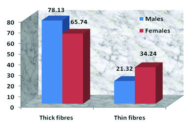
The proportion of thick and thin fibres among males and females were compared. It was found that there was statistically significant difference in proportion of thick and thin fibers between male and female. Thick fibres in males were (78.13%) more than females (65.74%) and thin fibres were more in females (34.24%) than males (21.32%).
Conclusion
Picrosirius red and polarization microscopy may be used as a tool for gender differentiation. Yet the manner of death has to be considered during gender differentiation using this method, as in the present study out of 30 cases studied three cases of death due to debilitating diseases and poison consumption showed altered collagen distribution.
Birefringence, Collagen fibers, Forensic dentistry
Introduction
Forensic dentistry is defined as “the branch of forensic medicine which in the interest of justice deals with the proper handling and examination of dental evidence and with the proper evaluation and presentation of the dental findings” [1]. It plays a significant role in determination of the gender of victims whose bodies are mutilated past recognition due to major mass disasters. Forensics experts utilize skeletal remains, features of teeth such as morphology, crown size, root lengths etc., for establishing gender [2]. Dental forensic specialists have a key role in analysis of the identity of individuals in mass tragedies, mishaps or where the casualty’s bodies cannot be predicted by visual methods. Individual identification by dental features is broadly recognized.
Oral mucosa is composed of two tissue components, stratified squamous epithelium and connective tissue or lamina propria. Similar to other mucous membrane lamina propria of oral mucosa consists of cells, blood vessels, neural elements and fibres embedded in anamorphous ground substance. Type I collagen is the most predominant fibre component of the extracellular matrix along with collagen Type III and elastic fibers [3–5]. Electron microscopic studies have shown that Type I collagen fibres are coarse, composed of closely packed thick fibrils, whereas Type III collagen forms thin fibres and are composed of loosely disposed thin fibrils [6]. Thickness of skin varies with gender and age of the individual. Increase thickness of skin in men is due to the effect of androgens, whereas female hormone oestrogen has inhibiting effect of collagen synthesis [7]. Electron microscopic studies have been used to investigate the role of gender on fibril diameter, which revealed that both female mouse and rabbit collagen fibril showed marked decrease in mean collagen fibril diameter compared to males [8]. Skin collagen decreased with age and was less in the females at all ages [9].
Traditionally stains such as Van Gieson’s and various forms of trichrome stains were used for staining collagen fibres, but were not ideal [10]. Later Sirius red F3BA an elongated sulfonatedazo dye on interacting with collagen fibres [11] showed a spectrum of colours when viewed under polarized light depending on the fibre size, packing density and thus, showed clear orientation of collagen fibres. Numerous studies have demonstrated that Type I collagen fibres are thick, strongly birefringent, yellow or red fibres while Type III collagen fibres are thin, weakly birefringent and greenish in colour [6,12,13]. In the present study the above features of Type I and Type III collagen fibres were considered and an attempt was made to identify the difference between males and females which can be useful in the field of forensic science.
The aim of the study was to evaluate picrosirius red and polarization microscopy as a tool for gender differentiation. The birefringence pattern and distribution of thick and thin collagen fibres in post mortem oral tissues using picrosirius red and polarized light microscopy were evaluated.
Materials and Methods
This observational study was conducted on labial mucosal tissues of deceased individuals, collected from Department of Forensic Medicine, District Hospital, Tumkur, Karnataka, India, after obtaining written permission from the district surgeon and forensic expert, District Hospital, Tumkur. The study was carried out for a period of six months in the year 2013. The inclusion criteria were age between 20-40 years, post mortem period within 12th hour, while the exclusion criteria were death due to debilitating diseases, burnt and traumatized tissue, post mortem period after 12th hours, children and old age. Informed consent was obtained from the legal representative of deceased individual.
The study group included 30 cases, which comprised of 18 males and 12 females aged between 20 to 40 years. Labial mucosal tissues were obtained within 12 hours of death, which were later fixed in 10% formalin at 12th hour and were processed routinely. Each case was given a code so that the observer is blinded to the gender identification of the case.
Sections of 5 μm thickness were prepared from the paraffin embedded tissue blocks and were stained with picrosirius red. After deparaffinization and rehydration, the sections were incubated in 0.1% (w/v) Sirius red F3BA (C.I.35780) in saturated picric acid solution for one hour at room temperature; sections were then rinsed with distilled water followed by staining with Weigert’s haematoxylin. Differentiation was done in 1% HCl, followed by alkalization with tap water. The sections were then dehydrated and mounted in DPX [12,13].
To evaluate birefringence pattern of collagen fibers, the sections were examined under 40X magnification using Olympus polarized light microscope (BX43) along with image analysis system software (ProgRes, Speed XT core3). Randomly 100 fibers were examined in each tissue sample and were segregated into thick (1.6-2.4_m) and thin fibers (0.8_m or less). The obtained score were tabulated and the statistical analysis was done using Z-test to compare the proportion of thick and thin fibers among males and females. Oneway ANOVA was used to determine the difference in birefringence pattern of collagen fibers.
Results
The study included labial mucosal tissues obtained from 30 deceased individuals (18 males, 12 females) aged between 20 to 40 years. The picrosirius red stained sections under bright field microscopy showed collagen fibres stained deep red. On examining under polarization microscopy at a lower magnification it was observed that male (15) subjects showed more of yellowish orange birefringency (65.066%) [Table/Fig-1], while tissues from female (12) subjects showed a combination of yellowish orange (49.83%) and greenish yellow (29.17%) birefringence [Table/Fig-2]. At a higher magnification it was found that male subjects showed more of thick fibers with yellowish orange birefringency [Table/Fig-3] than female subjects [Table/Fig-4]. The observation of 100 fibers in each case according to their thickness and birefringence were tabulated and analyzed. Males and females showed varying predominance of thick yellowish orange fibers. Thin greenish yellow fibers were more in females compared to males. When the proportion of thick and thin fibers among males and females were compared [Table/Fig-5], thick fibers in males were (78.13%) more than females (65.74%) and the difference was statistically significant (p value 0.031). On comparing thin fibers it was found that they were more in females (34.24%) than males (21.32%) and the difference was statistically significant too (p value 0.023).
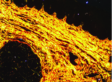
Labial mucosal tissue of male under polarized microscopy showing. predominantly yellowish orange birefringence (picrosirius red stain, 40X).
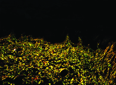
Labial mucosal tissue of female under polarized microscopy showing combination of yellowish orange and greenish-yellow birefringence (picrosirius red stain 40X).
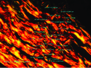
Labial mucosal tissue of male under polarized microscopy showing measurement of collagen fibers (picrosirius red stain, 400X).
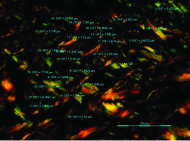
Labial mucosal tissue of female under polarized microscopy showing measurement of collagen fibers (picrosirius red stain, 400X).
Proportion of thick and thin fibers among males and females.
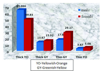
The birefringence of thick and thin fibers in males when observed, were predominantly thick yellowish orange fibers (65.07%) followed by thin greenish yellow fibers (17.60%), thick greenish yellow fibers (13.67%) and thin yellowish orange fibers (3.67%). In females when the birefringence of thick and thin fibers were observed it showed predominantly thick yellowish orange fibers (49.83%) followed by thin greenish yellow fibers (29.17%), thick greenish yellow fibers (15.92%) and thin yellowish orange fibers (5.08%). Statistical analysis with oneway ANOVA was highly significant (p-value <0.001) [Table/Fig-6].
Birefringence of fibers among males and females. (ANOVA analysis: p-value: 0.0000 Interpretation: Highly significant)
Discussion
Forensic dentist plays a pivotal role in forensic research and identification of humans globally. During extreme cases such as explosions, accidental cremations or a fire set deliberately to cover crimes destroys a body, precious little may remain which may help in human identification. Problematic issues that a forensic team faces during identification are deprived state of preservation of unidentified bodies and incomplete presence of remains which may delay the identification process. In such circumstances oral autopsy could help in proper visualization of teeth and its associated structure. Oral autopsy aids to make a suitable post-mortem dental record [14]. Oral mucosa is composed of collagen and it is the most abundant protein in the extracellular matrix of lamina propria. Majority are Type I (90%) and Type III (8%) [15].
Studies have shown that picrosirius red staining followed by polarizing microscopy selectively demonstrates collagen. According to Montes GS [6]: a) Type I collagen- closely packed thick fibres, strongly birefringent, yellowish or red fibres; b) Type III collagenloose network of thin fibres, weakly birefringent, greenish fibres.
In the present study we found 78.13% of Type I collagen and 21.32% of Type III collagen fibres in males, whereas in females it was 65.74 % and 34.24 % respectively. The increased Type I collagen fibres in males could be due to the effect of androgens. Usually the adrenal gland secretes the hormone Dehydroepiandrosterone (DHEA) which acts on the sebaceous glands thus, promoting procollagen synthesis and limits collagen degradation by synthesis of collagenase and Matrix Metallo Proteinases (MMPs) [16]. According to Liu X et al., Type I gene expression is more in males than females [17]. Studies have also shown that men usually have higher collagen density than females [7].
Circulating levels of DHEA are low in adult women which could be the result of lower skin collagen content [16]. According to Liu X et al., in cadavaric studies, width of anterior cruciate ligament fibers in females is significantly smaller than those in males [17] In the present study, females were of age group 20-40 years. Studies have shown that in premenopausal women increased Type III/Type I ratio is observed due to oestrogen effect which could be the reason for the increase in Type III collagen [18].
Out of 18 cases of males, 15 cases showed thick fibres predominantly yellowish orange in the range of 60-72% and thin greenish yellow fibres in the range of 18-22%. But, we found that three cases of males showed contradicting results in the collagen distribution with thick fibres predominantly yellowish orange in the range of 40-48% and thin greenish yellow fibres in the range of 26-32%. These values were almost similar to females who showed thick fibres predominantly yellowish orange in the range of 48-52% and thin greenish yellow fibres in the range of 26-32%. We assume this could be due to the cause of death which was not a parameter considered in our study. When the reason for death was scrutinized from the post mortem records, it was found that two cases had suffered from debilitating disease and one case was poison induced death. Studies have shown that malnutrition, preoperative illness, corticosteroids, immunosuppressants and smoking impairs collagen deposition which could have caused bias in results [19]. According to Karakiulakis G et al., and Jain KK, poison induced death causes histotoxic hypoxia which inhibits final step of oxidative phosphorylation and halts the aerobic metabolism and also increases the secretion of inflammatory cytokines, MPPs which could have caused the change in the birefringency of the collagen [20,21].
Limitation
The limitations of the present study are restricted number of subjects. The literature though has studies regarding differences in skin thickness with age, gender; doesn’t have studies stating difference in collagen between genders. Another limitation is that this method is time consuming and expensive as compared to other routinely used methods for gender determination. Although this method could be useful in gender differentiation in cases where gender identification by other methods may be difficult. However, further studies are required which should consider different age groups as well as reasons for death.
Conclusion
Small remains of human may help forensic dentists in establishing gender of the individual. Picrosirius red and polarization microscopy can be used as a tool for gender differentiation. Yet, the manner of death has to be considered during gender differentiation using this method, as in the present study out of 30 cases studied three cases showed altered collagen distribution due to debilitating diseases and poison consumption.
[1]. Pramod JB, Marya A, Sharma V, Role of forensic odontologist in post mortem person identificationDent Res J 2012 9(5):522-30. [Google Scholar]
[2]. Acharya AB, Sivapathasundharam B, Forensic Odontology. In: Shafer, Hine, levy.Shafers Text Book of Oral Pathology 2009 6th edNew DelhiElsevier:871-97. [Google Scholar]
[3]. Nanci A, TenCate’s Oral Histology Development, Structure, and function 2010 7th edHaryanaMosby Elsevier:319-55.Chapter 12, Oral Mucosa [Google Scholar]
[4]. Avery JK, Chiejo DJ, Essentials of Oral Histology and EmbryologyA clinical Approach 2007 3rd edMosby Elsevier:177-94. [Google Scholar]
[5]. Squier CA, Kremer MJ, Biology of oral mucosa and esophagusJ Natl Cancer Inst Monogr 2001 (29):7-15. [Google Scholar]
[6]. Montes GS, Junqueira LC, The use of picrosirius polarization method for the study of the biopathology of collagenMem. Inst. Oswaldo Cruz 1991 86(Suppl 3):1-11. [Google Scholar]
[7]. Howard D. Is a man’s skin really different? The International Dermal Institute. http://www.dermalinstitute.com/us/library/17_article_Is_a_Man_s_Skin_Really_Different.html (accessed on 26/11/2013) [Google Scholar]
[8]. Tzaphlidou M, Diameter distributions of collagenous tissues in relation to sex: A quantitative ultrastructural studyMicron 2001 32(3):333-36. [Google Scholar]
[9]. Sam Shuster, Black MM, McVitie E, The influence of age and sex on skin thickness, skin collagen and densityBr J Dermatol 1975 93(6):639-43. [Google Scholar]
[10]. Rich L, Whittaker P, Collagen and picrosirius red staining: A polarized light assessment of fibrillar hue and spatial distributionBraz J morphol Sci 2005 22(2):97-104. [Google Scholar]
[11]. Roush JK, Breur GJ, Wilson JW, Picrosirius red staining of dental structuresStain Technol 1988 63(6):363-67. [Google Scholar]
[12]. Koren R, Yaniv E, Kristt D, Shvero J, Veltman V, Grushko I, Capsular collagen staining of follicular thyroid neoplasms by picrosiriusred: Role in differential diagnosisActa Histochem 2001 103(2):151-57. [Google Scholar]
[13]. Allon I, Vered M, Buchner A, Dayan D, Stromal differences in salivary gland tumours of a common histopathogenesis but with different biological behavior: A study with picrosirius red and polarizing microscopyActa Histochem 2006 108(4):259-64. [Google Scholar]
[14]. Charan Gowda BK, Mohan CV, Hemavathi Oral autopsy: A simple, faster procedure for total visualization of oral cavityJ Forensic Dent Sci 2016 8(2):103-07. [Google Scholar]
[15]. Berkovitz BKB, Holland GR, Moxham BJ, Oral Anatomy Histology and Embryology 2009 4th edNew YorkMosby Elsevier:223Chapter 14, Oral mucosa [Google Scholar]
[16]. Farage MA, Miller KW, Zouboulis CC, Piérard GE, Maibach HI, Gender differences in skin aging and the changing profile of the sex hormones with ageJ Steroids Horm Sci 2012 3(2):1-15. [Google Scholar]
[17]. Liu X, Tingle T, Lintner DM, Lowe WR, Zhai QJ, Luo ZP, Differences in collagen gene expression in male and female anterior cruciate ligament injured athletesBiol Sport 2009 26(3):255-61. [Google Scholar]
[18]. Affinito P, Palomba S, Sorrentino C, Di Carlo C, Bifulco G, Arienzo MP, Effects of postmenopausal hypoestrogenism on skin collagenMaturitas 1999 33(3):239-47. [Google Scholar]
[19]. Lenhardt R, Hopf HW, Marker E, Akça O, Kurz A, Scheuenstuhl H, Perioperative collagen deposition in elderly and young men and womenArch Surg 2000 135(1):71-74. [Google Scholar]
[20]. Karakiulakis G, Papakonstantinou E, Aletras AJ, Tamm M, Roth M, Cell type-specific effect of hypoxia and platelet-derived growth factor-bb on extracellular matrix turnover its consequences for lung remodellingJ Biol Chem 2007 282(2):908-15. [Google Scholar]
[21]. Jain KK, Chapter 12, Carbon monoxide and other tissue poisonsText book of hyperbaric medicine 2009 5th ed.GermanyHog free:112-33.Available from http://www.hboorcca.com/pdf/information/Chp%2012%20Carbon%20Monoxide%20and%20Other%20Tissue%20Poisons.pdf (last accessed on 18/11/2016) [Google Scholar]