The main rationale of endodontic therapy is to eliminate microorganisms and their toxic products from the root canal system [1]. One of the major reasons attributed for the clinical failure of endodontic therapy, is apical microleakage [2]. In a root canal filled teeth, this leakage can occur at the various interfaces like endodontic sealer and dentin or sealer and canal filling material (gutta-percha) or through voids within the sealer [3]. Smear layer removal is considered one of the methods for improving the apical seal and for reducing micro leakage before root canal filling [4]. This is due to the fact that, smear layer removal allows better sealer penetration into the dentinal tubules and therein increases the bond strength of resin-based sealers to dentin [5].
For effective removal of the smear layer, combined application of sodium hypochlorite (NaOCl) and a chelating agent, such as Ethylenediamine Tetra-Acetic Acid (EDTA), is commonly recommended [6]. Organic acids such as citric acid may also be used for smear layer removal [7]. It has been suggested that the combined use of 10% citric acid and 2.5% NaOCl is a very effective approach for the smear layer removal [7]. MTAD, which is a mixture of a tetracycline isomer, citric acid, and a detergent, is also effective as a final rinse to remove the smear layer with minimal erosive changes to the surface dentin compared with EDTA [8].
The most commonly used material for root canal obturation is gutta-percha combined with a sealer; however, this standard approach does not provide a completely fluid-tight seal of the root canal system [4]. Resilon (Resilon Research LLC, Madison, Connecticut), a synthetic material was developed as an alternative to replace gutta-percha and traditional sealers for the obturation of endodontically treated teeth. The Epiphany obturation system comprises of three substances: the core material, the sealer, and its bonding agent [9]. Later, Epiphany Self-Etch (SE) resin endodontic obturation system (Pentron Clinical Technologies, Wallingford, Connecticut) has been marketed which consists of two components namely: Epiphany SE sealer and Resilon, the core material. The manufacturer states that with the addition of Epiphany SE sealer, no change in technique is required when transferring from conventional gutta-percha to the Epiphany system. Such resin-based adhesive material may have the potential to reduce the apical leakage of micro-organisms into the root canal system because of its significant dentinal tubule penetration, in addition to its adhesive nature.
It has also been suggested that, intra-canal irrigation with citric acid rather EDTA is recommended for achieving improved apical seal, in particular, when a resin-based sealer is employed for root canal obturation [10].
Until date, there are no published articles related to the effect of citric acid on the apical sealing ability of Resilon/Epiphany SE sealer. Hence, the purpose of the present study was to evaluate and compare the apical sealing ability of Resilon/Epiphany SE sealer following intra-canal irrigation with 17% EDTA, 10% citric acid, and MTAD as final irrigants in combination with NaOCl, using the dye leakage under vacuum method.
Materials and Methods
This was an in vitro, comparative, experimental study in which 55 human single rooted maxillary central incisors, along with straight roots extracted for periodontal reasons within the age group of 45-55 years were selected. Ethical clearance was obtained from the institutional review board of Manipal University, India. Radiographs of each specimen were obtained to confirm the presence of a single canal, mature root apex, resorptive defects (if any) or previous root canal filling. Teeth with caries, cracks, fractures, resorption, previous restorations and root dilacerations were excluded from this study. All specimens were cleaned carefully of debris and calculus with curettes and were stored in 0.2% sodium azide solution (Sigma Chemical Co, St. Louis, Missouri) at 4°C until their use. All the teeth were then decoronated at or near the cemento-enamel junction using a high speed diamond disc (Horico, Berlin, Germany) with an adequate cooling system and were standardized to the root length of 13 mm. The pulp tissue was removed using a barbed broach and the working length was established by placing a No. 15 size K-file (Mani Inc, Tochigi Ken, Japan) into each root canal until it was just visible at the apical foramen and by subtracting 1 mm from this estimated length. Cleaning and shaping was done using K-files (Mani Inc. Japan) by step back technique. Apically canals were enlarged up to size 40 and gates glidden drills (Mani Inc) No. 2-4 were used to enlarge the coronal third of the root canals. All canals were irrigated with 2 ml of 1.3% NaOCl (KMC Pharmacy, Manipal, India) between the uses of each file. Based on the final irrigation sequence, the samples were then divided randomly into three experimental groups of 15 each and two control groups of five each as follows:
Experimental Groups
Group I: 5 ml of 17% EDTA (Merck, Darmstadt, Germany) for 1 minute followed by 5 ml of 1.3% NaOCl for 1 minute.
Group II: 5 ml of 10% citric acid (KMC Pharmacy, Manipal, India) for 1 minute followed by 5 ml of 1.3 % NaOCl for 1 minute.
Group III: Biopure MTAD (Dentsply Tulsa Dental, Tulsa, Oklahoma) was used according to manufacturer’s instructions. 1.3% NaOCl followed by 1 ml of MTAD was placed in each canal for 5 minutes and then each canal was rinsed with 4 ml of MTAD.
The above mentioned irrigating solutions were introduced into the root canals by means of a 27-gauge stainless steel beveled needle. The needle was placed within 1 to 2 mm of working length in each canal. The root canals were finally rinsed with 5 ml of distilled water (KMC Pharmacy, Manipal, India) to terminate the action of irrigants and to remove any precipitate that might have been formed. The root canals in the experimental groups were then dried with sterile absorbent paper points (Dentsply-Maillefer, Ballaigues, Switzerland) and obturated with Resilon/Epiphany SE sealer (Pentron Clinical Technologies, LLC, Wallingford, Connecticut) using the following technique. Epiphany SE sealer was mixed on a mixing pad, and then a Resilon master cone (size 40/0.02) was lightly coated and placed into the canal to the working length. Lateral compaction with accessory Resilon cones was performed until the entire root canal was filled. Following removal of the excess obturating material with a heated instrument and vertical condensation using a plugger, the coronal surface was light-cured for 40-60 seconds to aid in adequate immediate seal.
Control Groups
Positive control group: Five roots were filled only with a single Resilon cone (size 35) without Epiphany SE sealer.
Negative control group: Five root canals were obturated using laterally compacted Resilon/Epiphany SE sealer.
Radiographs of all specimens were taken in buccolingual and mesiodistal directions to evaluate the quality of the root canal fillings. The root canal orifice was sealed with Fuji II glass ionomer cement (GC Corporation, Tokyo, Japan). All specimens were left to set in an incubator for seven days at 37°C and 100% humidity. The external surfaces of the experimental specimens and the samples from the positive control group were covered by two layers of nail varnish apart from the apical 3 mm around the foramen. The samples in the negative control group were entirely covered with two layers of nail varnish, including the apical 3 mm. The samples were immersed in 2% rhodamine B dye solution and placed under vacuum pressure of 75 Torr for 30 minutes and allowed to remain in the dye for seven days. This extremely versatile vacuum machine (Bright Line Vacuum Chamber Multiple 315n, Orved China) measuring size of 450x440x340 mm, was made up of stainless steel with an individual nozzle for containers, along with a pump dehumidification cycle H2Out, and an accessory easy vacuum pump (8 m3/h) which works under sensory system for creating the external vacuum. After exposure to the dye, samples were rinsed with running water to remove the dye and the nail varnish was gently removed with a #15 disposable scalpel blade (Lister, Kanpur, India). The roots were then longitudinally grooved using a diamond disc and split into halves using a chisel. Following the separation of the roots, the Resilon/Epiphany filling was removed. The sectioned halves of each root specimen in the respective experimental and control groups were labelled as follows: Group I – A; Group II – B; Group III – C; positive control – D for easy identification and better differentiation which were then subjected to linear dye penetration. The dye leakage was measured by an examiner who was unaware of the experimental groups using a x10 stereomicroscope (Zeiss, Thornwood, New York) from the apex to the coronal extent with an ocular built micrometer scale and the images were captured with a charged couple device camera. Then, the obtained results were statistically analyzed using a SPSS software (SPSS 11.5). Data were analysed by one-way ANOVA to determine whether there were significant differences between the experimental and control groups and pairs of groups were compared using the post hoc tukey test. The level of significance was set at p<0.05.
Results
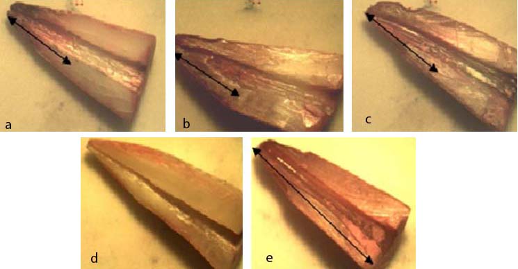
Stereomicroscopic images of dye penetration in the respective experimental and control groups were represented in [Table/Fig-1]. In the positive control group, dye leakage was observed through the entire length of the root canal whereas in the negative control group, no leakage was observed through the canal [Table/Fig-1]. The descriptive statistics of linear dye penetration values for all the groups are illustrated in [Table/Fig-2]. One-way ANOVA test revealed that there was a high significant difference observed in the mean apical leakage between the various experimental groups and control group i.e., between Group I (17% EDTA), Group II (10% citric acid), Group III (MTAD), and positive control group with corresponding F-value = 8.70 and p-value=0.005 (as displayed in [Table/Fig-3,4]. But there was no statistically significant difference (p>0.05) observed in the mean apical leakage among the three experimental groups [Table/Fig-4].
Extent of dye leakage observed in the root canals of various experimental and control groups: (a) Group I (EDTA + NaOCl); (b) Group II (Citric acid + NaOCl); (c) Group III (MTAD + NaOCl); (d) Negative control with no dye penetration; (e) Positive control with complete dye penetration.
Descriptive data of the experimental and control groups.
| GROUPS | N | Mean | Std. Deviation | Std. Error | 95% Confidence Interval for Mean |
|---|
| Lower Bound | BCC subtype |
|---|
| Group –I :17 % EDTA | 15 | 50.6667 | 16.13190 | 4.16524 | 41.7331 | 59.6002 |
| Group – II: 10 % Citric acid | 15 | 43.3333 | 11.44344 | 2.95468 | 36.9962 | 49.6705 |
| Group – III:MTAD | 15 | 42.0000 | 8.82367 | 2.27826 | 37.1136 | 46.8864 |
| Control group: (positive) | 5 | 100.0000 | 3.53553 | 1.58114 | 95.6101 | 104.3899 |
| Total | 4 | 50.8000 | 20.56250 | 2.90798 | 44.9562 | 56.6438 |
One-way ANOVA: Analysis of variance for leakage (leakage versus various groups).
| Dye leakage | (DF) | (SS) | (MS) | F-ratio | p-value |
|---|
| Between groups | 3 | 14101.333 | 4700.444 | 32.678 | .0005 |
| Within groups | 46 | 6616.667 | 143.841 | | |
| Total | 49 | 20718.000 | | | |
DF : Degree of freedom, SS : Sum of square, MS : Mean square.
Post hoc tukey tests depicting multiple intergroup comparisons of dye leakage (x 10 1 um).
| Groups ofmaterial(A) | Groups ofmaterial(B) | MeanDifference(A-B) | Std.Error | Level ofSignificance | 95% ConfidenceInterval |
|---|
| Lower Bound | Upper Bound |
|---|
| 10 % Citric acid | 17% EDTA | -7.3333 | 4.37935 | .349 > | -19.0065 | 4.3398 |
| MTAD | 1.3333 | 4.37935 | .990 > | -10.3398 | 13.0065 |
| Control group: (positive) | EDTA | 49.3333(*) | 6.19334 | .0005*** | 32.8250 | 65.8417 |
| Citric acid | 56.6667(*) | 6.19334 | .0005*** | 40.1583 | 73.1750 |
| MTAD | 58.0000(*) | 6.19334 | .0005*** | 41.4917 | 74.5083 |
(*) - significant mean difference at the 0.05 level.
(***) - very highly significant
(>) - no significance
Discussion
Citric acid as an intra canal irrigant, was investigated in concentrations ranging from 1-50% in endodontics [6]. It has been suggested that the combined use of 10% citric acid and 2.5% NaOCl was an effective approach for the smear layer removal [6]. However, there are no reports in the literature till date related to the effect of 10% citric acid in the apical sealing ability of Resilon/Epiphany SE sealer. Hence, the present study was carried out in an attempt to study the effect of the same.
MTAD has been reported to be effective in smear layer removal [8], in addition to the elimination of microorganisms that are resistant to conventional endodontic irrigants and medications [11] and providing sustained antimicrobial activity through the binding affinity of doxycycline for dental hard tissues [12]. However, the effectiveness of MTAD in complete removal of smear layer is enhanced when low concentrations of NaOCl (1.3%) was used as an intra canal irrigant before the final rinse of MTAD [8]. Therefore, in the present study, 1.3% NaOCl was employed in between the instrumentation as well as in combination with EDTA, citric acid, and MTAD as a final rinse.
According to the results obtained in the present study, there were no statistically significant differences in the mean apical leakage observed between the three experimental groups. These results can be attributed to the smear layer removal abilities of these three agents when used as a final irrigant [13]. The results obtained with Group I and Group II in this study were concomitant with that of other studies that reported a minor or no difference in smear layer removal with EDTA and citric acid [14,15]. This was well documented in a study conducted by Khedmat and Shoukouhinejad et al., [15] who reported no significant differences in the efficacy of smear clear [17% EDTA including a cationic (cetrimide) and an anionic surfactant], EDTA and citric acid in smear layer removal at the coronal, middle and apical thirds of the root canal. On the contrary, Reis et al., [16] showed that the chelating effect of 1, 5, and 10% citric acid is significantly more than 17% EDTA which is in accordance with Machado-Silveiro et al., [17] who showed that the decalcifying effect of 10% citric acid on dentin is more than 17% EDTA. The results obtained in this study with reference to Groups I and II were in contrast to the results of the study conducted by Yamada et al., who reported that EDTA is more effective in removing the smear layer than citric acid [18].
The results of the present study also demonstrated that there was no significant difference between Group I and Group III when used as a final irrigant, which is concomitant with the results of other studies [19,20]. These findings were in contrast to the results of other studies which have been shown that MTAD was superior to EDTA in removing the smear layer in the apical third of the canals [21]. The factors to be considered in obtaining these conflicting results in various studies where different irrigation regimens of EDTA, citric acid, and MTAD were used can be attributed to: the type of teeth used, the concentration of irrigation solutions, the order of irrigation solutions used, time span of irrigation, and the criteria considered by the investigators as being effective and successful in addition to the diversity of methodologies used to assess leakage.
In the present study, the validity of experimental model was justified, since positive control specimens revealed complete dye penetration of the root canal whereas the negative control teeth demonstrated no dye penetration. Hence, there was a high significant difference observed in the mean apical leakage between the experimental groups and the positive control group.
The results of the present study were concomitant with the results of previous studies which have proved that smear layer removal improves the apical seal [22,23]. It has been suggested that resin–based sealers have superior apical sealing ability with the removal of the smear layer [22]. Economides et al., in a study compared the microleakage of two sealers, Fibrefill (resin-based sealer) and calciobiotic root canal sealer (CRCS; calcium hydroxide-based sealer), with and without the presence of smear layer and reported that microleakage values were less when the smear layer was removed [23]. Farhad et al., in a bacterial leakage study, compared the coronal sealing ability of three sealers such as, AH26, Roth’s 801, pure Zinc oxide eugenol (ZOE) sealer and concluded that these sealers exhibited better coronal seal against bacterial ingress following smear layer removal [24]. The advantage of resin-based sealers like AH26, Epiphany SE over ZOE-based sealers is that they cannot only lock into open dentinal tubules but also exhibit adhesion to the exposed dentinal surfaces.
However, a perfect seal is difficult to achieve with bonded root fillings for a number of reasons. This was also evident in the present study, where all the roots obturated with Resilon/Epiphany SE sealer (except that of the negative control group) showed leakage. The extremely high configuration factor (C-factor) of the root canals might have resulted in maximizing the polymerization shrinkage stress of adhesive systems [25]. Another possible explanation that can be attributed to the leakage in the experimental groups might be due to the collapse of demineralized collagen matrices left in the root canal walls following irrigation with various chelating agents which might have prevented sealer infiltration and formation of high quality layer bonding [26]. In addition, polycaprolactone (the raw material of Resilon) is biodegradable under microbial attack [27] in contrast to gutta-percha which is relatively an inert material. These factors are to be considered while employing Resilon/Epiphany SE sealer in clinical practice where the human oral cavity is comprised of a wide variety of microbial flora.
Different methods have been employed to assess the apical sealing ability of endodontic filling materials such as: dye leakage [28], bacterial leakage [29], electrochemical method [30], fluid filtration [31], radioisotope labelling [32] and scanning electron microscopic analysis [33]. Among these techniques, Grossman’s dye penetration method is very simple and easy to perform [34]. This passive method implies the significant phenomenon of capillarity, where the root apex is submerged in dye (e.g., eosin, methylene blue, black India ink, Procion brilliant blue, Rhodamine B etc.,) that penetrates through any available space between the canal walls and the root canal filling material [35]. Then, the specimens are sectioned transversely or longitudinally or cleared following which, the effective linear dye penetration is evaluated [36]. Hence, this dye penetration methodology was employed in the current study to evaluate the apical seal of the root canal because of its sensitivity, ease of use, convenience and cost effectiveness [37].
However, the validity of dye leakage studies has been questioned because of the possible effect of entrapped air on ingress of dye solution [38] which may falsify the depth of dye penetration and it is not any more a commonly used method [39]. To overcome these problems, dye penetration under vacuum or centrifugation techniques are preferred [40]. Hence, in the present study, dye leakage under vacuum was performed for assessing the apical seal of the specimens. Rhodamine B dye was used in the present study because it presents greater diffusion in human dentin than methylene blue [41]. The molecules of Rhodamine B dye are nanometric and are optimal to simulate bacterial enzymes and their toxins to assess microleakage. Other factors favouring the use of Rhodamine-B dye in leakage studies include: small particle size, water solubility, ease of visualization, better diffusability into dentinal tubules and hard tissue non-reactivity [42].
In the present study, apical enlargement of the canals was done unto 40 size hand K-file. This is due to the fact that increasing the size of apical file to #40 could reduce the bacterial count significantly more than smaller sizes as shown by Siqueira et al., in a study [43].
Limitation
The samples used in this study were single-rooted maxillary central incisors with relatively straight canals. Thus, our results may be limited to only such clinical cases. Further studies can be conducted to evaluate the post obturation apical seal of multirooted teeth in curved canals with Resilon / Epiphany SE sealer following 17% EDTA, 10% citric acid, and MTAD as final irrigants. Different leakage methods can also be employed, as this may provide further information about the sealing ability, particularly when there is lack of standardized method for estimating leakage in vitro.
Conclusion
Within the limitations imposed by this in vitro study, it could be concluded that there was no significant difference in the apical sealing ability of Resilon/Epiphany SE with 17% EDTA, 10% citric acid, and MTAD solutions when used as a final irrigant in combination with NaOCl.
DF : Degree of freedom, SS : Sum of square, MS : Mean square.
(*) - significant mean difference at the 0.05 level.
(***) - very highly significant
(>) - no significance