Expect the Unexpected: Mycobacterial Infection in Post Total Knee Arthroplasty Patients
Vikram Kishor Kandhari1, Mohan M Desai2, Roshan N Wade3, Surendar S Bava4
1 Senior Resident, Department of Orthopaedics, Seth G.S. Medical College and K.E.M. Hospital, Mumbai, Maharashtra, India.
2 Professor and Head of Unit, Department of Orthopaedics, Seth G.S. Medical College and K.E.M. Hospital, Mumbai, Maharashtra, India.
3 Assistant Professor, Department of Orthopaedics, Seth G.S. Medical College and K.E.M. Hospital, Mumbai, Maharashtra, India.
4 Associate Professor, Department of Orthopaedics, Seth G.S. Medical College and K.E.M. Hospital, Mumbai, Maharashtra, India.
NAME, ADDRESS, E-MAIL ID OF THE CORRESPONDING AUTHOR: Dr. Vikram Kishor Kandhari, Plot No. 5/5a, Pande Layout, Khamla, Nagpur-440025, Maharashtra, India,
E-mail: dr.vikramkandhari@gmail.com
Orthopaedic Surgeons rarely encounter mycobacterial infections in Post Total Knee Arthroplasty (TKA) patients. We present series of two cases to create awareness among clinicians to expect the unexpected. Tuberculosis typical/ atypical is a hidden culprit in catch clinical situations when chronic infection is Suspected, but the lab investigations are negative in persistently symptomatic patients. In such situations clinicians should suspect atypical or complex mycobacterial infections and evaluate the patients accordingly. Clinical suspicion, evaluation, isolation and treatment of atypical or complex mycobacterial infections with sensitive chemotherapy, leads to complete resolution of infection and full functional rehabilitation.
Mycobacterium abscessus, Imipenem, Joint replacement
Case Report
Case-1: A 76-year-old female having knee osteoarthritis in both limbs was operated with bilateral TKA 18 months back. She had undergone hemimandibulectomy of the left side for oral cancer about 10 years prior, but was not on immunosuppressive therapy. Patient had an uneventful intra and postoperative course.
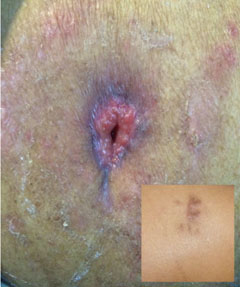
At six months post surgery, patient developed right gluteal abscess. There was no local site tenderness, warmth or induration. An experienced general surgeon did incision and drainage for the collection. Analysis of collection drained did not show presence of any infecting organism on routine microbiological analysis. After the procedure patient completed three weeks course of oral co-amoxyclav, twice a day therapy; but there was no relief in her symptoms. Contained abscess had turned into an active draining sinus in right gluteal region [Table/Fig-1]. Persistent discharge from local site for one month and lack of expected wound healing with a persistent non healing sinus, with discolouration of the surrounding skin, led us to investigate further. Repeat investigations revealed White Cell Count (WBC) was 7,500 CU/dL, Erythrocyte Sedimentation Rate (ESR) of 75 mm/hr, C-reactive protein (CRP) 10 mg/ dL, haematocrit (Hct) of 32 % and haemoglobin (Hb) of 10.5 gm/dL. Though microscopy with Ziehl-Neelsen(ZN) stain on discharge from sinus was negative, but culture with Bactec-MGIT 960 was positive and Mycobacteriumabscessus was identified. On automated BACTEC-MGIT sensitivity testing, it was found to be sensitive to clarithromycin, linezolid and imipenem. The local wound status of the patient improved and ESR dropped to 20 mm/hr with daily three drug oral regime including clarithromycin, linezolid and faropenem for nine months. Presently patient has completed nine months of three drug therapy, is asymptomatic for the gluteal abscess and is walking full weight bearing with the help of walking stick.
Non healing discharging sinus with skin discolouration in right gluteal region. Inset shows healed sinus.

Case-2: A 55-year-old female underwent bilateral TKA for rheumatoid arthritis four and half years back. Pre and postsurgery patient was managed with Disease Modifying Anti-Rheumatoic Drugs (DMARDs). At three years postoperative patient developed painful knee effusion of the left knee with terminal restriction of knee range of motion. The effusion was not associated with fever or other constitutional symptoms. On further investigating, patient had a WBC of 6,400 /dL, ESR of 84mm/hr, CRP 12 mg/ dL, Hct 36 %, Hb of 11.0 gm/dL. Radiologic evaluation revealed well-fixed total knee arthroplasty implant in proper alignment. Therapeutic arthrocentesis fluid analysed failed to isolate any infecting organism. Patient developed re-effusion with painful terminal restriction of knee range in same knee 15 days after arthrocentesis.
Unilateral involvement and persistent effusion led us to investigate patient for possible tubercular aetiology. Repeat arthrocentesis fluid was sent for microscopy and culture, to rule out tubercular pathology. Mycobacteriumtuberculosis complex was isolated on BACTEC–MGIT 960 culture. Patient was put on daily antitubercular regime consisting of four drugs (rifampicin, isoniazid, ethambutol, pyrazinamide) in intensive phase for two months, followed by two drugs (rifampicin, isoniazid) for 10 months, according to drug sensitivity testing. Knee effusion resolved and pain disappeared gradually over a period of one month after start of antitubercular therapy and did not recur. In two months ESR became 16 mm/hr. Presently patient is not on antitubercular medications and is free of her complaints.
Discussion
Infection after Total Knee Arthroplasty (TKA) has been well studied and reported [1,2]. These infections can either be intra or extra-articular and can involve bone or soft tissues [3–5]. In course of treating them, we may encounter frustrating clinical instances, when it becomes difficult to reach correct microbiological diagnosis. In such clinical situations, mycobacterial infections are a hidden culprit [5]. Due to rarity of such instances and lack of suspicion on part of clinician, these infections are not well understood and do not have standardized diagnostic criteria or management protocols [6]. Experience of managing such infections is limited to isolated case reports or to case series. Thus, reporting any experience of clinical encounter with these infections is essential. We present two such cases, encountered during follow up of postoperative TKA patients.
After TKA chronic underlying infection is an important underlying cause of morbidity in ailing patients [7]. Early recognition of infection is the key to limit joint and tissue damage, and achieve desired functional results.
Approximate infection rate following TKA is >2% [7]. Majority of these infections are caused by S. aureus and rarely by organisms Enterococcus, Corynebacterium, Listeria, Brucella and Mycobacteria (0.06%) [8]. All put together they form <5% of the total number of infections in TKA or <0.04-0.06% after TKA (0.04 – 0.06% i.e., < 4 – 6 cases/ 10,000 TKAs) [8].
Lack of experience due to rarity of such infections, makes it difficult for clinician to suspect and investigate for mycobacterial infections. Typical mycobacteria may be isolated from Air, while atypical can be isolated from air, water and soil [9]. There are certain documented risk factors for such infections; trauma, local injection, previous surgical procedure or immunocompromised state [6]. Both our patients were at risk of such infections; one was earlier treated for mandibulectomy for an oral cancer and the other was a known case of arthritis and was on management with DMARDs, however clear source of infection could not be identified.
Identifying certain clinical pointers also aids to suspect and investigate presence of mycobacterial infections. These include late onset unremitting symptoms, presence of discolouration of local area involved and unexpected failure of conventional treatment of suspected infection. Mycobacterial infections especially with atypical mycobacteria tend to be less pathogenic, but are morbid as they are undiagnosed for long duration and do not respond to the conventional management [6]. Thus, in such catch clinical situations, presence of risk factors with one or more clinical pointers should prompt the clinician to suspect and investigate for the mycobacterial infections i.e., to Expect the Unexpected! This will place the clinician at therapeutic advantage and help in early eradication and complete functional rehabilitation. Thus, suspicion of mycobacterial infections in such catch clinical situations is the key to diagnosis. Cheung I and Wilson A also concluded that a high index of suspicion on part of clinician is a must for diagnosis [10].
Correct tissue diagnosis and isolation of causative organism is essential for patient management. Samples can be sequentially evaluated with microscopy using special stains like ZN staining or LED microscopy and cultured in LJ medium or specialized liquid media- BACTEC-MGIT 960. Using BACTEC-MGIT 960 is advisable, as its relative positivity rate is approximately 60% and 67.5% higher than LJ medium and LED fluorescence microscopy respectively [11].
No consensus exists in literature on the number of drugs to be used in combination therapy and the duration for which it is to be continued, but it is essential to choose the drugs based on the sensitivity profile of isolated organism [6]. Progressive reduction of biochemical markers like ESR and CRP along with decrease in patient complaints of pain, restriction of range of motion, decrease in swelling, healing of sinus are a clinical proof of effective management. Duration of therapy can be decided based on patient profile but literature recommends minimum 6-9 months of continuous chemotherapy [12]. Prolonged multidrug medical therapy based on the sensitivity profile was main part of effective treatment armamentarium for treating mycobacterial infections.
Conclusion
Clinical suspicion is the key to diagnosis of mycobacterial infections in postoperative TKA patients. Prolonged treatment with sensitive multi drug antitubercular therapy is an effective mode of treatment. Surgical management can supplement, but not replace medical management of these infections. Clinicians should evaluate postoperative TKA patients with suspected infection, but no response to conventional management, for possible mycobacterial infections.
[1]. Chun KC, Kim KM, Chun CH, Infection Following Total Knee ArthroplastyKnee Surg Relat Res 2013 25(3):93-99. [Google Scholar]
[2]. Babkin Y, Raveh D, Lifschitz M, Itzchaki M, Wiener-Well Y, Kopuit P, Incidence and risk factors for surgical infection after total knee replacementScand J Infect Dis 2007 39(10):890-95. [Google Scholar]
[3]. Bloom AW, Brown J, Taylor AH, Pattison G, Whitehouse S, Bannister GC, Infection after total knee arthroplastyJ Bone Joint Surg [Br] 2004 86-B:688-91. [Google Scholar]
[4]. De Carvalho Jr LH, Temponi EF, Badet R, Infection after total knee replacement: diagnosis and TreatmentRev Bras Ortop 2013 48(5):389-96. [Google Scholar]
[5]. Manning BT, Lewis N, Tzeng TH, Saleh JK, Potty AG, Dennis DA, Diagnosis and Management of Extra-articular Causes of Pain After Total Knee ArthroplastyInstr Course Lect 2015 64:381-88. [Google Scholar]
[6]. Uslan DZ, Kowalski TJ, Wengenack NL, Virk A, Wilson JW, Skin and soft tissue infections due to rapidly growing mycobacteria: comparison of clinical features, treatment, and susceptibilityArch Dermatol 2006 142:1287-92. [Google Scholar]
[7]. Edwards J, Peterson KD, Mu Y, Banerjee S, Allen-Bridson K, Morrell G, National healthcare safety network (NHSN) report: data summary for 2006 through 2008, issued December 2009Am J Infect Control 2009 37(10):783-805. [Google Scholar]
[8]. Aggarwal VK, Bakhshi H, Ecker NU, Parvizi J, Gehrke T, Kendoff D, Organism profile in periprosthetic joint infection: pathogens differ at two arthroplasty infection referral centers in Europe and in the United StatesJ Knee Surg 2014 27(5):399-406. [Google Scholar]
[9]. Brown-Elliott BA, Wallace RJ, Clinical and taxonomic status of pathogenic nonpigmented or late-pigmenting rapidly growing mycobacteriaClin Microbiol Rev 2002 15:716-46. [Google Scholar]
[10]. Cheung I, Wilson A, Mycobacterium fortuitum infection following total knee arthroplasty: A case report and literature reviewThe Knee 2008 15:61-63. [Google Scholar]
[11]. Hasan M, Munshi SK, Momi SB, Rahman F, Noor R, Evaluation of the effectiveness of BACTEC-MGIT 960 for the detection of mycobacteria in BangladeshInternational Journal of Mycobacteriology 2013 2:214-19. [Google Scholar]
[12]. Eid AJ, Berbari EF, Sia IG, Wengenack NL, Osmon DR, Razonable RR, Prosthetic joint infection due to rapidly growing mycobacteria: report of 8 cases and review of the literatureClin Infect Dis 2007 45:687-94. [Google Scholar]