Ball head implant, Bone anchorage, Maxillary protraction, Correction of class III
Dear Editor,
Skeletal Class III malocclusion can be presented with maxillary retrognathism or mandibular prognathism or combination of both. Maxillary hypoplasia is characterized by deficiency of skeletal height, width and anteroposterior relationship [1], which requires correction. The challenge starts when the treatment of maxillary hypoplasia is done in a growing child. Face mask therapy has been regarded as an appliance of choice to treat growing Class III patients with mild to moderate maxillary hypoplasia [2].
Elastic traction of the maxilla is done by an acrylic plate supported on the maxillary dentition and palate. The supporting intra oral device may consists of acrylic occlusal bite block, labiolingual arch, quad helix appliances or Rapid Maxillary Expansion (RME) and hooks in between lateral incisors and canine. The side effects of maxillary dentition as an anchorage unit for protraction could lead to labio version of maxillary incisors, extrusion of maxillary molars, counter clockwise rotation of palatal plane and clockwise rotation of mandible [3].
As a result, to provide a direct transmission of orthopaedic force to the circummaxillary suture, orthodontic mini screws should be used as skeletal anchorage for maxillary protraction [4].
Mini screws were introduced as absolute anchorage devices in orthodontic treatment [5]. Initially mini implants have been demonstrated to be biologically compatible to provide anchorage when subjected to orthodontic force in animal models [6] and human cases [7]. Later mini implants were shown to provide absolute anchorage when subjected to orthopaedic force in animal models [8].
Experiments with osseointegerated Branemark-style implants for maxillary protraction in monkeys had shown good skelatal results [9]. Later the potential of Temporary Anchorage Device (TAD) in protraction of maxilla was demonstrated. Most of the above procedures require surgical intervention for inserting the TAD or bone plates which is associated with discomfort and delayed healing. There is no implant which can be directly placed and force applied through elastics.
The new innovative design is being presented that helps to overcome all these problems. The following is the description of new designdual ball headed mini implant.
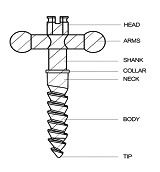
Parts of Dual Ball Head Implant [Table/Fig-1]
Head
Arms
Shank
Collar
Neck
Body
Tip
Parts of the ball headed mini implant.
Head [Table/Fig-2]
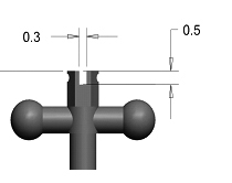
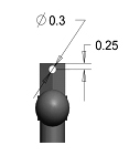
Head is hexagonal in shape. The hexagonal shape provides good resistance on inserting the implant. A slot is provided on the head to hold the orthodontic arch wire. The slot helps in holding the arch wire in some cases when it is used along with the fixed appliance therapy. The depth of the slot is 0.5 mm and the width of the slot is 0.3 mm. A hole is provided on the head to engage the ligature wire [Table/Fig-3]. Diameter of the hole is 0.3 mm and the distance from head to centre of hole is 0.25 mm.
Arms [Table/Fig-4,5]
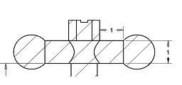
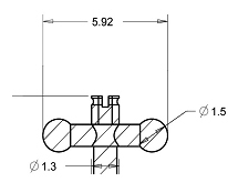
There are two arms attached on either side of the shank with the ball head. Arms assist in holding the extra oral elastics and the ball head helps in preventing slippage of elastics. Length and diameter of the arm is 1 mm. Diameter of the ball is 1.5 mm. Total length of attachment is 5.92 mm.
Shank [Table/Fig-6]

Shank prevents the gingival impingement of ball head by keeping it away from gingiva. Length of the shank is 1.5 mm and the diameter of the shank is 1.1 mm.
Collar [Table/Fig-7]
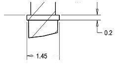
Collar rests at the attached gingiva. It acts as a limit to which the implant should be inserted. Length of the collar is 0.2 mm and diameter of the collar is 1.45 mm.
Neck [Table/Fig-8]
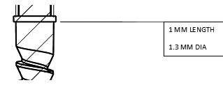
It is the non- threaded portion of the implant. It lies inside the soft tissue. The length of the neck is 1 mm. Diameter of the neck is 1.3 mm.
Body
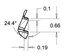
It is the threaded portion of the implant. Threads are of reverse buttress type which acts as a self drilling implant. It helps in the easy insertion of implant without any pre preparation for drilling.
The pitch width is 0.66 mm. Thread angle is 24.4o. Flank width is 0.1 mm. Thread width is 0.19 mm and total number of threads is 7 [Table/Fig-9].
Tip [Table/Fig-10]
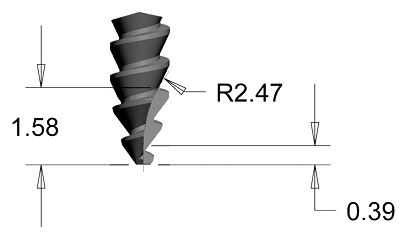
Tip has a cork screw design. Diameter of the tip is 1.2 mm, active tip end is 0.39 mm.
Notch is given on the tip for the self drilling of implant. Radius arc of the notch is 2.47o.
This type of implant assisted maxillary protrusion will require minimal anchorage preparation and maximum acceptable force limit (upto 500 grams) can be achieved with minimal side effects and good patient compliance. The purpose of this innovation was to attain the proper skeletal changes required rather than the dental changes. Clinical success of the ball headed mini implant is unknown since it has not been applied clinically in any of the cases and further research in this aspect is underway.
[1]. Enlow DH, Facial growth 1990 3rd edPhiladelphia, paWB saunders [Google Scholar]
[2]. Mcnamara JA, An orthopaedic approach to the treatment of class III malocclusion in young patientsJ Clinic Orthod 1987 21:598-608. [Google Scholar]
[3]. Itoh T, Chaconas SJ, Caputo AA, Matas J, Photoelastic effects of maxillary protraction on the craniofacial complexAm J orthod 1985 88:117-24. [Google Scholar]
[4]. Kokich VG, Shapiro PA, Koskinen Oswald R, Mottetl L, Clarren SK, Ankylosed teeth as abutments for maxillary protraction: A case reportAm J Orthod 1985 -88.:303-07. [Google Scholar]
[5]. Leung MT, Lee TC, Rabie AB, Wrong RW, Use of mini screws and mini-plates in orthodonticsJournal of Oral and Maxillofacial Surgery 2008 66:1461-66. [Google Scholar]
[6]. Block MS, Hoffman DR, A new device for absolute anchorage for orthodonticsAm J Orthod Dentofacial Orthop 1995 107:251-58. [Google Scholar]
[7]. Higuchi KW, Slack JM, The use of titanium fixtures for intraoral anchorage to facilitate orthodontic tooth movementInt J Oral Maxillofac Implants 1991 6:338-44. [Google Scholar]
[8]. Turley PK, Shapiro PA, Moffett BC, The loading of bioglass coated aluminium oxide implants to produce sutural expansion of the maxillary complex in the pigtail monkey (Macaca nemestrina)Archs Oral Biol 1980 25:459-69. [Google Scholar]
[9]. Smalley WM, Shapiro PA, Hohl TH, Kokich VG, Branemark PL, Osseointegrated titanium implants for maxillofacial protraction in monkeysAm J Orthod Dent-ofacial Orthop 1988 94:285-95. [Google Scholar]