A Rare Case of Twinning Involving Primary Maxillary Lateral Incisor with Review of Literature
Santosh Hunasgi1, Anila Koneru2, Vardendra Manvikar3, M Vanishree4, Rudraraju Amrutha5
1 Professor, Department of Oral and Maxillofacial Pathology, Navodaya Dental College and Hospital, Raichur, Karnataka, India.
2 Reader, Department of Oral and Maxillofacial Pathology, Navodaya Dental College and Hospital, Raichur, Karnataka, India.
3 Assistant Professor, Department of Oral and Maxillofacial Pathology, Navodaya Dental College and Hospital, Raichur, Karnataka, India.
4 Professor and Head, Department of Oral and Maxillofacial Pathology, Navodaya Dental College and Hospital, Raichur, Karnataka, India.
5 Assistant Professor, Department of Oral and Maxillofacial Pathology, Navodaya Dental College and Hospital, Raichur, Karnataka, India.
NAME, ADDRESS, E-MAIL ID OF THE CORRESPONDING AUTHOR: Dr. Santosh Hunasgi, Professor, Department of Oral and Maxillofacial Pathology, Navodaya Dental College and Hospital, Raichur, 584103, Karnataka, India.
E-mail: drsantosh31@gmail.com
Twinning is referred to the development of two separate teeth that arose from the complete separation of one tooth bud. To the best of our knowledge very few cases of twinning in primary or permanent dentition have been previously reported. Here, we report an additional case of twinning involving primary maxillary left lateral incisor and a literature review of clinical and radiographic findings of previous reported cases of gemination and twinning is also discussed. A six-year-old male patient reported to the dental clinic with the complaint of decay in the left front teeth region of the upper jaw. On clinical examination, dentinal caries was observed on the labial surface of primary maxillary left lateral incisor. The tooth showed a deep groove present in relation to the labial surface and incisal edge and continued cervically as a shallow groove. The patient had normal compliment of teeth for his age. The intra-oral periapical radiograph of the maxillary anterior region revealed large crown and a radiolucent notch was observed in relation to the incisal edge of the maxillary left primary lateral incisor. Relatively one pulp chamber and two root canals were observed in relation to the primary maxillary left lateral incisor, which was suggestive of a case of twinning involving primary maxillary left lateral incisor. This present case is the first case report of twining seen in primary dentition.
Anomaly, Double tooth, Gemination, Root canals, Twinning
Case Report
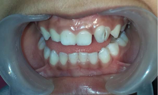
A six-year-old male patient reported to the dental clinic with the complaint of decay in the left front teeth region of the upper jaw. On clinical examination, dentinal caries was observed on the labial surface of primary maxillary left lateral incisor. The tooth was larger in the mesiodistal dimension and there was a deep groove present in relation to the labial surface and incisal edge and continued cervically as a shallow groove. The patient had normal compliment of teeth for his age [Table/Fig-1,2].
Clinical photograph showing primary maxillary left lateral incisor which is larger in mesiodistal dimension and the patient has a normal compliment of teeth for his age.
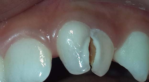
Clinical photograph of primary maxillary left lateral incisor showing a deep groove present in relation to the labial surface and incisal edge. The groove continued cervically as a shallow groove and dentinal caries was observed on the labial surface.
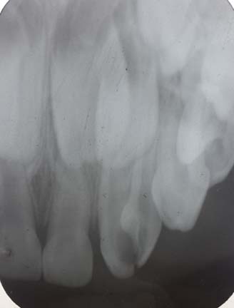
Periapical radiograph of the maxillary anterior region showed a large crown and a radiolucent notch in relation to the incisal edge of the primary maxillary left lateral incisor. Relatively one pulp chamber and two root canals were observed in relation to the primary maxillary left lateral incisor, which was suggestive of a case of twinning involving primary maxillary left lateral incisor [Table/Fig-3]. Since the patient was not concerned about the aesthetic problems, only the caries present in the labial groove was removed and filled with GC 2 restoration (Glass Ionomer Cement).
Intra-oral periapical radiograph of 51, 61, 62 and 63 revealed relatively one pulp chamber and two root canals seen in relation to the primary maxillary left lateral incisor.
Discussion
Developmental dental disorders may range from abnormalities in the demarcation of the dental lamina and anomalies of tooth germs (number, size and shape) to abnormalities in the growth of the dental hard tissues (structure). Dental anomalies are not only congenital but they may also be idiopathic, inherited or acquired [1].
The terms “double tooth”, “fused teeth”, connoted teeth”, conjoined teeth”, “double formations” are commonly used to exemplify gemination and fusion. These anomalies together are primary developmental disorders of the teeth. Double teeth are two separate teeth exhibiting merger by dentin and perhaps their pulps. The unification may be the result of fusion of two adjacent tooth buds or the unfinished splitting of one into two [2,3].
Formerly, gemination was defined as an attempt of a single tooth bud to split, with the ensuing development of a tooth with a bifid crown and typically a general root and root canal. On the other hand, fusion was considered as the union of two normally separated tooth buds with the consequential development of a joined tooth with confluence of dentin. Finally, Concrescence was the union of two teeth by cementum without confluence of the dentin. All the definitions were confusing and open to debate. Few authors have recommended that the conditions such as gemination, fusion and concrescence should be discontinued and a general term twinning to be used [1].
Twinning is considered when there is development of two separate teeth that arose from the complete division of one tooth bud [4]. Many case reports of gemination involving primary and permanent dentition have been reported in literature [5,6]. However, occurrence of twinning in primary or permanent dentition is very rare. Therefore, this article documents a case of twinning involving primary maxillary left lateral incisor and also reviews clinical and radiographic findings of previous reported cases of gemination and twinning.
Tannenbaum and Alling, in 1963 defined gemination as the formation of the equivalent of two teeth from the same follicle, with an attempt for teeth to be incompletely separate, this indicated clinically by a depression or groove which might demarcate two teeth. Radiographically, there appears to be only one root canal. However, if the attempt is completely separate and two root canals are seen radiographically, it is termed as twinning. In gemination and twinning, if the bifid tooth is counted as one entity, the total number of teeth in the dental arch is otherwise normal [7].
Gemination has a higher prevalence in deciduous teeth, with a higher frequency in anterior maxillary region with equal sex predilection. Hence it is most commonly seen in maxillary primary incisors but is rare in permanent dentition. This anomaly has a large bifid crown which is incompletely separated that has single pulp chamber and root canal. However, the prevalence of twinned tooth is more in permanent dentition than primary. Twinned tooth shows a low deep groove from incisal to gingival third and is considered when the division results in equivalent structures consequentially leading to one normal and one supernumerary tooth. Radiographically, in twinning there will be only one pulp chamber with two root canals. Complete case history, clinical examination, and radiographic investigation can provide the information required for the distinction between gemination and twinning [8]. The tooth involved in our case was primary maxillary left lateral incisor.
The English language literature was reviewed on cases of gemination and twinning and all the findings were tabulated in [Table/Fig-4] [2,4–6,8,9–17]. Among all the 15 cases reported, only two cases were diagnosed as true twinning along with our case which showed complete cleavage of tooth bud, and radiographically showing single pulp chamber and two separate root canals.
A review of clinical and radiographic features of gemination and twinning that were previously reported, together with the present case.
| Author | Year | Age/ Sex | Tooth involved | Radiographic Examination |
|---|
| Spuller RL et al., [9] | 1986 | 7yr/F | Permanent maxillary right central incisor | Single root canal and pulp chamber |
| Krishnan S et al., [4] | 1999 | 35yr/M | Permanent maxillary right lateral incisor | Presence of two separate root canals * |
| Turkaslan S et al., [10] | 2007 | 22yr/M | Bilateral Permanent maxillary central incisor | Each incisor had one root |
| Tarasingh P et al., [2] | 2010 | 2yr/M | Primary maxillary right central incisor | Presence of single pulp chamber and root canal |
| Tarasingh P et al., [2] | 2010 | 8yr/M | Right Primary maxillary central incisor | Single root & root canal |
| Sener S et al., [11] | 2012 | 17yr/M | Bilateral Permanent maxillary central incisor | The pulp chambers and root canals were large in both teeth, but there was only 1 of each |
| Sharada HL et al., [5] | 2013 | 23yr/M | Permanent maxillary lateral incisor | Incomplete cleavage of the lateral incisor with single pulp chamber and root canal. |
| Rao PK et al., [8] | 2013 | 25 yr/M | Bilateral maxillary central incisors | A radiolucent notch was observed in relation to the incisal edges of the central incisors. Relatively single large pulp chambers and root canals were observed in relation to permanent maxillary right and left central incisors. |
| Mazumdar P et al., [12] | 2013 | 6yr/F | Permanent maxillary right central incisor | Showed a solitary large pulp chamber as well as canal but the pulp chamber was found to be obstructed by pulp stone and there was evidence of mild periapical radiolucency |
| Shokri A et al., [13] | 2013 | 9yr/M | Bilateral Permanent maxillary central incisor | All the maxillary permanent central incisors showed one root with two separate canals having one orifice at the apical foramen. |
| Nandini DB et al., [6] | 2014 | 4yr/M | Maxillary left first premolar | A single root and two crowns |
| Mahendra L et al., [14] | 2014 | 27yr/M | Bilateral Permanent maxillary central incisor | Two completely separated roots were seen in the right maxillary incisors with central notching which was seen as a radiolucent line in the fused right maxillary incisor crown |
| Moushekhian S et al., [15] | 2014 | 27yr/F | Permanent left lateral incisor | Macro tooth with two canals that were seen to be united in apical area * |
| Neena IE et al., [16] | 2015 | 5yr/F | Primary maxillary right central incisors | Single pulp chamber and root canal |
| Rahman H et al., [17] | 2016 | 19yr/M | Mandibular left first premolar | Single root was normal but it was wider cervically and conical in shape |
| Present case | 2016 | 6 yr/ M | Primary maxillary right central incisor | Presence of two separate root canals * |
* Two cases showed twinning along with the present case.
The aetiology of gemination and twinning remains unknown; however some of the interferences which occur during morphodifferentiation of the tooth germ are [6,14]:
Hereditary or congenital diseases;
Nutritional deficiency;
Local traumas;
Infectious/inflammatory processes;
Ionizing radiation is also considered;
Endocrine influences;
Excessive ingestion of medicines;
A group of molecular signalling pathways are concerned in the usual development of the tooth germ. These include components of the hedgehog, Wnt, MSX-1 and MSX-2, TNF, BMP Families, FGF, which provides a valuable basis of applicant genes that may potentially participate a function in human tooth formation. However, if inappropriately regulated by various causes mentioned above can lead to the formation of gemination/ twinning or fusion or additional teeth [3].
The problems, mostly if the anterior teeth are involved, differ from spacing problems, tooth malalignment, aesthetic problems, arch asymmetry, periodontal involvement and impeding the eruption of the adjacent tooth. Occurrence of deep groove in some cases of gemination and twinning makes them vulnerable to periodontal diseases and caries. When these defects are extremely deep and lengthen to gingival third, the probability of bacterial plaque build up in this area is high. Stringent oral hygiene practice is advised to preserve periodontal health [2,8]. In our patient, the anomaly caused a deep labial groove and was involved with caries; no serious periodontal disease was evident. The caries was removed and filled with GC 2 (Glass Ionomer Cement).
The anomalies of primary dentition are strongly associated with anomalies in the permanent dentition. Consequently, before time diagnosis of the anomaly has a significant importance and it must be followed by cautious clinical and radiographic comments that will allow interference at suitable time.
Conclusion
The present case is rare because it resembles to canine with deep labial groove and causing diastema between left maxillary primary central incisor and lateral incisor. Many cases of Gemination involving primary and permanent have been reported in literature. However, the present case of twining involving primary dentition is extremely rare where the crown is bifid with two root canals. In the literature review, only two cases of twining have been previously reported with the addition of present case.
* Two cases showed twinning along with the present case.
[1]. Neville BW, Damn DD, Allen CM, Bouquot JE, Oral and maxillofacial pathology 1995 1st edPhiladelphiaW.B Saunders Company:362-384. [Google Scholar]
[2]. Tarasingh P, Balaji K, Gemination in primary teeth – A Report of Two Clinical CasesAnnals and Essences of Dentistry 2010 2(2):48-51. [Google Scholar]
[3]. Yuen S, Chan JCY, Wei S, Double primary teeth and their relationship with the permanent successors: A radiographic study of 376 casesPediatric Dentistry 1987 9(1):42-48. [Google Scholar]
[4]. Krishnan S, Wadhwani KK, Case of Twinning in Permanent lateral incisorEndodontology 1999 11:57-59. [Google Scholar]
[5]. Sharada HL, Deo B, Briget B, Gemination of a Permanent Lateral Incisor- A Case Report with Special Emphasis on ManagementJ Int Oral Health 2013 5(2):49-53. [Google Scholar]
[6]. Nandini DB, Deepak BS, Selvamani M, Puneeth HK, Diagnostic Dilemma of a Double Tooth: A Rare Case Report and ReviewJ Clini Diagn Res 2014 8(1):271-72. [Google Scholar]
[7]. Tannenbaum KA, Alling EE, Anomalous tooth development: Case reports of germination and twinningOral Med and Oral Path 1963 16:883-87. [Google Scholar]
[8]. Rao PK, Veena KM, Chatra L, Shenai P, Twin tooth on either side: A case report of bilateral geminationAnn Med Health Sci Res 2013 3:271-73. [Google Scholar]
[9]. Spuller RL, Harrington M, Gemination of a maxillary permanent central incisor treated by autogenous transplantation of a supernumerary incisor: Case reportPediatric Dentistry 1986 8(4):299-302. [Google Scholar]
[10]. Turkaslana S, Gokceb HS, Dalkız M, Aesthetic Rehabilitation of Bilateral Geminated Teeth: A Case ReportEuropean Journal of Dentistry 2007 1:188-91. [Google Scholar]
[11]. Sener S, Unlu N, Basciftci FA, Bozdag G, Bilateral geminated teeth with talon cusps: A case reportEuropean Journal of Dentistry 2012 6:440-44. [Google Scholar]
[12]. Mazumdar P, Das UK, Rahaman SM, Endodontic Management of Geminated tooth: A case reportInternational Journal of Scientific and Research Publications 2013 3(2):1-4. [Google Scholar]
[13]. Shokri A, Baharvand M, Mortazavi H, The largest bilateral gemination of permanent maxillary central incisors: Report of a caseJ Clin Exp Dent 2013 5(5):e295-97. [Google Scholar]
[14]. Mahendra L, Govindarajan S, Jayanandan M, Shamsudeen SM, Kumar N, Madasamy R, Complete Bilateral Gemination of Maxillary Incisors with Separate Root CanalsCase Reports in Dentistry 2014 :1-4. [Google Scholar]
[15]. Moushekhian S, Shiehzadeh M, Shammas A, Treatment Plan and Clinical Management of a Geminated Maxillary Lateral Incisor: A Case ReportJ Dent Mater Tech 2014 3(2):87-90. [Google Scholar]
[16]. Neena IE, Sharma R, Poornima P, Roopa KB, Gemination in primary central incisorJ Oral Res Rev 2015 7:55-57. [Google Scholar]
[17]. Rahman H, Chandra R, Gaur TK, Singh S, Tripathi S, Jain A, An Unusual Case of Gemination in Mandibular Second Premolar: A Case ReportResearch & Reviews: Journal of Dental Sciences 2016 4(2):19-22. [Google Scholar]