Variants of Talon Cusp (Dens Evaginatus)
Mythri Sarpangala1, Ashwin Devasya2
1 Senior Lecturer, Department of Periodontology, Kannur Dental College, Anjarakkandy, Kannur, Kerala, India.
2 Senior Lecturer, Department of Paedodontics and Preventive Dentistry, Kannur Dental College, Anjarakkandy, Kannur, Kerala, India
NAME, ADDRESS, E-MAIL ID OF THE CORRESPONDING AUTHOR: Dr. Ashwin Devasya, Department of Paedodontics and Preventive Dentistry, Kannur Dental College, Anjarakkandy, Kannur-670612, Kerala, India.
E-mail: ashwindkumbla@gmail.com
Accessory cusp, Mesiodens, Primary dentition
Talon cusp is a well delineated additional cusp that is located on the surface of an anterior tooth and extends at least half the distance from the Cementoenamel Junction (CEJ) to the incisal edge. It was first described by Mitchell WH in 1982 [1,2]. This is a type of accessory cusp that projects from the lingual surface that resembles an eagle’s talon and hence, the name [3].
It could be due to an excess layering during morphodifferentiation of tooth development [4]. The occurrence is seen 1 to 6% in population, mainly in permanent dentition [1].
Talon cusp can be presented as exaggerated cingula, cusp like hyperplasia, accessory cusps, supernumerary cusps, interstitial cusps and palatal accessory cusps. It contains enamel, dentine and possible pulpal extension histologically and radiologically superimposed over the tooth on which it occurs. It is also seen in syndromes like Berardinelli-Seip, Mohr, Rubinstein-Taybi, Ellis-van Creveld, Sturge-Weber and incontinentia pigmenti achromians [5].
Based on the literature, occurrence of talon cusp has been uncommon and has many variants. Here we present and discuss the occurrence of five cases of talon cusp [Table/Fig-1].
Details of five cases of talon cusp.
| Age/Sex | Chief Complaint | intraoral Examination | Radiographic Examination | Diagnosis | Treatment Given |
|---|
| 10 years/male | Abnormally shaped upper front tooth | Maxillary right permanent lateral incisor had a projection on the labial surface [Table/Fig-2] | Not available | Facial talon cusp | The parents were assured that it is a rare occurrence and regular check up is needed. |
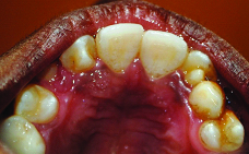
| 13 years/male | Irritation to the tongue | No signs of injury or presence of ulcers on tongue. Patient had no tongue thrusting habit. Mandibular left permanent central incisor had a sharp, prominent talon cusp [Table/Fig-3] | IOPA radiograph showed radiopacity on the lingual surface of mandibular left permanent central incisor [Table/Fig-4] | Lingual talon cusp | The projection was grinded gradually with topical fluoride application in multiple appointments. |
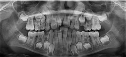
| Eight years/ male | Extra tooth in upper front tooth region | Mesiodens with a projection in the cingulum [Table/Fig-5] | OPG showed mesiodens in between the permanent central incisors [Table/Fig-6] | Talon cusp with mesiodens | Parents were explained that it is a rare occurrence and regular check-up is needed |
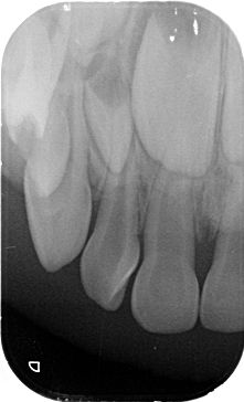
| Four years/male | Caries in upper tooth | Talon cusp in maxillary primary left lateral incisor [Table/Fig-7] | IOPA radiograph showed radiopacity on the lingual aspect of the crown [Table/Fig-8] | Talon cusp on primary teeth | Parents were explained that it is a rare occurrence and regular check-up is needed |
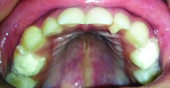
| 12 years/female | Deciduous canine was mobile | Maxillary left deciduous canine was mobile. Further examination showed that the patient had bilateral talon cusps in maxillary permanent lateral incisors [Table/Fig-9] | IOPA radiograph showed typical presentation of talon cusp, adiopacity in the lingual portion of the lateral incisors [Table/Fig-10] | Bilateral talon cusp | Asked to come for regular check-up |
Case 1: A 10-year-old male with facial talon cusp on maxillary right lateral incisor.
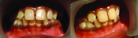
Case 2: A 13-year-old male patient with a complaint of irritation to the tongue due to the projection in mandibular left central incisor.
Case 2: IOPA showing a projection of tooth structure in the lingual surface of the lateral incisor.
Case 3: An eight-year-old male patient having talon cusp in mesiodens.
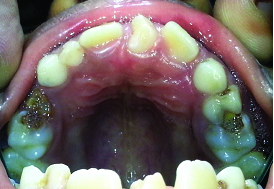
OPG showing the mesiodens in between the permanent central incisors.
Case 4: A four-year-old male child with talon cusp in maxillary left lateral incisor.
IOPA of talon cusp showing projection of enamel and dentine.
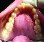
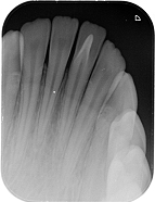
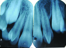
Case 5: A 12-year-old female with bilateral talon cusps on maxillary H lateral incisors.
IOPA of both maxillary lateral incisors showing H projection of tooth structure.
Discussion
Presence of talon cusp may cause difficulty in maintaining hygiene, aesthetics, occlusal interferences, carious developmental grooves, irritation to the tongue during speech and mastication, nursing difficulty and accidental cusp fracture [6]. The possible treatment modalities for this are; periodic observation, application of topical fluoride, sealing the fissures, aesthetic restorations, pulpectomy or root canal treatment and crowns based on the tooth affected and extraction, as a last resort [7]. Early diagnosis and proper preventive treatment is important.
To conclude, talon cusp is a rare morphological developmental anomaly that can occur both in primary and permanent dentition. The knowledge of different types, variety of presentations and occurrences can help in easy and early identification so that proper preventive and therapeutic treatments can be undertaken. In this case report, we discussed five cases of talon cusp hoping that it will help clinicians in identification, if encountered.
[1]. Neville BW, Damm DD, Allen CM, Bouquot JE, Abnormalities of TeethOral and Maxillofacial Pathology 2004 2nd edNew DelhiSaunders:77-78. [Google Scholar]
[2]. Mitchell WH, Letter to editor 1892 34:1036 [Google Scholar]
[3]. Mellor JK, Ripa LW, Talon cuspA clinically significant anomaly 1970 29:225-28. [Google Scholar]
[4]. Tratman EK, An unrecorded form of simplest type of the dilated composite odontome 1949 86:271-75. [Google Scholar]
[5]. Davis PJ, Brook AH, The presentation of talon cuspDiagnosis, clinical features, associations and possible etiology 1986 160:84-88. [Google Scholar]
[6]. Yoon N, Chussid S, Dental management of a talon cusp on a primary incisor 2007 29:51-55. [Google Scholar]
[7]. Thakur S, Gupta R, Thakur NS, Gupta M, Facial talon cusp on permanent maxillary canineA rare dental anomaly 2013 2:324-27. [Google Scholar]