In any natural disaster, criminal case investigation or when the body is burnt or destroyed the body remains degraded and other biological tools such as DNA analysis, fingerprint etc., fail to assess the identification of the body, forensic radiology plays an important role in personal identification as well as gender determination [1]. Structures in human skull such as frontal sinus, nasal septum, vascular groove patterns, sella turcica etc., can act as useful indicators because of their uniqueness in every individual. Frontal sinus is a cavity which is lobulated. It is bilaterally situated in between the external and internal cortical surface of the frontal bone. It may vary person to person or even in monozygotic twins. The frontal sinuses are not even visible at the time of birth. [2,3] They gradually develop at the age of two years and the development completes at the age of 20 years.
The nasal septum pattern is also useful and unique tool. In each individual many types of patterns present such as Straight (S’), Left deviated (L’) or Right deviated (R’), Sigmoid type (Si), Reverse sigmoid type (RSi) and Others (O) (epsilon and reverse epsilon type; rare types) [4]. The present study ascertains the unique role of frontal sinus and nasal septum for individual identification and sexual dimorphism.
Materials and Methods
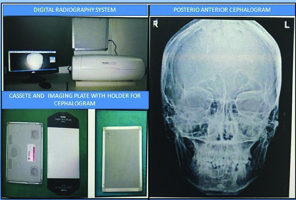
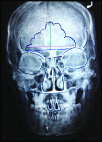
The observational study was proposed and approved by the Institutional Ethical Committee and was conducted for a period of one year. A total of 80 individuals, 40 males and 40 females, between the age ranges of 18-30 years were selected for the present study, from the outpatient department visiting the Department of Oral Medicine, Diagnosis and Radiology at Pacific Dental College and Hospital, Udaipur, Rajasthan, India. The sample size was calculated using previous studies [1,2]. The selected individuals were included in the study and had their Posterio Anterior (PA) cephalograph performed after taking their written consent and necessary radiation protection measures were undertaken according to the standard protocol. The study employed PA cephalogram as it demonstrates adequate and standardized frontal sinus morphology and nasal septum pattern with minimal distortion on a single radiograph. The radiographs were performed on Xtropan 2000 OPG X-ray machine with cephalography attachment and KODAK CR 7400 digital radiography system. The resultant images were obtained on a 20x25 cm sized Kodak Dry View Laser Imaging film through Carestream DRY VIEW 5700 Laser Imager. Each radiograph was subjected to tracings of frontal sinus outline and nasal septum, from which the dimensions and patterns of frontal sinus and nasal septum deviations were recorded, for both the genders [Table/Fig-1,2].
Materials used in the study.
Tracing of frontal sinus and nasal septum.
Healthy adult male and female individuals were included. Individuals uncooperative, unstable or unable to undergo cephalography procedure, individuals with any history of trauma or surgery of skull in past or present, individuals with apparent maxillofacial deformities or asymmetry, individuals with any ailment of paranasal sinuses, history or clinical features of any other systemic or congenital disorder, pregnant individuals, radiographs with artifacts, improper orientation and poor quality radiographs demonstrating unilateral/bilateral aplasia of frontal sinus were excluded.
Frontal Sinus Evaluation
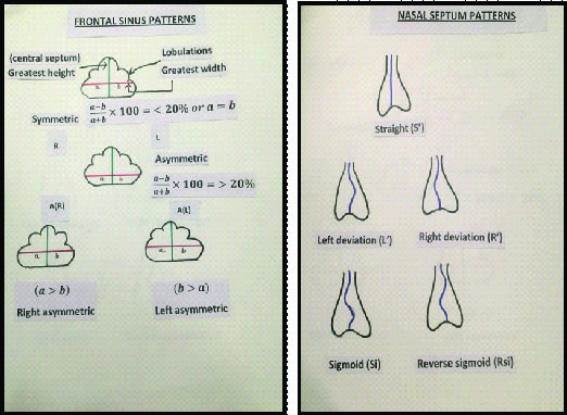
Determination of frontal sinus asymmetry: The maximum horizontal dimension was measured from the central septum on both sides. The difference in the right and left side dimensions was divided by the greatest dimension and multiplied by 100. If the percentage obtained was more than 20% then it was asymmetrical for the respective site side [1].
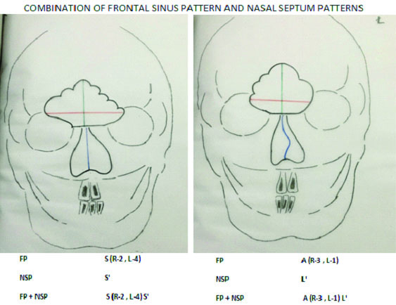
The patterns of nasal septum were also traced from the radiographs and classified according to the deviations in the septa as : Straight (S’), Right deviation (R’), Left deviation (L’), Sigmoid (Si), Reverse sigmoid (RSi) and Others (O). The patterns were tabulated and categorized for all the individuals and also separately for both the genders. The various possible combinations of resultant pattern of FP and NSP were evaluated [Table/Fig-3,4].
Frontal and nasal sinus patterns used in the study.
Combination of frontal sinus pattern and nasal septum patterns.
Statistical Analysis
The resultant data for the dimensions, patterns of FP, NSP and combination patterns (FP+NSP) for all the individuals and both the genders were subjected to statistical analysis to determine the significance of dimensions of frontal sinus and particular patterns of frontal sinus and nasal septum in personal identification and gender determination. The statistical analysis was carried out with the help of SPSS software.
Student’s t-test: Unpaired t-test or independent samples t-test were executed to compare means between the two unrelated groups. The frequencies (n) of the FP and NSP and combination of these patterns were tabulated separately for individuals and both the genders. The percentage was calculated and the data were further subjected to chi-square test as follows to evaluate the statistical significance of difference in frequencies between subgroups.
The significance of occurrence of a particular entity was expressed as p-value<0.05 was considered as significant.
Results
Out of 80 individuals, the mean corrected height of frontal sinus with SD in males was 20.87±4.65 mm and in females it was 15.36±5.07 mm. After intercomparison of mean and SD values of maximum corrected height of frontal sinus in both the genders, the p-value was found to be 0.000 (p<0.001) which was highly significant [Table/Fig-5].
Mean and SD values of maximum corrected height of frontal sinuses in males and females.
| Study Group | N | Mean | Std. Deviation (SD) | t | Significance |
|---|
| Male | 40 | 20.8685 | 4.65495 | 5.068 | p=<0.001 |
| Female | 40 | 15.3550 | 5.06724 |
Student t-test was applied
Out of 80 individuals the mean corrected width of frontal sinus with SD in males was 61.66±8.38 mm and in females it was 36.67±6.59 mm. After intercomparison of mean and SD values of maximum corrected width of frontal sinus in both the genders, the p-value was found to be 0.000 (p<0.001) which was highly significant [Table/Fig-6].
Mean and SD values of maximum corrected width of frontal sinuses in males and females.
| Study Group | N | Mean | Std. Deviation (SD) | t | Significance |
|---|
| Male | 40 | 61.6642 | 8.37867 | 15.832 | p=<0.001 |
| Female | 40 | 36.6677 | 6.58874 | | |
Student t-test was applied
The study revealed that out of 80 individuals, 62 (77.5 %) subjects had bilateral symmetric pattern of frontal sinus and 18 (22.5%) subjects had asymmetric pattern of frontal sinus. Out of the total 18 patients demonstrating asymmetric FP, 10 (55.56%) subjects had right asymmetric FP and 08 (44.44%) subjects had left asymmetric FP [Table/Fig-7].
Frontal sinus patterns (FP) distribution in total individuals.
| FP | N | % |
|---|
| Symmetry (S) | 62 | 77.50 |
| Asymmetry (A) | 18 | 22.50 |
| Right Asymmetry A(R) | 10 | 55.56 |
| Left Asymmetry A(L) | 8 | 44.44 |
The study revealed that out of 80 individuals, Straight NSP was demonstrated in 33 (41.25%) subjects, NSP with left deviation was demonstrated in 17 (21.25%) subjects and right deviated NSP was demonstrated in 22 (27.5%) subjects. Further, five (6.25%) subjects exhibited sigmoid and three (3.75%) subjects exhibited reverse sigmoid NSP [Table/Fig-8].
Distribution of nasal septum patterns (NSP) in all the individuals.
| NSP | N | % |
|---|
| Straight (S’) | 33 | 41.25 |
| Left Deviation (L’) | 17 | 21.25 |
| Right Deviation (R’) | 22 | 27.50 |
| Sigmoid (Si) | 5 | 06.25 |
| Reverse Sigmoid (RSi) | 3 | 03.75 |
| Total | 80 | 100.00 |
After combining the frontal sinus and nasal septum patterns (FP+ NSP) in all the 80 individuals separately, there were nine classifiable patterns in 26 individuals (12 males and 14 females), each of which had common representations in more than one individual. Besides these patterns, there were unique patterns in 54 individuals each of which was represented in only one individual and couldn’t be found in any other subject.
The study revealed that each of S(R-2, L-3)L’, S(R-3, L-1)L’ and S(R-4, L-3)S’ patterns were exhibited by two individuals of which one (50%) was a male and one (50%) was a female subject, each of S(R-1, L-1)S’, S(R-2, L-3)R’ and S(R-3, L-3)S’ patterns were exhibited by three individuals of which one (33.33%) was a male and two (66.67%) were females, each of S(R-2, L-2)S’ and A(R-3, L-1)L’ patterns were exhibited by four individuals, of which 03 (75%) were males and one (25%) was a female,) S(R-2, L-4)S’ pattern was exhibited by three individuals all of whom were females (100%), 54 individuals demonstrated unique combinations of FP and NSP of which 28 (51.85%) were males and 26 (48.15%) were females [Table/Fig-9].
Distribution of combined (FP+NSP) patterns in all the individuals and both the genders.
| Classification of Combined Patterns | Male | Female | Total |
|---|
| N | % | N | % | N |
|---|
| S(R-2, L-3)L’ | 1 | 50.00 | 1 | 50.00 | 2 |
| S(R-1, L-1)S’ | 1 | 33.33 | 2 | 66.67 | 3 |
| S(R-2, L-2)S’ | 3 | 75.00 | 1 | 25.00 | 4 |
| S(R-2, L-3)R’ | 1 | 33.33 | 2 | 66.67 | 3 |
| S(R-3, L-1)L’ | 1 | 50.00 | 1 | 50.00 | 2 |
| S(R-2, L-4)S’ | 0 | 0.00 | 3 | 100.00 | 3 |
| S(R-4, L-3)S’ | 1 | 50.00 | 1 | 50.00 | 2 |
| S(R-3, L-3)S’ | 1 | 33.33 | 2 | 66.67 | 3 |
| A(R-3, L-1)L’ | 3 | 75.00 | 1 | 25.00 | 4 |
| Unique | 28 | 51.85 | 26 | 48.15 | 54 |
(S-symmetric, A-asymmetric, R’-right deviated, L’- left deviated)
Discussion
Forensic identification of an individual based on finger prints, autopsy or DNA typing has been a time tested criterion. But this has its own limitations due to degradation of soft tissues with time [1].
As hard tissues or body parts are normally resistant to putrefaction for years, they can serve as good alternate material for forensic investigation. Radiographic evaluation of various skeletal structures including skull is a potentially useful procedure for identification either in human remains or in living person [3]. In 1897 Zuckerkandl first established the uniqueness of frontal sinus in individuals, other researchers have also studied and worked to establish uniqueness and reliability of frontal sinus morphology for human identification in forensic science. Frontal sinus patterns when combined with nasal septum patterns may also be useful for identification and gender determination of an individual [1]. The present study was undertaken with the intention of frontal sinus morphology and nasal septum pattern with minimal distortion on a single radiograph.
In the present study, the comparison of mean maximum height of frontal sinuses in 40 males and 40 females revealed that males had mean maximum height of 20.87±4.65 mm, which was highly significant greater than mean maximum height of 15.36±5.07 mm in females. The study also compared the mean maximum width of frontal sinuses in 40 males and 40 females and revealed that males had mean maximum width of 61.66±8.38 mm, which was highly significant greater than mean maximum width of 36.67±6.59 mm in females. This suggested that mean maximum height and width of frontal sinus evaluated through skull radiographs is greater in males as compared to that in females and can be used as a criterion for determination of gender in forensic investigation. These findings in the present study were in accordance with those stated in the study done by Yoshino M [5].
Hsiao TH et al., conducted a similar study but employing lateral cephalometric radiographs and showed that the mean values for all linear measurements and proportional measurement of frontal sinus in males were larger than in females [6]. This is in similarity to the findings from the present study that was done employing PA cephalogram instead. Camargo JR et al., through their study on the forensic importance of radiographic morphology of frontal sinus employing Caldwell technique [7], stated that the mean dimensions of the frontal sinus in the men were consistently greater than in the women.
The present study differed from that done by Taniguchi M et al., in various aspects [4]. They demonstrated that, 43.1% cases had symmetry of frontal sinus and 56.6% had asymmetrical patterns which are contrary to findings of our study (77.5% symmetrical and 22.5% asymmetrical patterns of frontal sinus). Besides this, our study was antemortem, employing PA cephalogram of 80 individuals with equal number of males and females from Indian population between age 18-30 years and excluding cases with frontal sinus aplasia, whereas, their study was a postmortem skull radiograph study employing 209 forensic autopsy cases of Japanese population between 18-91 years of age and including frontal sinus aplasia cases. The results of the present study exhibiting insignificant difference in frontal sinus pattern also differed from those of the study done by Christensen AM for assessing the variation in individual frontal sinus outlines using Elliptic Fourier Analysis (EFA), which revealed a quantifiable and significant difference between the shapes of individual’s frontal sinus outlines [8].
The present study revealed that, out of 80 individuals 33 (41.25%) had straight nasal septum, 17 (21.25%) had left deviated, 22 (27.5%) had right deviated, 05 (6.25%) had sigmoid and 03 (3.75%) had reverse sigmoid pattern of nasal septum. These findings were different from the study conducted by Taniguchi M et al., which demonstrated straight nasal septum in 13.4%, right deviation in 35.3%, left deviation in 37.6%, sigmoid in 6% and reverse sigmoid in 6.3% individuals [4].
In the present study, PA cephalogram of all the 80 individuals were simultaneously evaluated for the combination of frontal sinus and nasal septum patterns (FP+NSP). It was found that there were 09 classifiable patterns in 26 (32.5%) individuals; there was unique unclassifiable patterns in 54 (67.5%) individuals, each of which were represented in only one individual and couldn’t be found in any other subject. These findings from the present study were similar to those of similar study by David MP et al., done on 50 individuals, which demonstrated eight classifiable patterns in 17 (34%) of individuals and unique patterns in 33 (66%) individuals [1].
In year 2015, Verma P et al., has conducted a study [9]. A total of 149 PA cephalograms were taken and frontal sinus pattern and nasal septum patterns were evaluated and the found frontal sinus symmetry in 78.5% individuals, asymmetry in 7.3% and bilateral aplasia was found in 8.7% individuals. The mean height and width was more in male individual. The straight nasal septum pattern was seen more whereas, in present study the straight nasal septum pattern was seen more almost in 41.25% individuals followed by left, right deviated, sigmoid and reverse sigmoid.
In year 2016, Gopal SK et al., has conducted a retrospective study on 80 full skull and patterns of frontal sinus and nasal septum were evaluated [10]. They concluded that frontal sinus symmetry was observed in 43.75% individuals whereas, in 48.75% individuals asymmetry was detected, which is in accordance with our study. Whereas, straight nasal septum was seen in 33.75% individual followed by right deviation in 30%, left deviation in 22.5%, sigmoid was in 5% and reverse sigmoid in 8.75% individuals.
The present study has certain limitations such as this technique needs standardized parameters for measurement of the dimension of frontal sinus with minimal chances of error. The nasal septum patterns and frontal sinus patterns can be affected by genetic factors as well as growth hormone.
Conclusion
From the ongoing discussion it can be concluded the radiographic evaluation of frontal sinus dimensions, frontal sinus patterns, nasal septum deviations and the combination (FP+NSP) patterns is one of the aids for personal identification and gender determination in forensic investigations by using a single radiograph with minimal distortion. Present study also suggests that it is a reliable, easily reproducible and cost effective method. Hence, further studies with a larger sample size are required to evaluate the strength of the findings of the present study.
Student t-test was applied
Student t-test was applied
(S-symmetric, A-asymmetric, R’-right deviated, L’- left deviated)