Trichoadenoma of Nikolowski- A Rare Tumour with Unusual Presentation Over Vulva
Ashok Sangwaiya1, Shilpa Bairwa2, Shivani Kalhan3, Puja Sharma4, Rahul N Satarkar5
1 Assistant Professor, Department of Pathology, Shaheed Hasan Khan Mewati, Government Medical College, Nalhar, Nuh, Haryana, India.
2 Demonstrator, Department of Pathology, Shaheed Hasan Khan Mewati, Government Medical College, Nalhar, Nuh, Haryana, India.
3 Professor, Department of Pathology, Shaheed Hasan Khan Mewati, Government Medical College, Nalhar, Nuh, Haryana, India.
4 Associate Professor, Department of Pathology, Shaheed Hasan Khan Mewati, Government Medical College, Nalhar, Nuh, Haryana, India.
5 Associate Professor, Department of Pathology, Shaheed Hasan Khan Mewati, Government Medical College, Nalhar, Nuh, Haryana, India.
NAME, ADDRESS, E-MAIL ID OF THE CORRESPONDING AUTHOR: Dr Ashok Sangwaiya, Assistant Professor, Department of Pathology, Shaheed Hasan Khan Mewati, Government Medical College, Nalhar, Nuh-122107, Haryana, India.
E-mail: ashoksangwaiya25602@gmail.com
Trichoadenoma is a rare benign, slowly growing, cutaneous tumour of the hair follicle first described by Nikolowski in 1958. It presents as a non-specific nodule over the face or buttocks. However, unusual sites such as the neck, upper arm, thigh, shoulder, and shaft of the penis may also be affected. The tumour is less mature than a trichofolliculoma and is more differentiated than a trichoepithelioma with a differentiation towards the infundibular portion of the pilosebaceous canal. Histologically, it consists of numerous infundibulocystic structures present throughout the dermis, few of which are lined by eosinophilic epidermal cells with attempted glandular formation and contain laminated keratin, without evidence of hair follicle formation. We report a case of trichoadenoma over the vulva of 25-year-old female showing typical histological features.
Follicular tumour, Horn cyst, Skin malformation, Trichoepithelioma, Trichofolliculoma
Case Report
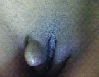
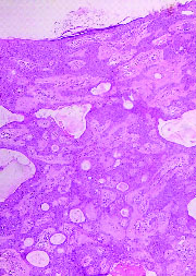
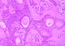
A 25-year-old married female presented with asymptomatic, single skin coloured nodule measuring 15mm x 8mm over the right labia majora since 8 months [Table/Fig-1]. There was no history of exposure to sexually transmitted diseases and discharge from the lesion. Physical examination of the patient was otherwise normal. The results of laboratory investigations including complete blood counts and urine microscopy were within normal limit. Serological tests for Syphilis, HIV, HbsAg and HCV were non-reactive. The lesion was excised under local anaesthesia and sent for histopathological examination. Gross examination revealed a skin covered specimen measuring 18mm x 10mm x 4mm. The whole tissue was processed in a single block and microsections were examined. Light microscopic examination revealed stratified squamous epithelium with underlying dermis. There were numerous infundibulocystic structures present throughout the dermis along with few solid nests of cells in between [Table/Fig-2]. Few of these cysts were lined by eosinophilic epidermal cells with attempted glandular formation and contained laminated keratin [Table/Fig-3]. There was no evidence of hair follicle formation and atypia. Considering these histological features a diagnosis of trichoadenoma was made. The case is being followed regularly and there has been no recurrence so far.
Photograph showing single skin coloured nodule over the right labia majora.
Photomicrograph showing numerous infundibulocystic structures present throughout the dermis along with few solid nests of cells in between (H&E, 10X).
Photomicrograph showing cysts lined by eosinophilic epidermal cells with attempted glandular formation and containing laminated keratin (H&E, 40X). (Images left to right)
Discussion
Trichoadenoma, first described by Nikolowski in 1958 is a rare, asymptomatic and slow growing benign cutaneous tumour that arises from the cells of pilous follicle. Some authors describe it as a neoplastic process while benign malformation by others [1–3]. Clinically, it presents as a non-specific, solitary grayish nodule, which may measure upto 1.5 cm in diameter, over the face (57.5% cases) and buttocks (24% cases). It may also present as a chronic discharging nodule or as an ulcerated growth. Some unusual sites such as the neck, upper arm, thigh, shoulder and shaft of penis have also been reported [4].
Trichoadenoma is found mostly in adults. The mean age of the reported cases is about 43 years with no sex predilection [5]. Though, it is a tumour of adulthood, infants as old as 20 months have also been reported to present with this tumour [4].
Trichoadenoma, in the case we are discussing, presented as a skin coloured nodule over vulva which is a rare site. Only single case of trichoadenoma over vulva has been reported in the literature by Mahajan SR et al., [4].
On light microscopy, trichoadenoma characteristically consists of numerous keratinous horn cysts in the dermis without hair shaft formation, surrounded by eosinophilic epithelial cells which resemble the eosinophilic cells seen in trichoepithelioma. The central cystic cavity shows epidermoid keratinization and resembles the cross section of infundibular portion of pilosebaceous canal. Solid epithelial islands of eosinophilic epithelial cells without central keratinization can also be seen [6].
The histogenesis of trichoadenoma remains unclear. However, it shares histological features with trichoepithelioma. The tumour is thought to differentiate on the lines of infundibular portion of the pilosebaceous unit owing to the presence of keratinized epidermoid cells in the cyst wall with keratohyaline granules [5,7]. Trichoepitheliomas are uncommon tumours which develop from undifferentiated germinative cells of the follicular-sebaceous-apocrine unit. Microscopically, they have nests of basaloid cells showing follicular differentiation which are surrounded by tumour stroma. The latter is seen traversing collagen upto the upper reticular dermis [8]. At the other end, the trichofolliculoma is a benign hamartoma that develops at any age, typically affecting the face and represents abortive differentiation of cutaneous pluripotent stem cells towards hair follicles. Histologically, it consists of a centrally located, keratin filled cystic cavity which may be unilocular or multilocular with hair shaft fragments, lined by infundibular squamous epithelium with prominent granular layer. Variable number of sebaceous glands bud out surrounds secondary and tertiary hair follicles and branch radially into a fibrotic stroma [6]. The tumour is thought to differentiate on the lines of follicular infundibulum and the follicular bulge region which is supported by its keratin profile expression [4].
Conclusion
Trichoadenoma is a mystical follicular tumour. Though more commonly it affects the face and buttocks, newer cases with unusual locations are being reported. Trichoadenoma over vulva in a 25-year-old female is an unusual presentation with typical histological findings. Since only one case at this site has been reported in the literature till date, therefore, the present case is being documented. This report emphasizes the need for histopathological study of all unusual skin lesions of vulva for correct diagnosis.
[1]. Friedhofer H, Sa AJDA, Mesquita MCDP, Landman G, Mesquita ADPA, Eyelid trichoadenoma: surgical treatment associated with aesthetic blepharoplastyRev Bras Cri Plast 2012 27(1):160-64. [Google Scholar]
[2]. Taylor RS, Perone JB, Ker H, Kaddu S, Appendage tumours and hamartomas of the skin. In: Wolff K, Goldsmith LA, Katz SI, Gilchrest BA, Paller AS, Leffel DJ editorsFitzpatrick’s Dermatology in General Medicine 2008 7th edNew YorkMcGraw Hill:1068-82. [Google Scholar]
[3]. Ackerman AB, Reddy VB, Sayer HP, Neoplasms with follicular differentiation 2001 2nd edNew YorkArdor Scribendi [Google Scholar]
[4]. Mahajan SR, Shah CA, Shah MM, Bilimoria EF, A rare case of trichoadenoma over vulvaIndian J Sex Transm Dis 2015 36(1):83-85. [Google Scholar]
[5]. Lee JH, Kim YY, Yoon SY, Lee JD, Cho SH, Unusual presentation of trichoadenoma in an infantActa Derm Venereol 2008 88:291-92. [Google Scholar]
[6]. Swaroop DSK, Ramakrishna BA, Bai SJ, Shanthi V, Trichoadenoma on NikolowskiInd J PatholMicrobiol 2008 51:277-79. [Google Scholar]
[7]. Pavithran K, Vijyalakshmy A, Trichoadenoma of NikolowskyInd J Dermatol 1996 41(3):106-07. [Google Scholar]
[8]. Sangwaiya A, Sharma J, Sharma S, Munghate A, Samal S, Sen R, Multiple familial trichoepithelioma with an adjacent basal cell carcinoma, transformation or collision- a case report and review of literatureInd J Dermatol 2015 60(3):280-83. [Google Scholar]