Oral health of the child includes inter-relationship with developmental, environmental and genetic factors [1] and any imbalance among these factors can affect the long term quality of child’s oral health. Early childhood caries commonly affects the maxillary primary incisors leading to gross damage of coronal tooth structure. Early loss of primary anterior teeth can lead to space loss, lacking pre-maxillary development, masticatory deficiency, phonetic challenges, aesthetic problems, malocclusion, development of para-functional habits and development of psychological problem [2].
As parents prefer to retain their child’s natural teeth with restoration rather than extraction [3] and aesthetic rehabilitation of primary teeth improves the self esteem of the child [4] the paradigm of treatment options of severely mutilated primary teeth has been shifted from extraction to restoration. Aesthetic restoration of primary anterior teeth is often a special challenge to paediatric dentist, because of earlier pulpal involvement in primary teeth and endodontic intervention further reduces the tooth structure leads to compromised retention of restoration [5]. Restoring a severely mutilated primary anterior tooth requires more skilful techniques, as it has a reduced coronal tooth structure.
Direct restorative techniques do not always give an appealing result for primary teeth due to short narrow crowns, inability to withstand the occlusal forces and difficulty in bonding techniques due to aprismatic structure [6,7]. Gaining intracanal retention is one of the treatment modalities to restore a severely mutilated primary anterior teeth, but is often a challenging task, because the intracanal posts are non-resorbable, the depth of placement of post is limited to 3 mm and the tooth treated with intracanal post should shed in a timely manner to allow the eruption of permanent successor [5,8–11].
Debonding is one of the common problems encountered during the placement of post due to inadequate adhesion to root canals [12]. This debonding leads to extraction of involved tooth due to irreversible failure. Several root surface treatment techniques have been employed to improve the adhesion and retention of post to the root surface and studies have proved that these root surface treatment improves the retention of post to the root surface in grossly damaged teeth. But most of these studies were limited to permanent teeth [13,14]. So, the present study was conducted to evaluate the effect of different root surface treatment on the shear bond strength of intracanal post in primary teeth.
Materials and Methods
The study protocol was approved from institutional review board, Saveetha Dental College, Chennai, Tamil Nadu, India. Freshly extracted 20 maxillary primary anterior teeth due to caries and serial extraction were selected for this study. According to the ISO/TS 11405, the teeth were cleaned and pumiced immediately after extraction and stored in distilled water for a week [15].
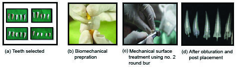
The crown of each tooth was sectioned transversally 2 mm above CEJ [Table/Fig-1a]. Access cavity preparation was done using size no. 6 round bur by orienting the bur perpendicular to the long axis of the tooth. The working length was determined visually (by subtracting 1 mm from the apex) using 15 size K-file (K-files 21mm Limas K; MANI, Tochigi, Japan) and biomechanical preparation was done till size 40 using Hedstorm files (H-files 21 mm Limas K; MANI, Tochigi, Japan) [Table/Fig-1b]. Copious irrigation with 1% NaOCl (sodium hypochlorite) and 0.9% saline was done. Then the root canals were dried with paper points and obturated using metapex (Meta Biomed). Obturation was done 3 mm short from the CEJ and radiographs were taken using Photostimulable Phosphor Plate (PSP) digital sensor system to confirm the level of obturation. The tooth was mounted in cold-cured acrylic resin blocks and then the specimens were randomly divided into four groups.
Procedural steps in the preparation of post placement.
Group 1 (Control): No surface treatment of root dentin.
Group 2 (Chemical): Chemical surface treatment was done using 10 ml of 2% chlorhexidine solution for 10 minutes chlorhexidine (Asep-RC Anabond Stedman Pharma Research, Adyar, Chennai, India) to increase the bond strength to root dentin and to remove the residual bacterial load [16–18].
Group 3 (Mechanical): Mechanical surface treatment was done by creating a mushroom shaped undercut below CEJ using no. 2 size round bur [Table/Fig-1c] [19].
Group 4 (Chemical+Mechanical): Both mechanical and chemical root surface treatments were done.
Number 2 serrated parallel glass fibre post (Reforpost glass fiber RX, Metal-reinforced glass fiber Angelus, Londrina, PR, Brazil) with 1.2 mm diameter and 21 mm length, consists of a 0.2 mm stainless steel reinforcement was used. Both the root canal walls and the fibre post was etched with 37% phosphoric acid for 15 seconds [20,21], followed by rinsing and gentle drying with air stream. The post was then luted using dyad flow composite (the first self-adhering composite powered by OptiBond™) and light cured for 30 seconds. The placement of post was confirmed radiographically [Table/Fig-1d].
Shear Bond Strength: The shear bond strength was evaluated by placing the tooth in an Instron UTM-3382. The force was applied with a sharp blade perpendicular to long axis of the tooth to deliver the shearing force at 1000 N at a speed of 2 mm/min. Shear bond strength was measured in terms of MPa, F/A (force per unit area).
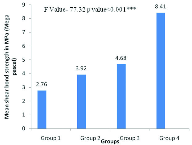
Comparison of mean shear bond strength of different groups after different root surface treatments.
p<0.05- Significant* p=0.001-Highly Significant** p<0.001-Very highly Significant*** p-value was evaluated using one-way ANOVA
Statistical Analysis
Data was collected and tabulated. Statistical analysis was done using SPSS software version 20.0. One-way ANOVA and Post-hoc Tukey tests were performed to determine its statistical difference.
Results
[Table/Fig-2] shows the comparison of shear bond strength of different groups. Post-hoc analysis with Tukey test was done to evaluate the highly significant group [Table/Fig-3]. It was evident that, the Group 4 in which both chemical and mechanical surface treatments were done exhibited higher shear bond strength (p<0.001), followed by the mechanical root surface treated Group 3 (p=0.001) and chemical root surface treated Group 2 (p=0.054) when compared with the control group.
Intergroup comparison of shear bond strength of post to root dentin after different root surface treatments.
| Groups | Group 4 | Group 3 | Group 2 | Group 1 |
|---|
| Group I | <0.001*** | 0.001** | 0.054 | - |
| Group II | <0.001*** | 0.424 | - | - |
| Group III | <0.001*** | - | - | - |
| Group IV | - | - | - | - |
***p<0.001-Very highly significant, **p = 0.001-Highly significant, *p<0.05-Significant Post-hoc Tukey test
Discussion
Endodontically treated grossly decayed primary anterior teeth often require intracanal retention due to lack of adequate coronal tooth structure for direct restorative techniques. Several factors affect the retention of posts like length, diameter, design and different surface treatments. However, these factors affecting the retention of post were evaluated only in permanent teeth. Hence, the present study was under taken to evaluate the effect of different root surface treatment on the retention of fibre post to root dentin.
In the present study, the depth of the post was limited to about 3 mm from CEJ, as the placement of post to the depth of 3 mm will not interfere with normal physiologic resorption [8–11] and it was confirmed radiographically using PSP digital sensor system. As the Reforpost RfX has a 0.2 mm of stainless steel metal reinforcement, it is easy to visualize the length of Reforpost RfX than the regular radiolucent glass fibre post. The width of the post was limited to one third of the root diameter to preserve the radicular dentin. Regardless of the surface treatment and the post system used, the teeth should be first treated endodontically and obturation should be 3 mm short of CEJ. In this study, irrigation was done using 1% NaOCl, as it has similar pulp dissolving capacity compared to 5.25% NaOCl and reduced cytotoxicity compared to 5.25% NaOCl [22,23] and subsequent irrigation with 0.9% saline was done before final irrigation with 2% chlorhexidine. Interaction between NaOCI and chlorhexidine leads to the formation of Parachloroaniline (PCA), which is a carcinogenic agent [24]. In the present study metapex obturation was preferred over Zinc Oxide Eugenol (ZOE) obturation, as eugenol present in the ZOE affects the polymerization of composite resin [25]. Due to the presence of hydroxyl group in the eugenol, which protonize the free radicals and inhibit the degree of polymerization of composite resin material, which was used for luting the post [26,27].
In the present study, the fibre post and the root wall were etched with 37% phosphoric acid for 15 seconds. As the etching of glass fibre post removes the epoxy layer of glass fibre post without damaging the fibre integrity and provides additional site for micromechanical retention of flowable resin [21]. Since studies have stated that, flowable composite does not compromise the bond strength [28,29], in the current study dyad flow composite was used as a luting agent. As the dyad flow is a self adhering composite, it does not require application of bonding agent. This is an added advantage and shortens the procedural time while treating paediatric patients. The reason for the evaluation of shear bond strength was due to the fact, that shear forces are predominant in the anterior region at the dentin-post interface and most of the failures occur due to high shear forces [30,31].
In the present study, teeth treated in Group 2 exhibited higher shear bond than the control group. Loss of bond strength is one of the major problems with adhesive resin restorations due to degradation of hybrid layer at dentin adhesive interface [32]. Human dentin matrix consists of Matrix Metallo Protenaeses (MMPs) like MMP-2, MMP-8, and MMP-9 and these MMPs cause degradation of collagen fibrils and premature loss of bond strength. Chlorhexidine shows an inhibitory effect to MMP-2, MMP-8, and MMP-9 and prevent degradation of collagen fibrils at dentin resin interference [33]. Irrigating the root dentin with chlorhexidine gluconate solution increases the bond strength to root dentin due to MMP inhibitory activity, which improves the integrity of the hybrid layer and thereby, improves the bond strength [33]. In addition chlorhexidine has shown promising antimicrobial action which can last upto 30 days when either added to restorations or used as intra canal irrigant [33,34]. Hence, copious irrigation of the root canal with 2% chlorhexidine after acid etching and just before the placement of post improves the bond strength and also eliminates residual bacterial load, as chlorhexidine also possess antibacterial activity [17–19]. Santos JN et al., stated that that there was no statistical difference between 2% chlorhexidine gel and liquid in bond strength [35], so in the present study 2% chlorhexidine solution was used.
In the current study, teeth treated in Group 3 exhibited higher shear bond strength than the Group 2 and control group. The higher shear bond strength could be due to mechanical interlocking of post to the root dentin by the creation of mushroom shaped undercut using size no. 2 round bur and the success of this technique had been reported in the study done by Judd PL et al. In addition the author also had stated the risk of lateral root perforation with this technique [19]. However, in the present study, care was taken to prevent lateral root perforation by orienting the bur perpendicular to the long axis of the tooth. The similar technique was explained in the study done by Memarpour M et al., in primary teeth using short composite post [36].
In the current study, teeth treated in Group 4, in which both chemical and mechanical surface treatments were done exhibited highest shear bond strength than all other test groups and control group. As there are no studies in literature by combining both the techniques in assessing the shear bond strength in primary teeth, the present study also was done as a new attempt to evaluate the shear bond strength of the post by combining both mechanical and chemical surface treatments to the root dentin of primary teeth. The highest shear bond of this group is due to both mechanical interlocking of the post to the root dentin by the creation of mushroom shaped undercut and the preservation of collagen degradation by the application of 2% chlorhexidine to the root dentin.
Limitation of the present study is that, only in vitro study has been carried out to prove the effectiveness of bond strength. So, further clinical trials are needed to prove the retention of post in clinical scenario.
Conclusion
Mechanical and chemical surface treatments together led to a improved shear bond strength and increased the retention of the post to the root surface. These mechanical undercut and chemical treatments should be considered as a treatment option when planning for post placement in primary teeth, as it improves the shear bond strength of post to the root dentin.
***p<0.001-Very highly significant, **p = 0.001-Highly significant, *p<0.05-Significant Post-hoc Tukey test