Cleaning and shaping of root canals is an indispensable part of endodontic treatment. But, it has been reported to cause the undesirable dentinal defects in form of cracks, craze lines or fractures [1–9]. Canal preparation involves removal of dentin and can compromise the strength of the roots [10,11]. The smaller, less pronounced defects like craze lines or cracks gradually propagate into vertical root fracture, under repeated stresses during various endodontic or restorative procedures [11]. Vertical root fracture can be a fatal consequence as the tooth has to be ultimately salvaged.
Stresses are generated on root canal walls during instrumentation. These are more in apical direction and along canal walls [12]. Hence, apical extent of instrumentation of canal can be crucial in terms of occurrence of cracks. Instrumentation length might influence crack formation and needs evaluation.
Rotary NiTi file systems have revolutionized canal preparation by making it a faster procedure with lesser procedural errors like canal transportation, straightening or perforations [13–16]. Despite these advantages, they might result in more apical dentin removal as they prepare canals to greater tapers of 4%, 6% or 8% compared to 2% tapers created by hand files [17]. This might compromise the strength of roots [10]. Also, these instruments by virtue of their design features [18] and the fact that they have more rotations inside the canal [19], can generate increased friction and stresses within the canal and thus, can influence the crack development.
The aim of this study was to evaluate the occurrence of apical cracks with stainless steel hand files, rotary NiTi RaCe and K3 files at two different instrumentation lengths.
Materials and Methods
The present in vitro study was conducted in Department of Conservative Dentistry and Endodontics, AECS Maaruti College of Dental Sciences, Bengaluru, Karnataka, India.
Sixty mandibular premolars with straight roots, extracted for orthodontic or periodontal reasons were selected. Teeth with straight roots and completely formed apices were used. Teeth with anatomic irregularities, caries involving the roots, or presence of fracture lines on root surface were excluded. The teeth were stored in distilled water throughout the experimental procedure.
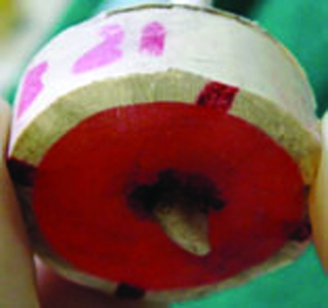
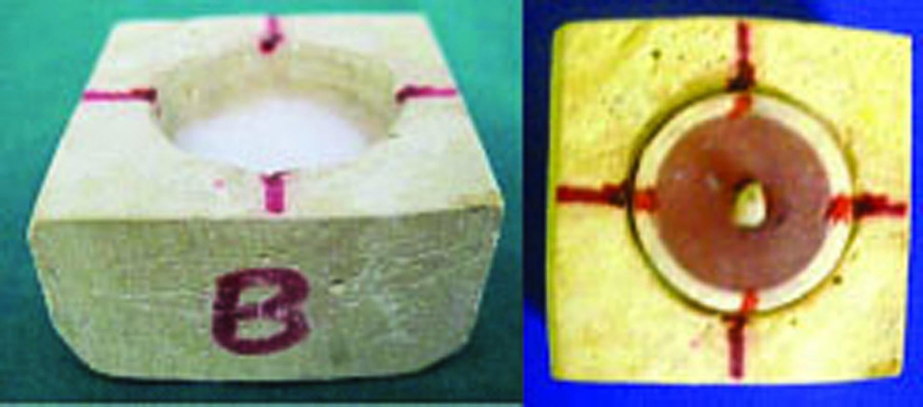
The teeth were embedded in cylindrical tube filled with autopolymerising resin (DPI-RR coldcure), and a simulated periodontal ligament was represented by hydrophilic vinyl polysiloxane impression material (Reprosil, Dentsply, USA). The apical 2 to 3 mm of the root was exposed to allow image recordings [Table/Fig-1]. The teeth were decoronated 2 mm above the proximal Cemento-Enamel Junction (CEJ) to gain an access into the root canal and to provide a stable reference point. A customized jig cuboidal in shape with central space for specimen placement was fabricated with dental stone [Table/Fig-2]. Reference markings were made both onto the jig and specimens. Specimens were placed in a way that the markings on them coincided with that of jig.
Specimen after periodontal ligament simulation with apex exposed for recording images.
Customised jig for placement of specimens under stereomicroscope.
The specimens were randomly divided into six groups of 10 each. A size 10 K-file was inserted into the canal, until the tip of the file became visible at the apical foramen. Rubber stopper was then adjusted till the reference point. The length was measured from tip of the file to the rubber stopper, which was taken as the Working Length (WL). The root apices were stained using India ink and preoperative images were obtained in apical, mesial and distal aspects under stereomicroscope (Olympus SZX16) at 100x magnification.
In Group I and II coronal flaring was done using Gates Glidden drill #1, 2 and 3. Root canal preparation was carried out with the stainless steel hand K-files using step back technique to a master apical size of #40.
In Group III and IV the root canal preparation was carried out using rotary NiTi RaCe files while in Group V and VI NiTi K3 files were used. Canal preparation was done in a crown down manner to a master apical file size of #40 with a taper of 4%. Coronal flaring was done using the orifice shapers of respective groups while instrumentation sequence followed for remainder of canal was:
#40/0.06 taper – in the middle third
#35/0.06 and #40/0.04 taper – in the apical third
Groups I, III, V were instrumented till working length while groups II, IV and VI were instrumented 1 mm short of working length (WL-1). All the canals were irrigated with 3% sodium hypochlorite followed by recapitulation with a size 10 K-file after each file change. Finally, canals were rinsed with distilled water.
After completion of the root canal preparation, images of root apices were obtained with stereomicroscope at 100x magnification in the same view as obtained preoperatively. Preoperative and postoperative images were compared for the occurrence of cracks by three independent observers and the majority of two was considered for scoring.
Statistical Analysis
The presence of crack was scored as 1 and the absence was scored as 0. Descriptive statistical analysis was carried out. Since the measurements were categorical, results were expressed in percentage and were assessed at 5% level of significance.
Chi-square test was carried out to find out the level of significance on a categorical scale between two or more groups.
A crack was defined as any visible discontinuity on an external surface of root apex which was not present preoperatively.
Results
Total number of cracks observed in each group is summarized in [Table/Fig-3].
Incidence of cracks in individual groups.
| Number of cracks (n=10) | Percentage of cracks | Significance (p) value at WL and WL-1 |
|---|
| Group I (K-files WL) | 4 | 40% | p=0.628 |
| Group II (K-files WL-1) | 2 | 20% |
| Group III (RaCe WL) | 6 | 60% | p=0.057 |
| Group IV (RaCe WL-1) | 1 | 10% |
| Group V (K3 WL) | 4 | 40% | p=0.087 |
| Group VI (K3 WL-1) | 0 | 0% |
* Descriptive statistical analysis. Chi-square test was carried out to find out the level of significance between two or more groups.
Among groups instrumented with K-files (Group I and II), more cracks were seen at WL (40%) than at WL-1 (20%). But, there was no significant difference in number of cracks formed at two working lengths (p=0.628).
Cracks were seen in 60% and 10% of teeth at WL and WL-1 respectively with RaCe files and in 40% of teeth at WL with K3 files. When instrumented at WL-1 with K3 files no cracks were seen. For groups instrumented using RaCe and K3 files (Group III and IV, Group V and VI) there was significant difference in number of cracks at the two instrumentation lengths. Significantly more number of cracks were formed when instrumentation was carried till WL than WL-1. (p=0.057 for Groups III and IV, p=0.087 for Groups V and VI).
Among the three instrumentation systems used, cracks were seen in 30% of teeth instrumented with K-files, 35% of teeth instrumented with RaCe files and 20% of teeth instrumented with K3 files. There was no statistical significance among three instrumentation systems in the formation of apical cracks (p=0.563) [Table/Fig-4].
Incidence of cracks among three instrumentation groups irrespective of instrumentation lengths.
| Hand K-files (Group I + Group II) (n=20) | Rotary RaCe files (Group III +Group IV) (n=20) | rotary K3 files (Group V+ Group VI) (n=20) |
|---|
| Total number of cracks | 6 | 7 | 4 |
| Percentage of cracks | 30% | 35% | 20% |
p=0.563. Descriptive statistical analysis. Chi-square test was carried out to find out the level of significance between two or more groups.
Discussion
Mandibular premolars were chosen for the study because majority of them have single root and single canal. Teeth were embedded in cylindrical tube filled with autopolymerising resin and simulated periodontal ligament represented by light body silicone impression material [20]. Silicone being rubber like elastomeric impression material allowed the root to move in a limited space mimicking physiologic tooth movement and avoided external reinforcement. Customized jig facilitated orientation of specimens at exactly same angulations in apical, mesial and distal aspects every time. Thus, preoperative and postoperative images of root apices could be recorded at exactly same position for comparison of occurrence of cracks.
Presently, there is no consensus about the optimum size of apical preparation [21]. We tried standardizing it to closest dimensions possible in all the groups. All the groups were prepared to final apical size of #40. Hand files produced preparations with 2% taper while rotary files RaCe and K3 produced preparations with 4% apical taper.
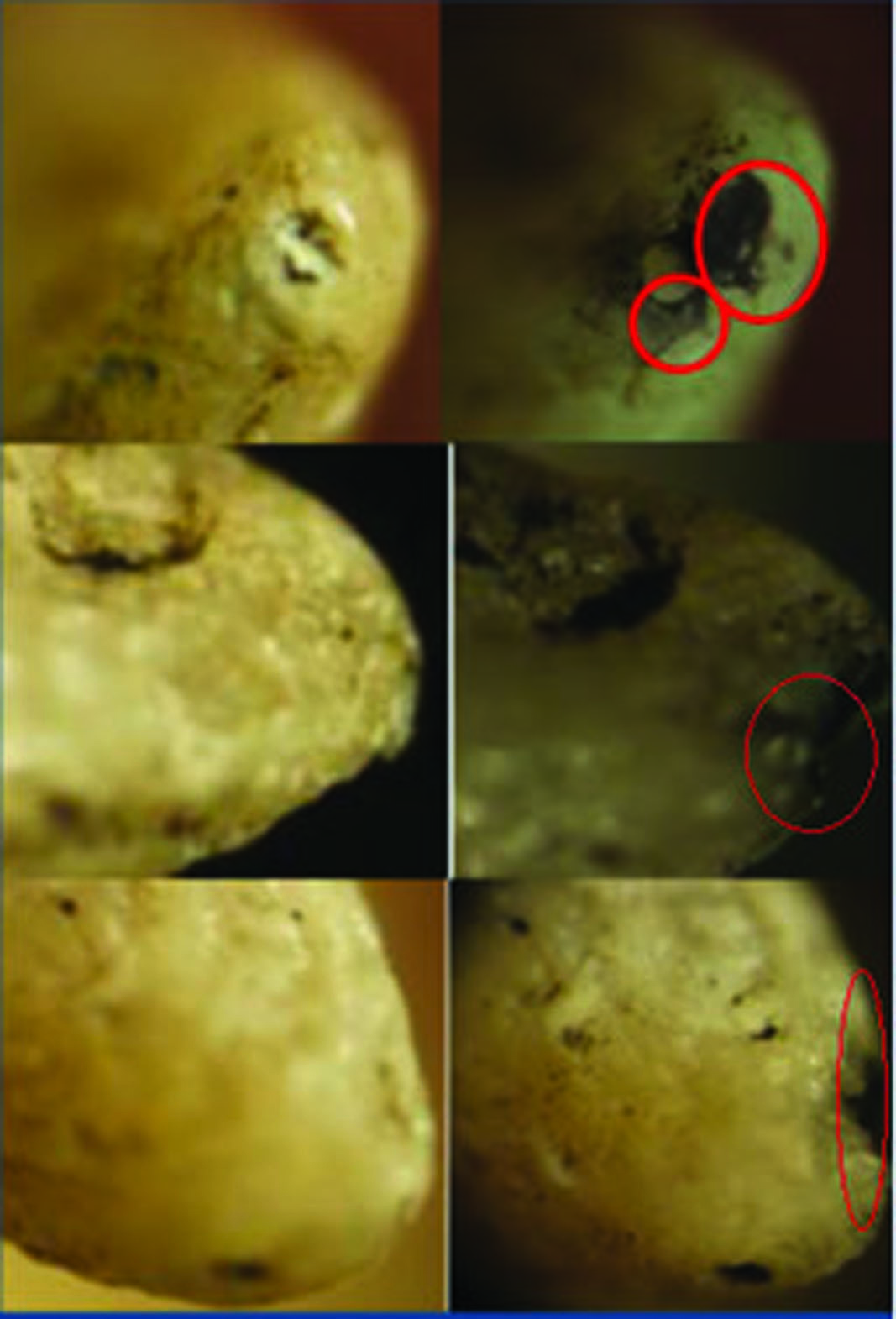
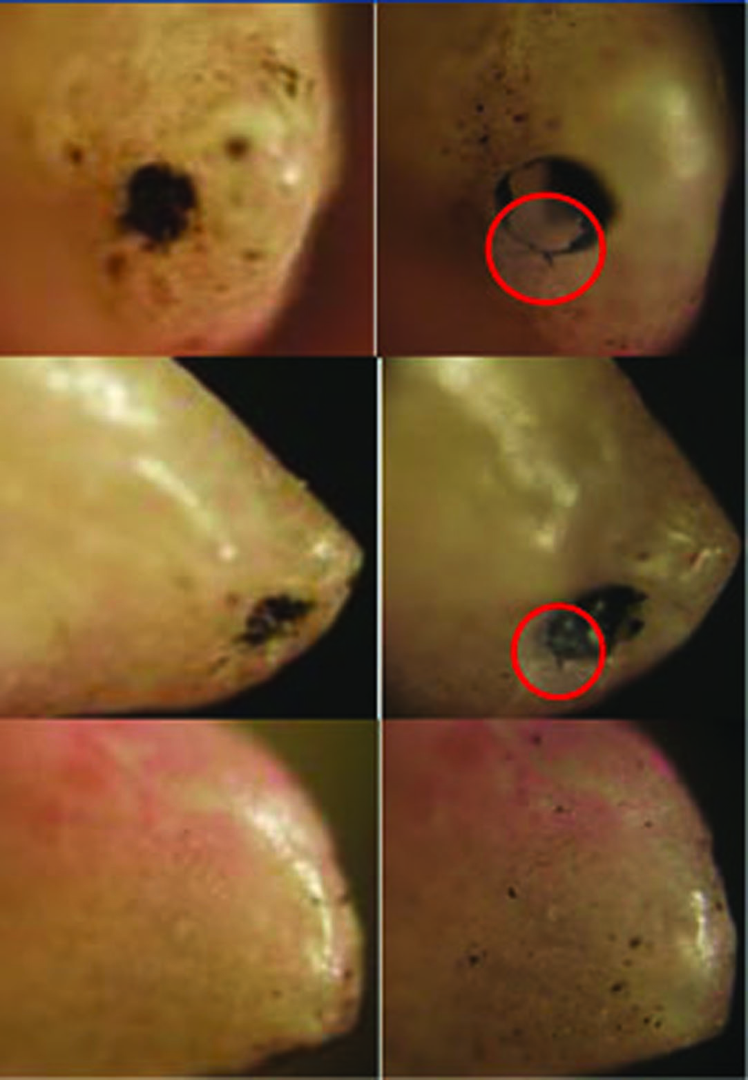
In Group I and II, instrumented with K-files, cracks were seen in 40% of the teeth at WL and 20% of the teeth at WL-1 [Table/Fig-5]. Statistical analysis showed no significance in the number of cracks formed when instrumented to WL or WL-1. Though straight roots were used in this study, it is known that the apical foramen is often eccentrically located from anatomical or radiographic apex in 68 to 80% of the teeth [22]. These deviations in apical foramen indicate that even in straight roots, canals can curve apically and are not perfectly straight. In fact, all root canals have some degree of curvature [23]. Stainless steel K-files are stiffer files and thus, can cause more stresses compared to the more flexible rotary NiTi files. This could be the reason for statistically non significant results with stainless steel hand files.
Representative pre and postoperative images of cracks formed after instrumentation with K- files at WL-apical, mesial and distal aspects respectively.
In studies by Adorno CG et al., on mandibular premolars, cracks were seen in 70%, 60% at WL and 40%, 20% at WL-1 [1,2]. Final apical size of preparation was #60. In these studies the total number of cracks was higher at both instrumentation levels than in our study owing to larger apical preparation.
Liu R et al., in their study on mandibular premolars found cracks in 5% of teeth at WL and none of the teeth showed crack at WL-1 [8]. They used flexible hand-files which could have resulted in much lesser cracks than in this study.
In Group III and IV, instrumented with rotary NiTi RaCe files, cracks were seen in 60% of the teeth at WL and in 10% of the teeth at WL-1 [Table/Fig-6].
Representative pre and postoperative images of cracks formed after instrumentation with RaCe files at WL-apical, mesial and distal aspects respectively.
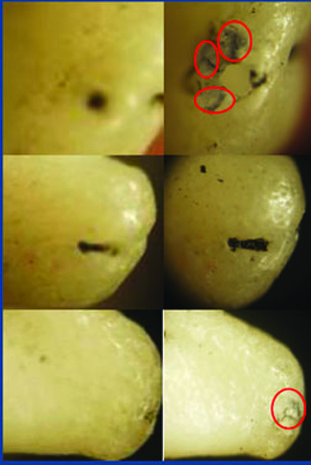
In Group V and VI, instrumented with rotary NiTi K3 system, cracks were seen in 40% of the teeth at WL [Table/Fig-7]. No cracks were seen when instrumented at WL-1.
Representative pre and postoperative images of cracks formed after instrumentation with K3 files at WL-apical, mesial and distal aspects respectively.
Liu R et al., found cracks in 70% of teeth at WL and 20% of teeth at WL-1 with K3 rotary files on mandibular incisors at apical preparation of #35/4% [8]. Adorno CG et al., found cracks in 66% of mandibular incisors at WL and 33% at WL-1 when canals were prepared with K3 files to apical size of #35/4 % [3]. Present study used mandibular premolars; hence, a direct comparison cannot be made.
In the groups instrumented with rotary files RaCe and K3, there was statistically significant difference in number of cracks formed at WL and WL-1. Our study is in accordance with previous studies [1–3,8] in that, they also found significant differences in number of cracks at two instrumentation levels. Lesser cracks occurred when instrumentation was done 1 mm short of WL.
Instrumentation length had a significant effect on apical crack formation with rotary NiTi files. The cracks originating at the root canal might depend on the level of file insertion. When the working length reached the apical foramen, there was a higher risk of producing cracks because of wedging force of the file.
The incidence of cracks for a particular system was calculated by taking sum of cracks at WL and WL-1 for the system. Nearly, 30% of teeth instrumented with K-files, 35% of teeth instrumented with RaCe files and 20% of teeth instrumented with K3 files showed apical cracks. There was no significant difference among the various systems used on apical crack formation.
RaCe files produced the highest number of cracks when instrumented till WL and K3 the lowest. The RaCe files have a sharp cutting edge with convex triangular cross-section. They have asymmetrical longitudinal design. A set of cutting edge alternates with the second set pitched at a different angle leading to two different cutting edges on the same file. This could cause stress concentration at specific points rather than distribute it along the entire length of file [24]. This concentration of stresses could have led to comparatively more cracks seen with this system.
Clinically, K3 feels stiffer and stiffer files are shown to generate higher stresses in the apical root dentin [12]. This raises the risk for dentinal cracks. But, comparatively lesser cracks were found using K3 files. The file’s metal core diameter does not increase at same rate as the external surface taper. The flutes surrounding the file become deeper over the length of the file. This might enhance the flexibility of the tip [25] leading to better distribution of stresses over the entire length of file. This could be the reason for lesser number of cracks with K3.
In a study done by Garg S et al., cracks were found in 10% and 16.7% of mandibular premolars prepared with K3 and easy RaCe files respectively. Reason for lesser number of cracks than in this study could be because the apical preparation was performed only till #25/4%, whereas, here apical it was done till #40/4% [9]. Nevertheless, more cracks were seen with RaCe files similar to this study.
In a study by Liu R et al., apical cracks were seen in 1.8% of teeth instrumented with hand files and 20.6% with rotary files [8]. In studies by Bier CAS et al., Yoldas O et al., Hin ES et al., and Garg S et al., no cracks were seen in teeth instrumented with hand files [4,5,7,9]. There was a significant difference in number of cracks formed with hand files and rotary files. Hand files caused minimal or no cracks.
On contrary to above studies, in our study we found no significant difference in number of cracks formed with either hand or rotary instruments and this is in accordance with studies done by Adorno CG et al., [1,2]. Difference could be because the above studies used flexible hand files [4–9] and present study and studies done by Adorno CG et al., [1,2] used rigid stainless steel hand files. Stiffer files can cause more stresses on root dentin compared to flexible files [18].
Majority of studies on apical crack formation have been performed in vitro either by directly observing apical regions of roots during various phases of preparation under stereomicroscope [1–3] or by sectioning of root apices at various levels and then observing under stereomicroscope [4–9]. Though, the former method enables comparison of cracks following each instrument change, but only the cracks which are on external surface of roots can be visualized. Their extension internally towards canal cannot be assessed. Another limitation with this methodology is lack of periodontal simulation in apical portion of roots which are left uncovered to enable the examination of cracks. The periodontal ligament might act as cushion and protect the apex against crack initiation in vivo. The latter method enables us to know crack extension from root surface towards canal or vice versa. But samples are destroyed after a single comparison and we cannot visualize the effect of each instrument in causation of cracks.
Majority of in vitro studies performed so far have used resin blocks with simulated periodontal ligament to mimic bony sockets. But they cannot completely reproduce viscoelastic properties of periodontal ligament and absorb stresses in a pattern similar to periodontal ligament [20]. A cadaveric study does not need periodontal simulation and creates environment similar to in vivo conditions. In a cadaveric study by Arias A et al., on mandibular incisors microcracks were found in 50% of control group with no preparation, 50% of teeth with hand ProFile GT files and 66% of teeth prepared with reciprocating file WaveOne [26]. A relationship between the shaping techniques (hand and reciprocating files) and the incidence of microcracks could not be seen compared with uninstrumented controls. But sample size was small with six teeth per group in the study. Though, the study environment was almost close to in vivo conditions, we need a study with larger sample size to negate results of various in vitro studies. Also, comparison of cracks at each instrument change is not possible.
A micro-CT study by De Deus G et al., showed no causal relationship between dentinal microcrack formation and canal preparation procedures with Reciproc, WaveOne, and BioRaCe systems [27]. The dentinal defects identified in postoperative cross-sections were also found in their corresponding preoperative images as well. Dentinal microcracks were found in 27.64% of the preoperative images which was not seen in control groups of previous studies [1–9]. Micro-CT imaging has a higher definition than stereomicroscopy and can get images of hundreds of slices per tooth. Conventional sectioning techniques allow the evaluation of only a few slices per tooth and there is possibility of missing several defects. But this was an in vitro study on extracted mandibular molar with periodontal ligament simulation.
A non-destructible and reproducible study methodology more closely simulating in vivo conditions needs to be devised to conclusively explain varying results among in vitro, cadaveric or micro-CT studies.
Conclusion
Within the limitation of this in vitro study, it can be concluded that preparation of apical portions with rigid stainless steel hand files can equally cause dentinal damage just like rotary files with no significant difference between the two. Also, there was no significant difference in cracks produced among the rotary systems used either RaCe or K3.
Lesser apical cracks were produced when instrumentation with rotary files was done 1 mm short of apical foramen. But rigid hand file showed no statistical significance at two instrumentation levels.
This implies that rigid hand files showed no reduction in number of cracks 1 mm short of apical foramen. So one must avoid using rigid hand files for apical preparation and instead use flexible hand files which are reported to cause no or less cracks in apical portion in preference to rigid hand files.
* Descriptive statistical analysis. Chi-square test was carried out to find out the level of significance between two or more groups.
p=0.563. Descriptive statistical analysis. Chi-square test was carried out to find out the level of significance between two or more groups.