Reports on hypersensitivity diseases in Nigerians are rare. We report the incidence of anaphylaxis in three siblings following fatal outcome in their mother. Urticarial rashes were noticed in three siblings’ resident in a South Western Nigerian town, one week before presentation at our facility. All the three siblings developed respiratory distress four days after the rash was noticed. Onset of respiratory distress made the family seek care at a private hospital, where they were admitted and treated with intravenous aminophylline and ceftriaxone.
The mother of the children had experienced the same symptoms earlier also. She took treatment and died in the same private hospital, where her children received care. Death of the mother and worsening respiratory distress in the children made the father effect transfer of the children to the paediatric emergency unit of Ladoke Akintola University of Technology Teaching Hospital, Osogbo. The three children made a slow but uneventful recovery after instituting appropriate management for anaphylaxis and acute respiratory distress syndrome. The cases are discussed with a view to create awareness amongst health practitioners about the occurrence of anaphylaxis in our society. The need for prompt recognition and appropriate management, when confronted with this disease is also underscored.
Allergic, Childhood, Diseases
Case Report
A 14-year-old boy was referred from a private hospital to Ladoke Akintola University of Technology Teaching Hospital (LAUTECH) Osogbo, on account of wheal like pruritic erythematous rashes observed a week prior to presentation. He gave a history of fever and dry cough noticed four days prior to presentation and difficulty with breathing a day prior to presentation.
He initially presented at a private hospital, where he was managed with intravenous ceftriaxone and aminophylline. His mother died of similar symptoms in the same hospital, about five hours before his referral to the paediatric emergency unit. Death of the mother and worsening respiratory distress in the child necessitated referral from the private hospital to the paediatric emergency unit of LAUTECH, Osogbo for treatment.
There was no history of toxin or poison ingestion, facial swelling, bee sting or oliguria. Similar history of rashes, cough and dyspnoea were also obtained in his two younger siblings. However, detailed enquiry did not reveal anything ingested, contacted, inhaled or otherwise exposed to by the mother or any of the three siblings. There was no past history of similar rashes or other forms of rashes, atopy, or asthma in any member of the family.
His 11-year-old brother and three-year-old sister were also admitted for similar symptoms on the same day, the index case was admitted. They presented with similar history of sudden onset of urticarial rashes, cough and fever of one-week duration. Breathlessness was noticed 24 hours prior to presentation. Other symptoms in the brother included colicky abdominal pain, frequent passage of stools, facial and lip swelling all noticed one day prior to presentation.
On examination, the index patient was acutely ill looking with a temperature of 39.2OC. He weighed 35 kg and had no peripheral oedema. There was no dehydration, palor, icterus, or cyanosis. Respiratory system examination revealed nasal flaring, subcostal and intercostal recession and a respiratory rate of 90 cycles per minute. Percussion notes were resonant and breath sounds were vesicular of normal intensity. Rhonchi were auscultated diffusely over all thoracic fields. Pulse rate was 140 beats per minute and normal volume pulses were palpated. No abnormalities were elicited on abdominal and the neurological examination.
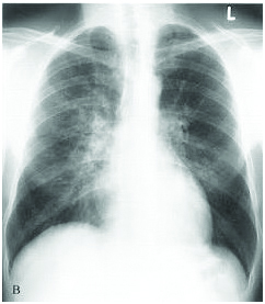
A diagnosis of anaphylaxis with acute respiratory distress syndrome was made to rule out asthma and pneumonia. A Packed Cell Volume (PCV) of 26% was recorded at admission and the electrolytes, urea and creatinine results were normal. Saturation oxygen pressure at admission was 87% and it normalized to between 98-99% on day 5 at discharge. Some of the necessary investigations such as IgE assay, tryptase and blood culture could not be carried out because the father was financially constrained [Table/Fig-1]. Chest radiograph of the index patient showed features of acute respiratory distress syndrome i.e., hyperinflation, batwing shadows and Kerly B lines [Table/Fig-2].
Results of conducted investigations.
| Index Case | Sibling-1 | Sibling-2 |
|---|
| Full blood count on admission |
| PCV | 26% | 32% | 35% |
| WBC | ND | ND | 23,900 |
| Neutrophils | ND | ND | 70% |
| Eosinophils | ND | ND | 01% |
| Lymphocyts | ND | ND | 29% |
| Electrolytes urea and creatinine on admission |
| Na+ | 136 | 136 | 140 |
| K+ | 3.7 | 4.5 | 4.2 |
| Cl- | 105 | 105 | 96 |
| HCO3- | 20 | 18 | 24 |
| Urease | 3.4 | 1.7 | 2.8 |
| Creatine | 71 | 48 | 59 |
| Blood culture |
| Blood culture | N.D | N.D | N.D |
| Immunoglobulin E assay |
| IgE | N.D | N.D | N.D |
| Tryptase | | |
| Tryptase | N.D | N.D | N.D |
| Pulse oximetry in room air |
| At admission (day 1) | 87% | 63% | 86% |
| At discharge (day 5) | 98-99% | 97% | 98-99% |
| Chest radiograph on admission |
| Chest Radiograph (on day of presentation) | All the three radiographs showed evidence of cardiomegaly, Kerly B lines and bat wings shadow suggestive of acute respiratory distress syndrome/pulmonary oedema |
*N.D: Not done
Chest radiograph of the index patient showing features of acute respiratory distress syndrome i.e., hyperinflation, batwing shadows and Kerly B lines.
The patient was commenced on oxygen, 5 mg of intravenous epinephrine and 5 mg of nebulized salbutamol and these drugs were administered as required till resolution of hypoxaemia and respiratory distress. Daily oral administration of prednisolone (35 mg), ranitidine (35 mg) and Loratidine (10 mg) were observed for five days. An uneventful clinical improvement was maintained over the five days of admission. The patient was discharged in a satisfactory condition and scheduled for a follow up review in a week post discharge. The patient, however, failed to keep the follow up appointment.
Clinical findings and management of the other two siblings were similar to that of the index case. Resolution of symptoms was also similar with both siblings who were discharged in a clinically satisfactory condition on the fifth post admission day. Epinephrine auto-injectors were not available and or prescribed at discharge.
Discussion
Anaphylaxis is an acute severe potentially life threatening allergic reaction, with multi-systemic manifestations due to rapid release of inflammatory mediators [1]. The inflammatory mediators released by interaction of IgE receptors on the mast cells, basophils, the allergen and degranulation of these cells exert systemic effects and cutaneous manifestations such as angioedema and urticaria. Respiratory manifestations of inflammatory mediators include rhinorrhoea, laryngeal oedema and acute respiratory distress syndrome, while hypotension and shock are cardiovascular symptoms. Syncope and dizziness are known central nervous system manifestation of anaphylaxis [1,2].
Mediators of anaphylaxis can exert different influences on the Gastrointestinal Tract (GIT), skin, respiratory, cardiovascular, and central nervous system [1,2]. Gut manifestations of anaphylaxis include, crampy abdominal pain and diarrhoea while urticaria and angioedema are common cutaneous manifestations. Rhinorhea, laryngeal oedema, asphyxiation and acute respiratory distress syndrome are some of the respiratory manifestations of anaphylaxis. Cardiovascular manifestations include hypotension or shock. Seizures, syncope or dizziness are common central nervous system features of anaphylaxis.
Reports on anaphylaxis in African children are rare. The few available reports incriminate insect stings and drugs as causes of anaphylaxis [3–5]. The rarity of reports on allergy or anaphylaxis in Nigerian children may either be as a result of rarity of hypersensitive disease conditions or under-reporting or failure to recognize the conditions when they are present [6].
In the cases presented, identifying the allergen responsible for anaphylaxis was impossible. The financial constraints on the part of the father limited our capacity to investigate. Furthermore, the lack of competent diagnostic laboratories to run IgE assay and other allergen tests in most parts of Nigeria are some of the factors that militate against allergen identification. These limitations also show that allergic diseases in Nigeria are not given the attention they receive in developed countries.
Most cases of anaphylaxis in children in the developed countries have been documented to arise from food allergies [1]. Food allergies are, however, believed to be rare in Nigeria although there is no evidence for or against this [7]. It is not unlikely that the male sibling of the case presented might have started off as a food allergy based on the facial and lip swelling, crampy abdominal pains, diarrhoeal history and respiratory signs. The possibility of an inhaled allergen origin for the causation of the anaphylaxis cannot also be ruled out, in the case presented and female sibling who had predominantly respiratory symptoms.
All the cases presented were poorly managed prior to the presentation at the paediatric emergency. It is not surprising that the patients did not respond. Deaths could have occurred in the children, but for the timely institution of appropriate management. Prompt administration of appropriate drugs such as epinephrine, anti-histamine and oxygen can be life-saving in the management of anaphylaxis and acute respiratory distress [1,2]. Failure to administer oxygen and adrenaline, in these patients at the private clinic can probably be explained on the basis of failure of the health care providers to recognize the nature or severity of the disease. There is also a possibility that these drugs were not given because of unavailability. Studies from the United States of America, Pakistan and the United Kingdom show that prompt recognition and administration of appropriate drugs can be life-saving [8–10].
Conclusion
It is suggested that hypersensitivity disorders should be considered as a cause of acute illnesses in individuals with similar features. Similar diseases involving two or more members of a household should arouse a high index of suspicion and affected individuals should be referred promptly to facilities endowed with the appropriate resources for investigation and management. Availability and prompt administration of appropriate drugs is essential for good outcome. In addition, the diagnosis, clinical features and management of allergic diseases should feature more prominently in continuous medical sessions or programs in order to upgrade the knowledge and practice of medical practitioners on allergic conditions.
*N.D: Not done
[1]. Simons FER, Ardusso LRF, Bilo MB, El-Gamal YM, Ledford DK, Ring J, and for the World Allergy OrganizationWorld Allergy Organ J 2011 4(2):13-17. [Google Scholar]
[2]. Sampson HA, Leung DYM, Anaphylaxis. In: Kliegman RM, Behrman RE, Jenson HB, Stanton BF (eds.)Nelson’s Textbook of Paediatrics 2007 18th EditionPhiladelphiaW.B Saunders Company:983-85. [Google Scholar]
[3]. Vetter RS, Visscher PK, Camazine S, Mass envenomations by honey bees and waspsWest J Med 1999 170:223-27. [Google Scholar]
[4]. Nafiu OO, Olumese PE, Gbadegesin RA, Osinusi K, Intraosseous infusion in an emergency situation: A case reportAnn Trop Paediatr 1997 17(2):175-77. [Google Scholar]
[5]. Oyedeji OA, Musa TL, Adebami OJ, Oyedeji GA, Fatal scorpion sting in a childNig J Clin Pract 2014 17(1):112-14. [Google Scholar]
[6]. Mbugi EV, Chilongola JO, Allergid disorders in Africa and Africans: Is it primarily a priorityWorld Allergy Organ J 2010 3(5):175-81. [Google Scholar]
[7]. Kung SJ, Steenhoff AP, Gray C, Food allergy in Africa, myth or realityClin Rev Allergy Immunol 2014 46(3):241-49. [Google Scholar]
[8]. Umasunthar T, Leonardi-Bee J, Hodes M, Turner PJ, Gore C, Habibi P, Incidence of fatal food anaphylaxis in people with food allergy: A systematic review and meta-analysisClin Exp Allergy 2013 43(12):1333-41. [Google Scholar]
[9]. Khan NU, Shakeel N, Makda A, Mallick AS, Memon MA, Hashmi SH, Anaphylaxis: Incidence, presentation, causes and outcome in patients in a tertiary-care hospital in Karachi, PakistanQJM 2013 106(12):1095-101. [Google Scholar]
[10]. GK Tiyyagura, Arnold L, Cone DC, Langhan M, Paediatric anaphylaxis management in the prehospital settingPrehosp Emerg Care 2014 18(1):46-51. [Google Scholar]