Management of Traumatic Fibroma in a Patient with Cerebral Palsy Using 810nm Diode Laser
Raúl Vicente Perales-Garza1, Gerardo Daniel Sierra-Garcia2, Rosa Isela Sánchez Nájera3, áRaúl Vicente Perales-Perez4
1 Resident, Facultad de Odontologia, Universidad Autonoma de Nuevo Leon, Monterrey, Mexico.
2 Professor, Department of Advanced Odontology and Endodontics, Facultad de Odontologia, Universidad Autonoma de Nuevo Leon, Monterrey, Mexico.
3 Professor, Department of Advanced Odontology, Facultad de Odontologia, Universidad Autonoma de Nuevo Leon, Monterrey, Mexico.
4 Specialist, Department of Orthodontics, Center for the Study and Research in Orthodontics, Facultad de Odontologia, Universidad Autonoma de Nuevo Leon, Monterrey, Mexico.
NAME, ADDRESS, E-MAIL ID OF THE CORRESPONDING AUTHOR: Dr. Raúl Vicente Perales Garza, Facultad De Odontología, Universidad Autonoma De Nuevo Leon, Calle Dr. Eduardo Aguirre Pequeño s/n, Colonia Mitras Centro, Monterrey, Nuevo Leon, Mexico C.P. 64460.
E-mail: rag9015@hotmail.com
There are several treatment options for hyperplastic gingival lesions. Among these, diode lasers have the advantages of less bleeding, which is an important characteristic in mucosal lesions, a shorter procedure time, better healing, and less complications. We present the case of a 48-year-old male patient with a history of cerebral palsy and a presumptive diagnosis of traumatic fibroma. The entire lesion was removed in one session with no complications. No recurrence was observed at 3 months follow up. This procedure can be considered a good modality especially for physically challenged patients.
Diode laser, Dental surgery, Disabled person
Case Report
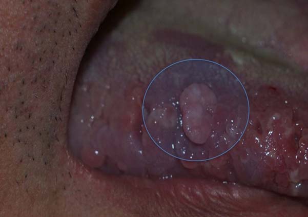
A 48-year-old male patient was referred because of a small wart-like lesion on the right lateral tongue that he noticed one year before. The patient had been diagnosed with cerebral palsy in childhood and was under treatment. On examination, the patient did not have difficulty with speech, hearing or sight; however, he did have muscle weakness in arms and legs, which made it difficult for him to move or walk alone. He needed help to move and attended a special needs school. This problem was caused by cerebral hypoxia at birth. In the oral cavity, we found a sessile mass measuring 5mm in the right lateral tongue located at the level of teeth occlusion [Table/Fig-1]. Additionally, the patient had signs of periodontal disease, caries, and dental malocclusion. Regarding extra-oral examination, the patient was dolicofacial with no cranial, facial or jaw deformities, no palpable lymph nodes or thyroid or muscle pain and also no pain in the temporomandibular joint was found. A diagnosis of traumatic fibroma because of chronic irritation due to biting was considered. Differential diagnoses included neurofibroma, lipoma, hemangiona, and pyogenic granuloma. These were ruled out because pyogenic granulomas have a smooth, wet surface that bleeds easily due to its abundance of blood vessels, lipomas are produced by the proliferation of subcutaneous tissue, and hemangiomas are found deep within the skin and their colour is red because of the abnormal build-up of blood vessels under its surface.
A 5mm sessile mass observed on the right lateral tongue.
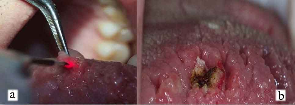
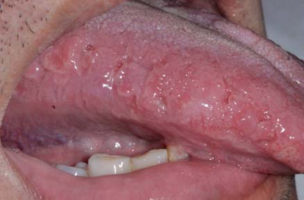
The decision to remove the tumour using a diode laser was taken to avoid the use of sharp instruments since the patient had problems of movement and posture. Spray anaesthesia with the Syrijet needleless injection system was also used. We decided to perform laser ablation with an 810nm Aurora diode laser (Premier Laser Services, Inc., San Diego, CA, USA) at 0.5W continuous wave mode. The laser was applied to the pedunculated base in sweeping motions and ablation of the mass was achieved in 2 minutes with no bleeding or the need of suturing [Table/Fig-2a, b]. The excised tumour was sent for histological examination. The histological findings showed normal and hyperplastic epithelial cells, focal hyperplasia and dense fibrous tissue without inflammatory cell infiltration [Table/Fig-3]. The patient was subsequently evaluated and after 10 days, showed satisfactory post-operative healing. Proper healing and no recurrence were found at a follow-up visit at three months [Table/Fig-4].
a) Laser ablation of the pedunculated tumour; b) Post-operative image.
Histological findings of traumatic fibroma. Focal hyperplasia and dense fibrous tissue with no inflammatory cells solid arrows indicate areas of hyperplasia. The dashed arrow fibrosis (H&E, x40).
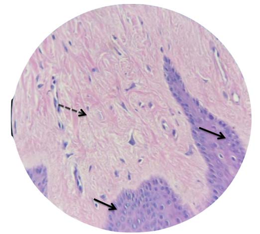
Image at 3 months follow-up with adequate healing and no recurrence.
Discussion
Researchers have investigated laser applications in dentistry since the development of the ruby laser in 1960 by Maiman. A laser is a device that emits light of various frequencies coherently; i.e., with all the waves in phase. It is capable of producing immense heat and power when focused at close range [1] and it can be used as an adjunct to other procedures or as a main form of treatment [2]. Different laser wavelengths are used because of their coagulation necrosis properties, the incision quality provided, and post-operative results [3].
Cerebral palsy is a permanent neuromuscular disorder caused by injury to the foetal or infant brain that begins in childhood and continues into adult life [4,5]. It is characterized by alteration of movement and posture that causes activity limitation. The disease can also lead to other health issues, including vision and dental problems [4]. Patients with cerebral palsy present a large number of oral manifestations and their physical or neurological limitations make the treatment problematic; therefore a multidisciplinary approach is necessary [6].
Among oral cavity lesions, traumatic fibroma is the most frequent tumour-like lesion in the oral cavity [7]. It is a common benign exophytic, reactive oral soft tissue lesion that develops due to an injury. Differential diagnosis include neurofibroma and soft tissue mesenchymal tumours. Recurrences are rare and can be caused by repetitive trauma. Traumatic fibromas do not carry a risk of malignancy and are most commonly found on the tongue, buccal mucosa, and lower labial mucosa [8,9]. The different treatments used for soft tissue lesions include scalpel excision, electrical surgery, and laser surgery [8]. In surgical procedures with high vascularity, tissue coagulation is performed using a diode laser. Diode lasers have a wavelength between 805 and 980nm and can be used to treat a variety of oral soft tissue lesions such as tumours, gingival hyperplasia, and haemangiomas [10]. Among its advantages are, suturing after surgery is usually not necessary, patients can be protected from high-risk infections, and there is less post-operative pain compared to traditional procedures; also lasers can induce suppression of bradykinin activity [11]. In this case, a diode laser was used because of the clinical situation of the patient who had difficulty in movement and posture making a traumatic surgical procedure difficult.
Conclusion
Physically challenged individuals of all ages can present with different pathologies that affect the oral cavity. The soft tissue diode laser can be considered a good treatment modality for lesions such as the fibroma presented herein, especially for these patients.
[1]. Kimura Y, Wilder-Smith P, Matsumoto K, Lasers in endodontics: A reviewInt Endod J 2000 33(3):173-85. [Google Scholar]
[2]. Roshkind DM, The practical use of lasers in general practiceAlpha Omegan 2008 101(3):152-161. [Google Scholar]
[3]. Cernavin I, Pugatschew A, de Boer N, Tyas MJ, Laser applications in dentistry: A review of the literatureAust Dent J 1994 39(1):28-32. [Google Scholar]
[4]. Rosenbaum P, Paneth N, Leviton A, Goldstein M, Bax M, Damiano D, A report: The definition and classification of cerebral palsy April 2006Dev Med Child Neurol Suppl 2007 109:8-14.Erratum in: Dev Med Child Neurol.2007;49(6):480 [Google Scholar]
[5]. Ican HN, Metin-Gürsoy G, Kale-Varlik S, Functional and fixed orthodontic treatment in a child with cerebral palsyAm J Orthod Dentofacial Orthop 2014 145(4):523-33. [Google Scholar]
[6]. Katz CR, Integrated approach to outpatient dental treatment of a patient with cerebral palsy: A case reportSpec Care Dentist 2012 32(5):210-17. [Google Scholar]
[7]. Lederman DA, Fornatora ML, Oral Fibromas and Fibromatoses [Internet] 2015 New York (NY)WebMD LLC[updated 2016 Apr 06; cited 2016 Jun 09] Available from: http://emedicine.medscape.com/article/1080948-overview#a2 [Google Scholar]
[8]. Bakhtiari S, Taheri JB, Sehhatpour M, Asnaashari M, Attarbashi Moghadam S, Removal of an extra-large irritation fibroma with a combination of diode laser and scalpelJ Lasers Med Sci 2015 6(4):182-84. [Google Scholar]
[9]. Rathva VJ, Traumatic fibroma of tongueBMJ Case Rep 2013 2013 [Google Scholar]
[10]. Trost L, Kaiser K, Emerging applications for the soft-tissue diode laser [Internet] 2015 [cited 2016 Jun 09] Tulsa (OK)PennWell Publishing CorporationAvailable from: http://www.dentistryiq.com/articles/wdj/print/volume-1/issue-3/science/emerging-applications-for-the-soft-tissue-diode-laser.html [Google Scholar]
[11]. Jayakumar PK, Paramasivam J, Jayakumar NK, Kumar GP, Muruppel AM, Management of a soft tissue tumor in a child with Worster Drought syndrome using 810nm diode laser - A case reportLaser Ther 2015 24:113-17. [Google Scholar]