An Uncommon Cause of Posterior Leg Pain– Ultrasound Image of Plantaris Tendinopathy
Chen-Yu Hung1, Ke-Vin Chang2
1 Clinician, Department of Physical Medicine and Rehabilitation, National Taiwan University Hospital Beihu Branch, Taipei, Taiwan.
2 Assistant Professor, Department of Physical Medicine and Rehabilitation, National Taiwan University Hospital Beihu Branch, Taipei, Taiwan.
NAME, ADDRESS, E-MAIL ID OF THE CORRESPONDING AUTHOR: Dr. Ke-Vin Chang, No.87, Neijiang St., Wanhua Dist., Taipei, Nil-10845, Taiwan.
E-mail: pattap@pchome.com.tw
Fibrillar pattern, Gastrocnemius muscle, Muscle junction
A 45-year-old male sprained his right middle calf during playing basketball 3 months ago. He did not notice swelling or ecchymosis over his painful leg. He complained of intermittent calf pain during ambulation. He visited a sport clinic where gastrocnemius muscle sprain was impressed. The initial sonographic examination was attempted to exclude the common diagnosis of tennis leg which revealed no muscle tear at the gastrocnemius-soleus junction. He was given analgesics (Naproxen 250mg twice a day for one week) and asked for rest with temporary cessation of sport activity for 3 months. However, his pain did not improve and was then referred for further diagnosis half a month later. The physical examination in our clinic revealed no limitation of knee and ankle range of motion, but tenderness over the mid-calf region. Another Ultrasound (US) examination was thus, arranged. At the painful side, the fibrillar pattern of the gastrocnemius and soleus muscles remained intact without fluid accumulation at the muscle junction [Table/Fig-1a]. The longitudinal view clearly revealed a focal enlarged linear structure of the plantaris tendon with anechoic effusion surrounding its distal portion [Table/Fig-1b]. The short axis view and dynamic examination also revealed focal swelling of the plantaris tendon ([Table/Fig-1c] and Supplementary Video-1 and 2). No tendon tear was seen while tracing distally to the calcaneal insertion. The Doppler mode did not reveal increased vascularity surrounding the swollen tendon ([Table/Fig-1d]). Because the patient did not complain of posterior knee pain, the US scanning did not extend to the muscle portion of the plantaris.
The panoramic (a), longitudinal (b) and transverse (c) ultrasound images with the Doppler mode (d) over the mid-calf of the painful side. The dashed square indicated the probe placement in the scanned region. Void arrow, effusion. Arrow, plantaris tendon. Asterisk, effusion.
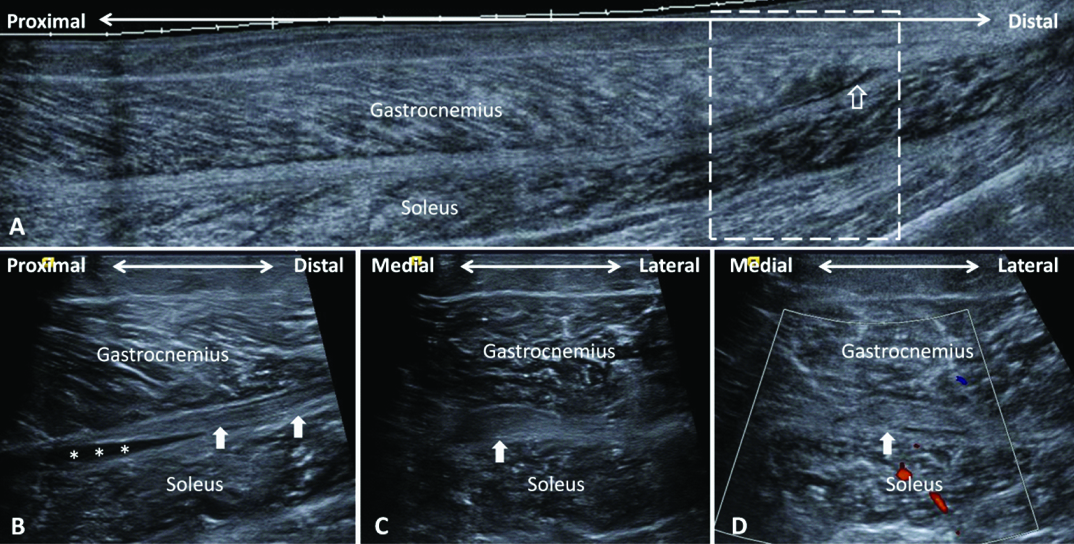
Based on an increase in thickness of the plantaris tendon with peritendinous fluid accumulation, grade II isolated plantaris tendinopathy was thus, diagnosed by US images [1,2]. The patient received physical therapy with US diathermy and transcutaneous electric stimulation focused at the area of plantaris tendon swelling and his symptoms gradually subsided after few weeks.
The differential diagnoses of calf pain after a sport injury, such as playing in basketball, jogging, tennis or other racquetball [3], include strain or tears of the triceps surae and Achilles tendons [4]. The triceps surae, the largest muscle bulk of the calf, is composed of the medial and lateral gastrocnemius and soleus muscles. The gastrocnemius muscle originates at the posterior aspects of the medial and lateral femoral condyle while the soleus muscle is attached to the proximal fibula and tibia [5,6]. These two muscles merge distally to form the Achilles tendon and insert on the calcaneus [7]. The plantaris muscle, a small rudimentary muscle, emerges from the lateral supracondylar region of the femur and courses deep to the lateral head of gastrocnemius [8]. The plantaris has a slender tendon which fuses with the Achilles tendon distally and inserts at the medial side of calcaneus [7]. Among all the musculotendinous injuries of the calf, rupture of the medial head of the gastrocnemius muscle at its musculotendinous junction, namely tennis leg, is the most common cause [4]. Soleus muscle tear is less common but it may lead to compartment syndrome if a huge hematoma is formed between the gastrocnemius muscle and the deep flexor hallucis longus, flexor digitorum longus and tibialis posterior muscles [5]. An isolated lesion of the plantaris tendon is rare and is often misinterpreted as tennis leg [8]. Lesions of the Achilles tendon happen most frequently at its middle third region where it is thinnest and most hypovascular [7]. It is of interest that the plantaris tendon often remains intact when the Achilles tendon is ruptured [9]. Unlike Achilles tendon tear, isolated plantaris tendon tear has a benign outcome and does not need surgical treatment [8].
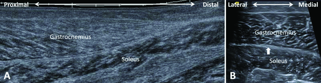
The clinical presentation of the above mentioned pathologies can be similar, making the decision of a specific management difficult [3,9]. The US examination can delineate the muscle fibers and explore collection of blood or effusion, thus, prompting the accurate diagnosis and proper management of a clinically ambiguous case [3,5,7]. In our case, the long-axis US image revealed intact fibrillar pattern of the gastrocnemius and soleus muscles without fluid accumulation at the muscle junction, indicating that it was not a myofascial injury between these two muscles. In a normal condition, the plantaris tendon in short-axis US image appears as an oval-shaped hyperechoic structure located between the medial gastrocnemius and soleus muscles [Table/Fig-2] [6]. It is often difficult to differentiate the tendon from the adjacent fascial planes if the tendon is not swollen. In our case, the short axis view examination revealed the focal swelling of the plantaris tendon and confirmed its anatomical position, which runs between the medial gastrocnemius and soleus muscles and was located medial to a normal Achilles tendon. In a recent study, a high proportion of the concomitant involvement of plantaris tendon were found in the patients with Achilles tendinopathy, in which the ultrasound examination with Colour Doppler revealed similar plantaris-like structures medial to the Achilles tendon with hypoechoic change and high blood flow [10]. In the cases series presented by Bianchi et al., five relatively distal plantaris tendon tears were documented with swollen and retracted torn tendon ends [8].
The panoramic (a) and transverse (b) ultrasound images over the mid-calf of the normal side. Arrow, plantaris tendon.
In our case, no tear was observed distal to the focal swelling of the plantaris tendon. So, the tendon swelling was a result of tendinopathy, not a proximal laxity caused by distal rupture. In conclusion, the slender character of plantaris tendon renders the diagnosis of plantaris tendon disorder challenging. The high-resolution US can well depict the tendon fibers of the plantaris tendon, thus, helping the diagnosis and the onward management.
Financial or Other Competing Interests
“Funding: The current research was supported by the research grant from National Taiwan University Hospital, Bei-Hu branch.”
[1]. Maffulli N, Sharma P, Luscombe KL, Achilles tendinopathy: aetiology and managementJ R Soc Med 2004 97:472-76. [Google Scholar]
[2]. Hung CY, Chang KV, Özçakar L, Wang TG, Chen WS, Can quantification of biceps peritendinous effusion predict rotator cuff pathologies?: A retrospective analysis of 1352 shoulder ultrasoundAm J Phys Med Rehabil 2016 95:161-68. [Google Scholar]
[3]. Shields CL Jr, Redix L, Brewster CE, Acute tears of the medial head of the gastrocnemiusFoot Ankle 1985 5:186-90. [Google Scholar]
[4]. Kane D, Balint PV, Gibney R, Bresnihan B, Sturrock RD, Differential diagnosis of calf pain with musculoskeletal ultrasound imagingAnn Rheum Dis 2004 63:11-14. [Google Scholar]
[5]. Hung CY, Chang KV, Ozcakar L, Ultrasound imaging of torn soleus musclePM R 2015 7:1106-07. [Google Scholar]
[6]. Shah JR, Shah BR, Shah AB, Pictorial essay: Ultrasonography in ‘tennis leg’Indian J Radiol Imaging 2010 20:269-73. [Google Scholar]
[7]. Daftary A, Adler RS, Sonographic evaluation and ultrasound-guided therapy of the Achilles tendonUltrasound Q 2009 25:103-10. [Google Scholar]
[8]. Bianchi S, Sailly M, Molini L, Isolated tear of the plantaris tendon: ultrasound and MRI appearanceSkeletal radiol 2011 40:891-95. [Google Scholar]
[9]. Helms CA, Fritz RC, Garvin GJ, Plantaris muscle injury: evaluation with MR imagingRadiology 1995 195:201-03. [Google Scholar]
[10]. Masci L, Spang C, van Schie HT, Alfredson H, How to diagnose plantaris tendon involvement in midportion Achilles tendinopathy - clinical and imaging findingsBMC Musculoskelet Disord 2016 17:97 [Google Scholar]