The chief goal of root canal therapy is to eradicate microorganisms from the root canal system and prevent recontamination. However, because of the presence of complex anatomy of the root canal system like the presence of lateral canals, ramifications and deltas, it is impossible to complete disinfection of the root canal using instrumentation alone [1,2] particularly in the apical third [3]. Therefore, instrumentation must be combined with adequate irrigation because it removes bacteria, debris and necrotic tissue which cannot be eliminated by instrumentation alone [4].
Studies had shown that the use of irrigants like NaOCl and EDTA are able to penetrate those areas which cannot be accessed mechanically by instrumentation [5] thereby killing microorganisms, flushing debris and removing smear layer from the root canal system [6]. There are many factors which influence the penetration of irrigants into the apical third of root canals which include: 1) final apical preparation size; 2) presence of vapour lock; 3) irrigant delivery method [7,8].
Currently different irrigation delivery devices and techniques are being used to improve the disinfection of the root canal system. Studies had shown that conventional needle irrigation does not allow the delivery of the irrigating solutions beyond the tip of the irrigating needle [9]. The Endovac system had significantly better debridement efficacy at 1mm from the working length when compared with needle irrigation.
Passive Ultrasonic Irrigation (PUI) system showed better irrigant penetration when compared with sonic irrigation [10]. Previous studies in literature have revealed that ultrasonic activation is associated with greater elimination of pulpal tissue remnants as well as debris from isthmi and fins of the canal [11]. Studies had shown that the Endovac system achieved better sealer penetration when compared with conventional needle irrigation [12].
However, the combined effect of Endovac and PUI on sealer penetration has not been studied. Therefore, the aim of the present study was to compare the effect of Endovac system, PUI and combination of Endovac and PUI on sealer penetration into dentinal tubules using confocal laser scanning microscope. The null hypothesis was that there is no significant difference in the percentage and maximum depth of sealer penetration between the Endovac, PUI and combination of Endovac and PUI.
Materials and Methods
This in-vitro study was conducted in the Department of Conservative Dentistry and Endodontics at G. Pulla Reddy Dental College and Hospital, Kurnool, Andhra Pradesh, India.
Forty eight human maxillary central incisors extracted due to periodontal reasons were used in this study. The presence of single canal was verified radiographically. Teeth with immature apex, radicular resorption or an endodontic filling were rejected. Teeth were immersed in 4% NaOCl for 2hrs and any visible calculus was removed ultrasonically.
Access cavity was prepared by using No. 2 endodontic access bur. Working length was established by inserting a size 10 K file until the file tip appeared at the apical foramen and then subtracting 1mm from this length. The coronal portion of the root canal was flared using Gates Glidden drills of size 1-3.
Biomechanical preparation was done using step-back preparation. The apical preparation was done up to size 40 K- file and then step back was done. Recapitulation was achieved to the estimated working length by using size 15 K-files.
During cleaning and shaping the protocol followed for irrigation included 3ml of 5.25% NaOCl, 3ml of 17% EDTA followed by 3ml of 5.25% NaOCl using a 27 gauge irrigation needle.
Experimental Groups
The specimens were broadly divided into three experimental groups based on the final irrigation technique used.
Group I [Apical negative pressure irrigation group (Endovac)]: In this group Endovac was used. Here the master delivery tip delivered 1ml of NaOCl into the access cavity; simultaneously the microcannula was passively introduced up to the working length under apical negative pressure for a period of 30 seconds.
Group II [Passive Ultrasonic Irrigation group (PUI)]: Ultrasonic irrigation was performed by using a stainless steel ultrasonic Irrisafe file of size 20 mounted on a Suprasson P5 booster ultrasonic unit. The file was kept 1mm short of working length with 1ml of NaOCl and was activated for a period of 30 seconds by using power setting of five.
Group III [Combination of apical negative pressure irrigation and PUI]: In this group the master delivery tip delivered 1ml of NaOCl into the access and simultaneously the microcannula was placed up to the working length under apical negative pressure for a period of 30 seconds. After 30 seconds of irrigation the microcannula was withdrawn from the canal and followed by ultrasonic irrigation, where the ultrasonic activation was achieved using Irrisafe file size 20 where the file was passively inserted 1mm short of the working length and activated for a period of 30 seconds with 1ml of NaOCl in the canal.
All the canals were dried with absorbent paper points and obturation was done by using AH plus sealer and gutta percha by using lateral compaction technique. For observation under confocal laser scanning microscopy, AH plus sealer was mixed with 0.1% fluorescent Rhodamine B isothiocyanate. Sealer was applied with gutta percha. Finally the access cavity was sealed with cavit and the teeth were stored at 37°C for 24 hours to allow the resin sealer to set.
All the specimens were sectioned perpendicular to the long axis by using water cooled, slow speed 0.3mm microtome saw. Sections were obtained at 1mm, 3mm and 5mm from the root apex. All sections were polished by using silicone carbide abrasive papers and the specimens were mounted onto glass slide and examined under Leica TSS-SPE confocal laser scanning microscope.
Confocal laser scanning microscope investigation: Here the method proposed by Gharib SR et al., was used to evaluate the images [13]. First, each image sample was imported into Photoshop then the circumference of the root canal wall was outlined and measured with a Photoshop measuring tool. The calculation of the percentage of sealer penetration was evaluated along the areas or circumference of the canal walls, and the sealer that had penetrated into the dentinal tubules was outlined and measured using the same method as proposed by Gharib SR et al., [13]. The outlined lengths were divided by the canal circumferences.
The point of deepest penetration was calculated from the canal wall to the point of maximum sealer penetration for measuring the depth of penetration.
Statistical Analysis
Statistical analysis was done by using two way ANOVA and Tukey’s post-hoc test to compare the percentage of sealer penetration and to measure the maximum depth of sealer penetration by using different irrigation techniques.
Results
Percentage of sealer penetration: The combination group resulted in higher percentage of sealer penetration at 1mm (27.47) and 3mm (38.09) from the working length than the Endovac and PUI group. There was no significant difference in sealer penetration at 5mm level between PUI and combination group (p-value <0.05) [Table/Fig-1].
Pair wise comparison of three groups (I, II, III) and three levels (1mm, 3mm, 5mm) with percentage of sealer penetration by Tukey’s multiple post-hoc procedures.
| Groups with levels | Group I with 1mm | Group I with 3mm | Group I with 5mm | Group II with 1mm | Group II with 3mm | Group II with 5mm | Group III with 1mm | Group III with 3mm | Group III with 5mm |
|---|
| Mean | 13.18 | 4.90 | 5.26 | 3.00 | 20.37 | 62.72 | 27.47 | 38.09 | 64.03 |
| SD | 0.65 | 0.76 | 0.67 | 0.37 | 2.10 | 2.93 | 2.79 | 4.71 | 2.55 |
| Group I with 1mm | - | | | | | | | | |
| Group I with 3mm | p=0.0001* | - | | | | | | | |
| Group I with 5mm | p=0.0001* | p=0.9999 | - | | | | | | |
| Group II with 1mm | p=0.0001* | p=0.3637 | p=0.1530 | - | | | | | |
| Group II with 3mm | p=0.0001* | p=0.0001* | p=0.0001* | p=0.0001* | - | | | | |
| Group II with 5mm | p=0.0001* | p=0.0001* | p=0.0001* | p=0.0001* | p=0.0001* | - | | | |
| Group III with 1mm | p=0.0001* | p=0.0001* | p=0.0001* | p=0.0001* | p=0.0001* | p=0.0001* | - | | |
| Group III with 3mm | p=0.0001* | p=0.0001* | p=0.0001* | p=0.0001* | p=0.0001* | p=0.0001* | p=0.0001* | - | |
| Group III with 5mm | p=0.0001* | p=0.0001* | p=0.0001* | p=0.0001* | p=0.0001* | p=0.1313 | p=0.0001* | p=0.0001* | - |
*p<0.05 indicates significant between them
Note: Red colored p-values are indicated comparison of 3 levels in each Group
Blue colored p-values are indicated comparison of Group I with II and II
Pink colored p-values are indicated comparison of Group II with III
Maximum depth of sealer penetration (in micrometers): The combination group resulted in significantly higher maximum depth of sealer penetration at 1mm (11598.40) and 3mm (20931.30) from the working length than the Endovac and PUI group. There was no significant difference at 5mm level between PUI and combination group (p-value>0.05) [Table/Fig-2].
Pair wise comparison of three groups (I, II, III) and three levels (1mm, 3mm, 5mm) with maximum depth of sealer penetration (in micrometers) by Tukey’s multiple post-hoc procedures.
| Groups with levels | Group I with 1mm | Group I with 3mm | Group I with 5mm | Group II with 1mm | Group II with 3mm | Group II with 5mm | Group III with 1mm | Group III with 3mm | Group III with 5mm |
|---|
| Mean | 5935.94 | 3775.31 | 3957.66 | 1410.94 | 9787.50 | 46318.75 | 11598.44 | 20931.25 | 48768.75 |
| SD | 881.43 | 233.49 | 256.58 | 468.15 | 1821.13 | 5408.89 | 5809.22 | 8182.13 | 5143.39 |
| Group I with 1mm | - | | | | | | | | |
| Group I with 3mm | p=0.8801 | - | | | | | | | |
| Group I with 5mm | p=0.9246 | p=0.9999 | - | | | | | | |
| Group II with 1mm | p=0.0621 | p=0.8150 | p=0.0001* | - | | | | | |
| Group II with 3mm | p=0.1962 | p=0.0019* | p=0.0001* | p=0.0001* | - | | | | |
| Group II with 5mm | p=0.0001* | p=0.0001* | p=0.0001* | p=0.0001* | p=0.0001* | p=0.9542 | | | |
| Group III with 1mm | p=0.0048 | p=0.0001* | p=0.0001* | p=0.0001* | p=0.0001* | p=0.0001* | | | |
| Group III with 3mm | p=0.0001* | p=0.0001* | p=0.0001* | p=0.0001* | p=0.0001* | p=0.0001* | p=0.0001* | - | |
| Group III with 5mm | p=0.0001* | p=0.0001* | p=0.0001* | p=0.0001* | p=0.0001* | p=0.1313 | p=0.0001* | p=0.0001* | - |
*p<0.05 indicates significant between them
Note: Red colored p-values are indicated comparison of 3 levels in each Group
Blue colored p-values are indicated comparison of Group I with II and II
Pink colored p-values are indicated comparison of Group II with III
However, the Endovac group showed significantly better sealer penetration at 1mm from the working length when compared with PUI.
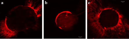
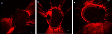
Representative pictures from each group are shown in [Table/Fig-3,4].
Sealer penetration at 1mm from the working length under confocal scanning laser microscopy; a) Apical negative pressure; b) Passive ultrasonic irrigation; c) Combination group.
Sealer penetration at 3mm from the apex under confocal scanning laser microscopy; (a) Apical negative pressure; b) Passive ultrasonic irrigation; c) Combination group.
Discussion
The main aim of root canal treatment is to seal the root canal system to prevent re-infection [14]. The components of root canal filling includes a hard core like gutta percha and a sealer to better adapt the root canal filling material to the canal wall [15]. The sealer root canal wall interface is essential for the sealing of the root canal system as the sealer can fill the irregularities within the root canal wall and the dentinal tubules which cannot be filled by gutta percha alone [16–18].
As discussed earlier, there are many factors which influence the penetration of irrigants in the apical third of the root canal. One of the factors is the final apical preparation size. An apical preparation of ISO size 40 is adequate to accommodate sufficient irrigant volume [19,20]. Therefore, in the present study apical preparation size was standardized to a size 40/0.02. A closed end model was used to simulate the clinical procedures by sealing the apical foramen with glue.
Presence of vapour lock is another factor which influences the penetration of irrigants in the apical third. Sel-D Saber and Hashem AA had reported that apical negative pressure and manual dynamic agitation resulted in better removal of the smear layer in the apical third [21]. This can be due to the fact that both these techniques are capable of reaching up to the full working length of the instrumented canals and therefore permit adequate irrigant replacement, which is not possible with conventional needle irrigation or ultrasonic agitation devices. Studies had shown that the ANP had significantly better debridement 1mm from the working length when compared with needle irrigation [22,23].
Another factor which influences the penetration of irrigants in the apical third is the irrigant delivery method. In the present study, PUI, Endovac and combination of Endovac and PUI was used to evaluate the sealer penetration in the apical third of the root canal.
Previous studies compared the conventional needle irrigation and Endovac system for sealer penetration in the apical third of the root canal and concluded that the percentage and maximum depth of sealer penetration using Endovac were significantly better than conventional needle irrigation at 1mm and 3mm from the working length [12,24]. However, none of the studies compared the Endovac, PUI and combination of Endovac and PUI for sealer penetration in the apical third of the root canal.
The results of the present study showed that the combination of Endovac and PUI resulted in maximum depth and percentage of sealer penetration at 1mm and 3mm level from the working length. These results are in accordance with previous studies which showed that the combination group was associated with the three dimensional penetration of irrigant up to the working length and into lateral canals [24], which has been further confirmed in the present study by observation of sealer penetration in the apical third of the root canal under confocal laser scanning microscope. Therefore, it is an indication that this three dimensional penetration of irrigant allowed complete debridement of the root canal or complete elimination of smear layer.
In the present study the Endovac group showed better sealer penetration at 1mm from the working length than at 3mm and 5mm. These results are in accordance with previous study that concluded that Endovac was significantly effective in removing debris from the root canals at 1mm short of the working length but is not significantly better at 3mm short of the working length [25].
The PUI group showed better sealer penetration at 3mm and 5mm from the working length. This might be due to placement of Irrisafe file 1mm short of the working length as per manufacturer’s instructions and as in most of the earlier studies [26,27]. This may be the reason for this group showing better sealer penetration at 3mm and 5 mm than at 1mm from the working length.
In order to analyze sealer penetration into the dentinal tubules several techniques were used like scanning electron microscopy [28,29], light microscopy [30]. In our present study confocal laser scanning microscope was used to analyze sealer penetration as it tends to produce fewer artifacts than the conventional methods.
The only group that was able to show better sealer penetration at 1mm and 3mm from the working length was the combination group. The reason might be, when the Endovac was used first, the negative pressure removed the debris from the main canal and ensured that sufficient irrigant reached the working length. Hence, adequate volume of irrigant was present when PUI was being used subsequently and enhances better debridement efficacy. This could significantly enhance better sealer penetration at 1mm and 3mm from the working length.
Limitation
Being an in-vitro study, further studies should be carried to know the clinical effectiveness when performed on patients.
Conclusion
Within the limitations of this study, it has been concluded that the combination group resulted in better sealer penetration at 1mm and 3mm from the working length than the Endovac and PUI group. However, the Endovac group showed significantly better sealer penetration at 1mm from the working length when compared with PUI. There is no significant difference in sealer penetration at 5mm level between PUI and combination group. Therefore the null hypothesis was rejected.
*p<0.05 indicates significant between them
Note: Red colored p-values are indicated comparison of 3 levels in each Group
Blue colored p-values are indicated comparison of Group I with II and II
Pink colored p-values are indicated comparison of Group II with III
*p<0.05 indicates significant between them
Note: Red colored p-values are indicated comparison of 3 levels in each Group
Blue colored p-values are indicated comparison of Group I with II and II
Pink colored p-values are indicated comparison of Group II with III