According to Buonocore MG (1955), acid etching has been a standard practice to remove smear layer for successful bonding [1]. The long standing bonding strength is due to the micromechanical bond formation. In 1965, Newman GV reported epoxy resin use for attaching brackets [2]. The overall treatment results improved with the bonding procedure by eliminating occupancy of inter-dental spaces by band, reduced gingival irritation, and facilitating plaque removal and reducing decalcification [3]. Since then a variety of bonding procedures and adhesives have been reported to improve the orthodontic bond strength [4,5].
Direct bonding of orthodontic brackets to the etched surface of enamel has many merits along with some demerits. The main problems are loss of surface enamel and demineralization near the bracket and conventional acid etching requires all the steps of enamel conditioning (acid etching, rinsing, drying, and application of bonding agent) to be properly carried out. The loss of surface enamel and subsurface enamel weakening, leading to detachment or fracture of enamel surface during debonding occurs due to a strong acidic conditioning liquid or prolonged etching [6]. It has been extensively reported that the use of SEPs produce a milder etch pattern than 37% phosphoric acid [7–14]. Although conventional acid etching of the enamel surface leads to more enamel loss than does the use of SEPs [11], the etching pattern observed with 37% phosphoric acid for 15 seconds seems more conservative than the typical honeycomb etched pattern observed when the enamel surface was etched for 30 seconds [7,12,14]. In any case, SEPs seem to produce a milder etching pattern than the phosphoric acid. However, due to some disadvantages in conventional technique like increased chair side time and uncontrolled demineralization of enamel surface, the self etching primer was formulated. Using a SEP, procedure of bonding is simplified by combining etching and priming processes into a single step procedure. Additional to saving time, minimum steps for bonding procedure might lead to minimum procedural errors.
The aim of this in-vitro study was to compare the SBS of orthodontic bracket bonded with SEP and conventional acid etching system, to study the surface appearance of teeth after debonding; etching with conventional acid etch and self-etch priming using stereomicroscope and to evaluate correlation between SBS and surface roughness.
Materials and Methods
The present in-vitro study was conducted in Department of Orthodontics, ACPM Dental College and Hospital Dhule, Maharashtra, India, during September-December 2015. In this in-vitro study freshly extracted 100 non-carious and un-restored maxillary first premolars extracted for orthodontic treatment were collected and stored in normal saline.
Premolars were selected on basis of non-carious, freshly extracted premolars with intact buccal surface, non-hypoplastic, non-fluorosed, no restorations, no cracks and no extraction forceps marks.
Excluded premolars were carious, hypoplastic, mottled, restored, had marks of extraction forcep and having cracks present on surface.
Teeth were fixed in acrylic resin up to the Cemento-Enamel Junction (CEJ) leaving crown surface exposed for bonding of brackets. Prior to bonding, cleaning and polishing of each tooth done with pumice and water paste application with the help of a rubber cup on a slow speed hand piece, rinsed with splash of water and dried with an oil and moisture free air jet stream.
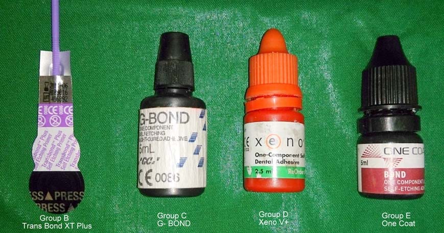
Bracket system 0.022″ slot (3M Unitek) was used in the study. The average bracket base surface area was found to be 10.037mm2. The self etch primer used in this study was - Transbond™ Plus (3M Unitek), Xeno V+ (Densply), G-Bond (GC) and One-Coat (Coltene). Bonding of all brackets was done with Transbond XT (3M Unitek). Self etching primer is shown in [Table/Fig-1]. Samples were divided into five different groups each containing 20 teeth.
Group A: Conventional acid etching – priming Transbond XT
Group B: Self etching primer - Transbond™ Plus (3M Unitek).
Group C: Self etching primer Xeno V+ (Dentsply).
Group D: Self etching primer G- Bond (GC).
Group E: Self etching primer One-Coat (Coltene).
Bonding Procedure
Group A: Conventional Group: Twenty teeth samples were etched with 37% phosphoric acid for 40 seconds, rinsed and air-dried. Transbond XT primer (3M Unitek) was applied to the enamel surface in the form of a thin layer and light curing was done for 10 seconds. Brackets were bonded onto the center of the buccal surface of teeth with Transbond XT (3M Unitek), a light cured composite adhesive. A sharp scaler used to remove the excess resin material before curing, care was taken not to disturb the bracket position; light curing of the adhesive done for a total of 40 seconds.
Group B: Etching and priming 20 teeth samples done with a self-etching primer i.e., TransbondTM Plus (3M Unitek), which contains both acid and primer, applied on the enamel surface of 20 teeth for 3 seconds and gently air dried with a jet free of oil and water vapour, as per manufacturer’s instructions. Mixing of two constituents causes activation and the resulting mixture is applied straightly on enamel surface. Gradual pinching of contents of a black (largest) storage space into a white (middle) storage space and then into a purple (smallest) storage space of the blister pack with uniform pressure. Bonding of brackets with TransbondTM XT adhesive (3M Unitek) and curing of the adhesive with light done for 40 seconds.
Group C: Etching and priming of 20 teeth samples were done with a self-etching primer i.e., Xeno V+ (Dentsply); it is applied on the enamel surface of 20 teeth for 3 seconds and gently air dried with a jet free of oil and water vapour, as per manufacturer’s instructions. Bonding of brackets with TransbondTM XT adhesive (3M Unitek) and curing of the adhesive with light done for 40 seconds.
Group D: Etching and priming of 20 teeth samples were done with a self-etching primer i.e., G- Bond (GC), it is applied on the enamel surface of 20 teeth for 3 seconds and gently air dried with a jet free of oil and water vapour, as per manufacturer’s instructions. Bonding of brackets with Transbond XT adhesive (3M Unitek) and curing of the adhesive with light done for 40 seconds.
Group E: Etching and priming of 20 teeth samples was done with a self-etching primer i.e., One-Coat (Coltene), it is applied on the enamel surface of 20 teeth for 3 seconds and gently air dried with a jet free of oil and water vapour, as per manufacturer’s instructions. Bonding of brackets with TransbondTM XT adhesive (3M Unitek) and curing of the adhesive with light done for 40 seconds.
The samples were kept in distilled water at 37°C for 24 hours.
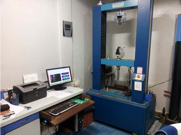
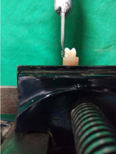
Bond Strength Testing: SBS test was done with a computerized, software based Universal Testing Machine (Star Testing Systems, India. Model No. STS 248) [Table/Fig-2]. The machine was set and calibrated as per manufacturer’s instructions. The acrylic block with the embedded tooth and it’s bonded bracket were positioned in clamp, so that the force was applied parallel to the tooth surface on top of each orthodontic bracket base during the shear strength test. The knife-edged blade was used to apply load at the bracket adhesive interface [Table/Fig-3]. The brackets were shear tested to failure using a load cell of 1000N and a crosshead speed of 5.0mm/min. The force magnitude to cause failure was recorded in Newton and conversion to force per unit area (MPa) done by dividing the measured force values by bracket surface area. Bracket mesh surface area was approximately 10.037mm2.
Universal testing machine (Star Testing Systems, India. Model No. STS 248).
Knife-edged blade was used to apply load at the bracket adhesive interface.
Adhesive Remnant Index: Examination of all the specimens after debonding done under the stereomicroscope (Magnum, Olympus, India Pvt., Ltd., New Delhi) at 40x magnification in order to assess adhesive remnants on tooth surfaces using the adhesive remnant index (Bishara SE et al.,) [15].
The ARI scale has a range of 5 to 1:
5 = no composite left over the enamel surface;
4 = less than 10% of composite left over the tooth surface;
3 = more than 10% but less than 90% of the composite left over the tooth surface;
2 = more than 90% of the composite left over the tooth surface;
1 = the entire composite, with an impression of the bracket base left on the tooth surface.
Surface Roughness Profilometry: Profilometry of etched enamel surface was done with a Profilometer (Mitutoyo, Japan, Model No. SJ210). Five representative samples from each group were taken to determine the surface roughness of enamel after application of conventional acid etch and self-etch primers. All etched teeth samples were rinsed thoroughly and air dried. After which, enamel surface roughness was measured with Profilometer.
Statistical Analysis
Statistical data for SBS of five groups is summarized as mean and standard deviation. The Analysis of Variance test (ANOVA) was used to determine the statistically significant differences of mean SBS between the five groups. To identify in between which group the statistically significant difference exists, Least Significant Difference (LSD) test was used as a part of Post-Hoc multiple comparison test and the five groups were compared in between each other. The difference of ARI score was compared in between the groups using Pearson’s Chi-square test.
Statistical Software: Data was analyzed using Statistical Package for Social Science (SPSS) Version 16, statistical software. Microsoft Word and Excel 2007 were used to generate graphs and tables. The statistical difference was considered to be significant if p<0.05 at 5% level of significance and p<0.01 as highly significant at 1% level of significance.
Results
[A] Shear Bond Strength: [Table/Fig-4] shows comparison of mean SBS among five groups, this shows that the SBS of the conventional group was significantly increased compared to self-etching primer groups.
Mean SBS in between five groups.
| Group | N | Mean (Mpa) | Std. Deviation (MPa) | Minimum | Maximum |
|---|
| A | 20 | 18.26 | 7.50 | 10.27 | 36.86 |
| B | 20 | 10.93 | 4.02 | 5.40 | 19.16 |
| C | 20 | 6.88 | 2.91 | 3.61 | 14.44 |
| D | 20 | 7.78 | 4.13 | 3.24 | 17.69 |
| E | 20 | 10.39 | 5.22 | 4.43 | 19.84 |
| ANOVA | Sum of Squares | df | Mean Square | F | p-value |
|---|
| Between Groups | 1607.137 | 4 | 401.784 | 16.044 | <0.01 |
Mean SBS of Group A was 18.26±7.5MPa, Group B was 10.93±4.02MPa, Group C was 6.88±2.91MPa while of Group D was 7.78±4.13MPa and Group E was 10.39±5.22MPa respectively.
There was statistically highly significant (p<0.01) difference of mean SBS in between the five groups.
So, that mean SBS of Group A> Group B> Group E > Group D > Group C. Indicating SBS of Group A was greatest while that of Group C was the least of all the five groups.
[B] Adhesive Remnant Index: Comparison between groups was done using Chi-square test. In conventional group ARI scores show that more than half of the adhesive was left over the tooth surface (score 1 to 3). In self-etching primer groups ARI scores show that there was no or slight amount of adhesive left over the tooth surface (score 4 and 5). Most of the failures in case of conventional group, occurred within the resin leaving more than half of the adhesive on the teeth, whereas in case of SEP they were between tooth surface and adhesive.
[Table/Fig-5] shows comparison of ARI score in between the groups. All 100% samples (20) of Group A and Group B had ARI score less than 4, while that was just 55% in Group D, 65% in Group E and 0% in Group C. ARI score of more than 4 was not in 80% samples of Group C, 25% samples of Group D and 35% samples of Group E. There was statistically highly significant (p<0.01) difference of percentage distribution of samples ARI scores in between the five groups.
Comparison of ARI score in between five groups.
| ARIScore | Group | Total |
|---|
| A | B | C | D | E |
|---|
| 1 | 8(40.0%) | | | | | 8 (8.0%) |
| 2 | 4(20.0%) | 11 (55.0%) | | 7 (35.0%) | 4 (20.0%) | 26 (26.0%) |
| 3 | 8(40.0%) | 9 (45.0%) | | 4 (20.0%) | 9 (45.0%) | 30 (30.0%) |
| 4 | | | 4 (20.0%) | 4 (20.0%) | | 8 (8.0%) |
| 5 | | | 16 (80.0%) | 5 (25.0%) | 7 (35.0%) | 28 (28.0%) |
| Total | 20(100.0%) | 20 (100.0%) | 20 (100.0%) | 20 (100.0%) | 20 (100.0%) | 100 (100.0%) |
[C] Profilometry Test: Surface roughness after etching with conventional acid etching was more than SEP, mean surface roughness of Conventional >TransbondTM Plus > One Coat > G-Bond >Xeno V+.
[Table/Fig-6] shows surface roughness (mean with SD) of teeth (n=5) in the groups, mean surface roughness of tooth in conventional group was 0.205μm, of TransbondTM Plus it was 0.191μm, in Xeno V+ it was 0.134μm, 0.142μm and 0.178μm in G-Bond and One Coat group respectively. There was statistically very highly significant (p<0.001) difference of surface roughness on tooth in between the five groups.
Surface roughness (mean with SD) of teeth in the groups done with ANOVA test.
| Group | Mean (μm) | SD (μm) | F | Sig. p-value |
|---|
| Group AConventional (n=5) | 0.205 | 0.0038 | 299.83 | <0.001 |
| Group BTransbondTM Plus (n=5) | 0.191 | 0.0036 |
| Group CXeno V+ (n=5) | 0.134 | 0.0033 |
| Group DG-Bond (n=5) | 0.142 | 0.0036 |
| Group EOne Coat (n=5) | 0.178 | 0.0052 |
[Table/Fig-7] shows correlation between SBS and surface roughness done with t-test. There was statistically very highly significant (p<0.001) mean difference in between surface roughness and SBS of conventional, TransbondTM Plus, Xeno V+, G-Bond and One Coat group.
Correlation between SBS and surface roughness done with t-test.
| Group | SBSMean±SD (Mpa) | Surface RougnessMean±SD (μm) | t | p-value |
|---|
| Conventional | 18.26±7.50 | 0.205±0.0038 | 5.296 | <0.001 |
| TransbondTM Plus | 10.93±4.02 | 0.191±0.0036 | 5.878 | <0.001 |
| Xeno V + | 6.88±2.91 | 0.134±0.0033 | 5.105 | <0.001 |
| G-Bond | 7.78±4.13 | 0.142±0.0036 | 4.067 | <0.001 |
| One Coat | 10.39±5.22 | 0.178±0.0052 | 4.309 | <0.001 |
Discussion
Several previous studies showed that the SBS of conventional system was notably more or similar to the self-etching primer and resin system Hitmi L et al., reported a value of control acid etched group was greater than SEP [16]. Cacciafesta V et al., Dorminey JC et al., Aljubouri YD et al., and Romano FL et al., reported that SEP was significantly less or similar than those bonded with a conventional etching process and SEP [17–20].
In this study, the bracket bonding with any of the four SEPs showed greater SBS values than those modestly required for orthodontic treatment [21,22]. Generally, the application of 37% phosphoric acid increases the bond strength [23]. In this context, the highest SBS value yielded by Group A (18.26MPa) was expected and this was appreciably greater than that of any other group. In the conventional acid etching (Group A) mean SBS was 18.26±7.50MPa. In the self-etching primer, TransbondTM Plus (Group B) mean SBS was 10.93±4.02MPa. In Xeno V+ (Group C) mean SBS was 6.88±2.91MPa. In G-Bond (Group D) mean SBS was 7.78±4.13MPa; In One Coat (Group E) mean SBS was 10.39±5.22MPa. There were no considerable differences among Groups B (Transbond Plus SEP) and E (One Coat). If main concern of clinicians is the bond strength in SEPs, TransbondTM Plus SEP might be routinely used for enamel conditioning. For the initial bonding, the application of this SEP (full mouth) is faster than that of the others; nevertheless, the other SEPs can be suitable alternatives.
Orthodontic bracket bonding on tooth is a temporary process; the bracket is removed after the active orthodontic therapy. Hence, debonding techniques are important and the amount of adhesive left over onto the enamel, additional to outcome of these procedures over enamel, etching process as well as method of removing bracket and teeth cleaning after debonding results in enamel damage [6]. Bond failure sites were characterized using the ARI. We found significant differences in the ARI scores. Analysis of teeth tested showed that there was significant difference in pattern of bond failure sites among the two groups. Maximum frequency of score was 4-5 in self-etch priming group and 1-2 in conventional groups i.e., majority of the failures in conventional group were at the bracket adhesive interface. Whereas, in self-etch primer failures at the enamel adhesive interface.
Surface roughness after etching with conventional acid etching was more than SEP, mean surface roughness of Conventional >TransbondTM Plus > One Coat > G Bond > Xeno V+. The surface roughness of Group A samples was much higher than the self-etch groups. The total depth of etching was not measured in this study. Surface roughness value was very small as compared to the values given for etching depth, which is loss of surface enamel. However, the strength value of bond will be dependent on the surface roughness than loss of enamel surface. After comparing SBS and surface roughness with t-test, there was statistically very highly significant (p<0.001) correlation in between surface roughness and SBS.
Bishara et al., compared 2 adhesive systems, 2-component acid etch primer and 1-component system; and found no significant difference between shear bond strength of the two; however, 1-component system performed better (t=0.681, p=0.501) [24].
The present study evaluated the use of different self-etching primer as compared with the conventional bonding procedure. The findings indicated that lower, but clinically acceptable, shear bond forces were achieved using the self-etch primer to bond orthodontic brackets to the enamel surface.
By reducing the number of steps during bonding, clinicians can save time and reduce the potential for error and contamination during the bonding procedure. Our results indicated that the new self-etch primers, containing both the enamel etchant and the primer, can be successfully used in bonding orthodontic brackets. The SBS of the new 1-component (no-mix) self-etch primer performed as well as the 2-component system but saved a step in the bonding procedure.
Limitation
Majority of the studies conducted on etching primer are in-vitro studies. Results of this study cannot be extrapolated to the intra-oral environment completely. So, further long term clinical studies are needed to be carried out before actually recommending the use of SEP in routine orthodontic bonding.
Conclusion
The conventional group SBS values (37% phosphoric acid) were significantly higher than other groups. All groups might show clinically useful SBS values and Transbond XT can be successfully used for bracket bonding after enamel conditioning with any of the SEPs tested. The SEPs used in Group C (Xeno V+) and D (G-Bond) had significantly lowered SBSs. Although the values might still be clinically acceptable, further studies are necessary to assess their effectiveness for bonding that produce stronger bonds. After etching with SEPs, it produces a lesser surface roughness on the enamel surface than conventional etching. However, statistical study shows that the bonding strength has a very high (p<0.001) correlation with surface roughness of enamel. So, excellent bond strength with minimal enamel loss can be achieved. SEP application reduces the amount of adhesive left over after debonding, and in turn reduces the invasive procedures needed to clean up the enamel surface. Understanding the advantages and disadvantages of every SEP can be supportive for obtaining better results when a gentler bonding procedure is used.