Jejunal Epiphany: Diverticulae, Enteroliths and Strictures
Priyank Pathak1, Babar Rehmani2, Navin Kumar3
1 Resident, Department of General Surgery, SRH University, Jollygrant, Doiwala, Dehradun, Uttarkhand, India.
2 Associate Professor, Department of General Surgery, SRH University, Jollygrant, Doiwala, Dehradun, Uttarkhand, India.
3 Assistant Professor, Department of General Surgery, SRH University, Jollygrant, Doiwala, Dehradun, Uttarkhand, India.
NAME, ADDRESS, E-MAIL ID OF THE CORRESPONDING AUTHOR: Dr. Priyank Pathak, Resident, Department of General Surgery, SRH University, Jollygrant, Doiwala, Dehradun, Uttarkhand, India.
E-mail: priyank56pathak@yahoo.co.in
Multiple jejunal diverticulae represent a rare entity and are usually asymptomatic. This case report is about one such jejunal diverticulae along with multiple enteroliths and jejunal strictures. All these three different findings in a short segment of jejunum is a very rare finding with all three variants seen in a segment of jejunum.
We herein present a case of a 45-year-old male, who presented with vague abdominal pain for one and half years associated with nausea and vomiting and altered bowel habits. Laparotomy revealed multiple large jejunal diverticulae compressing the bowel with multiple enteroliths and two strictures in a short segment of jejunum leading to intestinal obstruction. Patient underwent resection of the involved jejunal segment and then repair by anastomosis. Post-operative period was uneventful.
Enteroliths, Intestinal obstruction, Laparotomy
Case Report
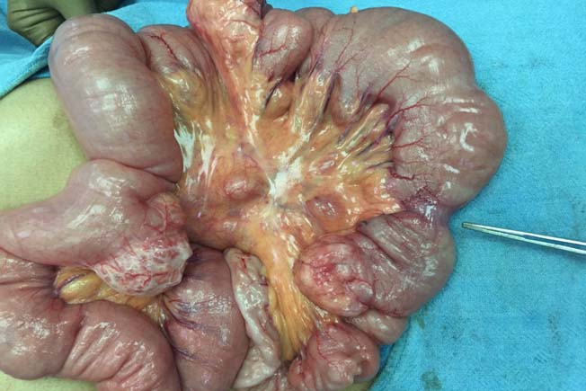
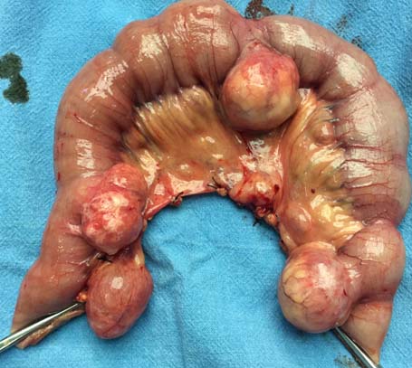
A 45-year-old male, chronic smoker, non alcoholic presented with left lower abdomen pain for one and half years, associated with nausea and altered bowel habits and tenderness in the left iliac fossa and left lumbar region. Patient was already receiving treatment for irritable bowel syndrome. Patient was advised to undergo Contrast Enhanced Computed Tomography (CECT) abdomen but the patient did not agree for the same and was lost to follow-up. After duration of 8 months, the patient again visited with similar complaints and this time with increased severity. Physical examination revealed non-distended soft abdomen with no visible peristalsis, periumbilical tenderness present, however, no lump was palpable. The bowel sounds were exaggerated. Digital rectal examination revealed normal findings. There was no history of any surgical interventions and any cardiovascular, renal and respiratory comorbidity. Haematological investigations revealed mild anemia while biochemical investigations including the renal function tests and liver function tests were within normal limits. Colonoscopy was normal study. A plain X-ray abdomen (erect and supine view) was done which showed multiple fluid levels and dilated jejunal loops. Ultrasound abdomen was also done which was only suggestive of fatty liver changes. In view of the clinical symptoms and radiological findings of partial bowel obstruction patient was planned for exploratory laparotomy. On exploratory laparotomy, an incidental finding of multiple large diverticulae, a total of six in number with few small diverticulae in the 100cm segment of jejunum, commencing about 50cm distal to the ligament of treitz [Table/Fig-1,2].
Shows multiple diverticulae in the region of jejunum with dilated bowel loops and the forceps pointing at the stricture.

Shows a resected specimen of jejunum with multiple diverticulae containing enteroliths, stricture adjacent to the diverticulae with dilated bowel loops.

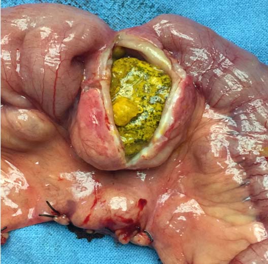
One of the diverticula showed an impassable stricture positioned immediately distal to it along with a large enterolith of about 3x2 cm and another passable stricture present about 15cm proximal to it [Table/Fig-3].
Shows a large enterolith with an impassable stricture in the anti mesenteric border.

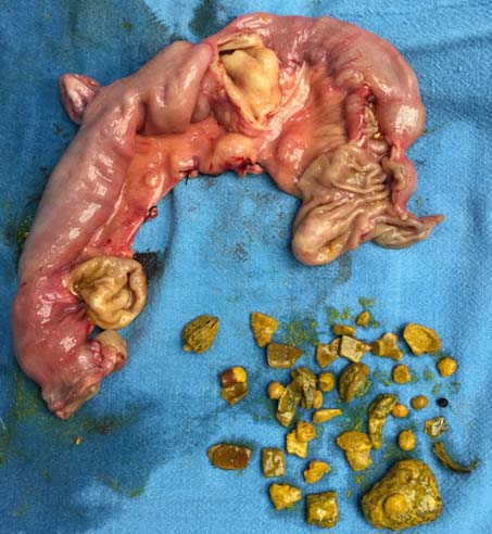
Mesenteric lymphadenopathy was also a feature. Since, both the stricture and the diverticular conditions could be the cause of the presenting symptom of the patient, resection of the entire diverticulae bearing segment of the jejunum with primary anastomosis by hand-sewn method was carried out. Multiple enteroliths along with fruit seeds kept alongside the resected segment were noted [Table/Fig-4]. The patient did not have any symptoms suggesting of complications and post-operative period went uneventful. Histopathological report of the resected segment and the strictured area revealed chronic inflammation with lymphocytic infiltration.
Shows multiple enteroliths along with fruit seeds kept alongside the resected segment.

Discussion
Acquired jejunal diverticulae are often a rare finding and are usually asymptomatic, but they can give rise to chronic complaints. As quoted by Markis K, the first case of jejunal diverticulosis was described by Sommering and then later by Sir Astley Cooper in 1807 [1]. Epidemiologically, more common in elderly males in 6th to 7th decade (58%) and commonly involve the proximal jejunum, in up to 75% cases, followed by distal jejunum and ileum [2]. Presentation is usually vague and must be taken into consideration in cases of unexplained malabsorption, anemia, chronic abdominal pain or discomfort [3]. As reported by Chowdhury G et al., a total of 34 cases related to small bowel obstruction due to enteroliths have been reported till 2009 [4]. The formation of enteroliths is mainly the result of dyskinesia and stasis of intestinal contents, leading to bacterial overgrowth, allowing precipitation and concretion of bile salts. The formation of enteroliths may be de novo or around fruit seeds and vegetable material and it was also the etiology in the present case [5]. Wilcox RD and Shatney CH in their study concluded that asymptomatic jejunal diverticulosis should not be resected and managed conservatively, and when symptoms are persistent or refractory to treatment, resection is imperative and will be required in up to 30% cases [6]. Impacted enteroliths may cause intestinal mucosal injury and can lead to intestinal gangrene [7]. Jejunum stricture formation immediately distal to a large impacted enterolith, can be due to the mucosal injury and the chemical effects of the outer shell of the enterolith. Similarly, few other case reports have been published in which the jejunal diverticulosis can lead to acute to partial intestinal obstruction and the patient may present with acute symptoms or varied delayed symptoms [8,9]. There is no data available, which directly relates stricture formation with enteroliths. The vice versa has been proven in many studies and articles, that the stricture formation, leading to stasis and henceforth making an ambient environment for enterolith formation [10].
Conclusion
In small intestinal obstruction in an elderly patient, diverticulosis as a cause of obstruction should not be forgotten. This case report also raises a concern about the impacted enteroliths leading to stricture formation and this area is yet to be explored.
Consent
Written informed consent was obtained from the patient for publication of this case report and any accompanying images.
Author’s Contribution
PP was part of the surgery, management of the patient, data collection, creating the manuscript. BR was the main operating surgeon and review of manuscript. NK was the 1st assistant in surgery, the postoperative management and making of manuscript.
[1]. Makris K, Tsiotos GG, Stafyla V, Small intestinal non-meckelian diverticulosis: clinical reviewJ Clin Gastroenterol 2009 43:201-07. [Google Scholar]
[2]. Singh S, Sandhu HP, Aggarwal V, Perforated jejunal diverticulum: A rare complicationSaudi J Gastroenterol 2011 17:367 [Google Scholar]
[3]. Evangelos F, Konstantinos V, Stavros M, Multiple giant diverticula of the jejunum causing intestinal obstruction: report of a case and review of the literatureWorld J of Emerg Surg 2011 6:8 [Google Scholar]
[4]. Chowdhury G, Kumar AS, Rahman A, Das BC, Uncommon cause (fecolith or enterolith) of small intestinal obstruction in the adultPulse 2009 3(1):35-37. [Google Scholar]
[5]. Bewes PC, Haslewood GA, Roxburgh RA, Bile-acid enteroliths and jejunal diverticulosisBr J Surg 1966 53:709-11. [Google Scholar]
[6]. Wilcox RD, Shatney CH, Surgical implications of jejunal diverticulaSouth Med J 1988 81:1386-91. [Google Scholar]
[7]. Nakao A, Okamoto Y, Sunami M, Fujita T, Tsuji T, Kurume. The oldest patient with gallstone ileus: report of a case and review of 176 cases in JapanMed J 2008 55(1-2):29-33. [Google Scholar]
[8]. Falidas E, Vlachos K, Mathioulakis S, Archontovasilis F, Villias C, Multiple giant diverticula of the jejunum causing intestinal obstruction: report of a case and review of the literatureWorld J Emerg Surg 2011 6:8 [Google Scholar]
[9]. Ferreira-Aparicio FE, Gutiérrez-Vega R, Gálvez-Molina Y, Ontiveros-Nevares P, Athie-Gútierrez C, Montalvo-Javé E, Diverticular Disease of the small bowelCase Rep Gastroenterol 2012 6(3):668-76. [Google Scholar]
[10]. Hofmann AF, Mysels KJ, Bile acid solubility and precipitation in vitro and in vivo: the role of conjugation, pH, and Ca2+ ionsJ Lipid Res 1992 33:617-26. [Google Scholar]