A Comparative Analysis of Caries Inhibitory Effect of Remineralizing Agents on Human Enamel Treated With Er:YAG Laser: An In-vitro Atomic Emission Spectrometry Analysis
Aswin Saseendran Nair1, R Krishna Kumar2, Sunil Thomas Philip3, Syed Shaheed Ahameed4, Sairaj Punnathara5, Joby Peter6
1 Senior Lecturer, Department of Paedodontics, Noorul Islam College of Dental Sciences, Aralumoodu, Neyyatinkara, Trivandrum, Kerala, India.
2 Professor, Department of Paedodontics and Preventive Dentistry, Rajah Muthiah Dental College and Hospital, Annamalai University, Chidambaram, Tamil Nadu, India.
3 Professor, Department of Paedodontics, Noorul Islam College of Dental Sciences, Aralumoodu, Neyyatinkara, Trivandrum, Kerala, India.
4 Senior Lecturer, Department of Paedodontics and Preventive Dentistry, Rajah Muthiah Dental College and Hospital, Annamalai University, Chidambaram, Tamil Nadu, India.
5 Senior Lecturer, Department of Paedodontics and Preventive Dentistry, Rajah Muthiah Dental College and Hospital, Annamalai University, Chidambaram, Tamil Nadu, India.
6 Reader, Department of Paedodontics and Preventive dentistry, Rajah Muthiah Dental college and Hospital, Annamalai University, Chidambaram, Tamilnadu, India.
NAME, ADDRESS, E-MAIL ID OF THE CORRESPONDING AUTHOR: Dr. Sunil Thomas Philip, Professor, Department of Paedodontics, Noorul Islam College of Dental Sciences, Aralumoodu, Neyyatinkara, Trivandrum-695123, Kerala, India.
E-mail: rubysunildr@rediffmail.com
Introduction
The tug of war to maintain tooth integrity is dependent on a ratio between demineralization and remineralization. Hence, demineralization should be retarded and remineralization should be enhanced to maintain a natural equilibrium in the oral cavity.
Aim
To compare in-vitro acid resistance of human enamel when using Casein Phosphopeptides Amorphous Calcium Phosphate (CPP-ACP) [GC Tooth mousse] cream, Casein Phosphopeptide Amorphous Calcium Fluoride Phosphate (CPP-ACFP) [GC Tooth mousse plus] cream, Er:YAG laser alone, combination of CPP-ACP with Er:YAG laser, CPP-ACFP with Er:YAG laser.
Materials and Methods
An in-vitro study was done on 100 specimens which were prepared from 50 human premolars to investigate the caries inhibitory effect of remineralizing agents and laser on enamel using an atomic emission spectrometry analysis. The enamel specimens were randomly allocated into 6 groups: Untreated (control); CPP-ACP (GC Tooth mousse); CPP-ACFP (GC Tooth mousse plus); Er:YAG laser treatment alone; CPP-ACP with Er:YAG laser; CPP-ACFP with Er: YAG laser. Then specimens were immersed individually in 5ml of acetate buffer solution (0.1mol/L, pH 4.5) and incubated at 37°C for 24 hours, to determine the acid resistance by analyzing the calcium release using atomic emission spectrometry. An ANOVA model was constructed (p-value 0.05), followed by post-hoc Tukey’s test for multiple pair wise comparisons of mean values.
Results
There was a significant difference among the various groups with respect to amount of calcium released (p<0.001). The lowest mean score of calcium release was observed for CPP-ACFP with Er:YAG laser followed by CPP-ACFP but the differences between these groups were statistically not significant (p>0.05). Similarly the differences between CPP-ACP with Er:YAG laser and CPP-ACP also were not significant (p>0.05). The highest mean score of calcium release was for Er:YAG laser and no significant statistical difference was noticed in comparison with control group (p>0.05).
Conclusion
Combination of CPP-ACFP with Er:YAG laser is more effective in decreasing enamel demineralization when compared with other groups.
Acid resistance, Enamel remineralization, Erbium laser, Tooth mousse
Introduction
The current concept of dental caries is that numerous episodes of demineralization and remineralization occur simultaneously in certain areas of incipient caries lesion [1]. The ideal requirement of a material to prevent caries is that it should release calcium and phosphate into the oral cavity and into enamel subsurface [2]. The anticariogenic effect of Casein Phosphopeptides Amorphous Calcium Phosphate (CPP-ACP) is that it localizes ACP at the tooth surface and dental biofilm (plaque) so that when there is acid challenge the ACP dissociates and release calcium and phosphate ions increasing degree of saturation with respect to hydroxyapatite thus preventing caries and promoting remineralization by diffusion of ions into subsurface lesion [3]. When fluoride was added to CPP-ACP the acid resisting capacity was improved by formation of stabilized novel Amorphous Calcium Fluoride Phosphate (ACFP) phase [4,5].
Laser irradiation must not ablate the surface, but should change the enamel morphology and physical properties including fusion, recrystallization and melting of enamel hydroxyapatite crystals decreasing enamel permeability [6]. Two explanations for caries prevention by lasers is organic blocking theory [7] and formation of microspaces and microfissures in lased enamel [8]. Most authors consider carbonate decomposition and water loss are responsible for caries prevention [9,10]. Hossain M et al., reported that during laser irradiation there is an increase in the calcium to phosphorous ratio causing caries inhibition [11]. Erbium-doped:Yttrium-Aluminium Garnet (Er:YAG) laser removed carbonate and modified the organic matrix thus inhibiting caries [12]. Er:YAG lasers have wave length of 2.94μm and is highly absorbed by water and hydroxyapatite and can reach temperature needed to alter enamel structure and make it less soluble [13]. The enamel morphological changes by Er:YAG laser depends on density of laser, wavelength, irradiation time, focal distance, water cooling and mode of application. Laser in combination with remineralizing agents and fluoride had an additive effect in remineralizing early caries lesions [14,15]. Laser irradiation induced micromorphological changes on enamel followed by application of CPP-ACFP caused incorporation of nanocomplexes on the tooth surface that act as a reservoir to replenish the soluble calcium and phosphate ions that has diffused into enamel sub-surface [14]. The present study was done to compare caries inhibitory potential of CPP-ACP, CPP-ACFP, Er:YAG laser, combination of CPP-ACP with Er:YAG laser and CPP-ACFP with Er:YAG laser.
Materials and Methods
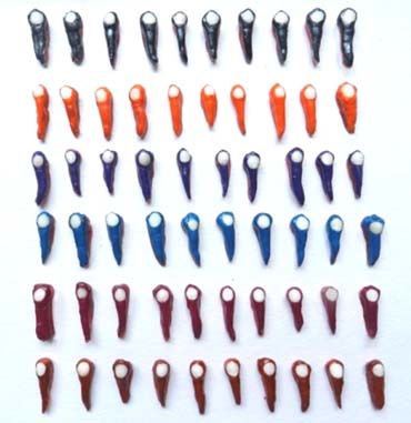
The present in-vitro study was done in the Department of Pedodontics and Preventive Dentistry, Rajah Muthiah Dental College, Chidambram, Tamil Nadu, India. Fifty human premolars free of caries or any other defects and which were extracted for orthodontic reasons were selected for the study. The armamentarium used were GC tooth mousse, GC tooth mousse plus, applicator tips, acid resistant nail varnish, acetone, diamond discs, distilled water. Teeth were cleaned and kept in 0.1% thymol solution for 1 month till use. Teeth were sectioned mesiodistally using water cooled diamond discs and two specimens was obtained from each tooth. The buccal halves were treated and lingual halves served as matched controls. The enamel surface of each specimen were painted with acid resistant nail varnish leaving a window of 3.5mm in diameter at the centre of buccal surface which was demarcated with adhesives [Table/Fig-1].
Prepared enamel specimens.
The surfaces were cleaned with cotton after removal of adhesives. The specimens were randomly divided into 6 groups (n=10 for each group)
Group 1 : Untreated (control)
Group 2 : Casein Phosphopeptide-Amorphous Calcium Phosphate (CPP-ACP)
Group 3 : Casein Phosphopeptide-Amorphous Calcium Fluoride Phosphate (CPP-ACFP)
Group 4 : Er:YAG treatment alone
Group 5 : CPP-ACP with Er:YAG laser
Group 6 : CPP-ACFP with Er:YAG laser
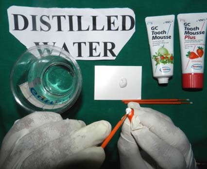
A sufficient amount of CPP-ACP cream on Group 2 and CPP-ACFP cream on Group 3 was dispensed on the buccal surface of the samples using an applicator tip [Table/Fig-2].
CPP-ACP and CPP-ACFP application.
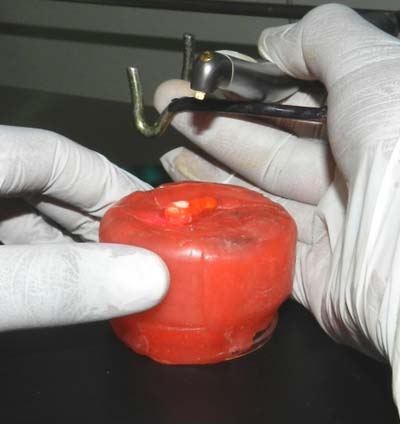
After 4 minutes treatment the cream was wiped off with a tissue paper. The process was repeated four times and then samples were washed using distilled water and then dried. The same procedure was done for Group 5 and Group 6 with CPP-ACP and CPP-ACFP cream. Er:YAG laser (Fotona Fidelis Plus III) using wave length of 2.94μm was used to irradiate the exposed surface of Groups 4,5 and 6. The parameters used were 1.4W power, 7Hz frequency, 200mJ pulse energy (fluence) with a non-contact handpiece, 0% air and 0% water coolant. Lasing was done from a fixed distance of 2.5cm from the tooth surface in a scanning style motion [Table/Fig-3].
Er:YAG laser irradiation.
The laser tip was perpendicularly placed to the enamel surface and subsequently the samples were irradiated by scanning in vertical as well as horizontal direction in order to promote homogenous irradiation and to cover entire area of the sample. To ensure consistent distance from the tooth surface, a custom made standardizing device was used. The Er:YAG irradiation was performed by hand, screening the enamel surface with a uniform motion for 30 seconds. In the combination groups (Group 5 and 6) the tooth surfaces were irradiated after treating with remineralizing agents and then washed and dried. These specimens were individually immersed in 5ml of acetate buffer solution (0.1mol/L, pH 4.5) and incubated at 37°C for 24 hours to imitate oral conditions. After acid treatment, the teeth were removed from the glass containers and the acetate buffer solution from each of the glass containers of test group and control group was collected and analyzed using Inductively Coupled Plasma-Atomic Emission Spectrometer (ICP-AES) to determine the parts per million of calcium ions of each demineralizing medium.
Analysis of variance (ANOVA) model was constructed (p-value of 0.05) to evaluate whether any significant difference exist in the mean value of calcium loss among the groups. A post-hoc Tukey comparison test was done to determine whether any statistically significant difference is noticed between the groups. The data collected from ICP-AES were statistically analyzed using SPSS 14 software.
Results
The mean value of calcium dissolution [Table/Fig-4] was least in the combination group of CPP-ACFP with Er:YAG laser (14.04 ppm) followed by CPP-ACFP (14.93 ppm) followed by combination group of CPP-ACP with Er:YAG laser (16.60 ppm) followed by CPP-ACP (17.25ppm) followed by laser group alone (18.37 ppm) and control group (18.82 ppm). The multiple comparison tests showed that CPP-ACFP, combination of CPP-ACFP with Er:YAG laser and CPP-ACP with Er:YAG laser showed a decrease in mean scores of calcium [Table/Fig-5] compared to control which was statistically significant (p<0.05). When Group 4: Er:YAG laser was used alone for irradiation there was no statistically significant difference in calcium dissolution when compared to the control group (p>0.05). CPP-ACFP with Er:YAG laser showed the lowest mean score of calcium release (highest acid resistance) followed by Group 3: CPP-ACFP but the differences between these groups were statistically not significant (p>0.05) followed by combination group i.e., Group 5: CPP-ACP with Er:YAG laser and Group 2: CPP-ACP group but these groups were statistically not significant (p>0.05). Although the difference between Group 1: control and Group 4: Er:YAG laser was not statistically significant (p>0.05), there was a decrease in calcium solubility after lasing.
ANOVA test for mean values of calcium release of each group in parts per million (ppm).
| Group | Mean | Std. Deviation | F-value | p-value |
|---|
| Group 1-CONTROL | 18.82 | 2.15 | 15.37 | 0.001* |
| Group 2-CPP-ACP | 17.25 | 0.83 |
| Group 3-CPP-ACFP | 14.93 | 1.64 |
| Group 4- LASER | 18.37 | 1.63 |
| Group 5- CPP-ACP+LASER | 16.60 | 1.43 |
| Group 6- CPP-ACFP+LASER | 14.04 | 1.06 |
| Total | 16.67 | 2.26 |
Tukey test for multiple comparison of calcium release among different groups.
| Groups | Comparison of Groups | p-value |
|---|
| Group 1-CONTROLGroup 2- CPP-ACPGroup 3- CPP-ACFPGroup 4- LASERGroup 5-CPP-ACP+LASERGroup 6-CPP-ACFP+LASER | 1 versus 21 versus 31 versus 41 versus 51 versus 6 | 0.200.001*0.980.01 *0.001* |
| 2 versus 32 versus 4 2 versus 5 2 versus 6 | 0.01 *0.570.930.001* |
| 3 versus 43 versus 53 versus 6 | 0.001*0.150.78 |
| 4 versus 54 versus 6 | 0.110.001* |
| 5 versus 6 | 0.001* |
*Significant
Discussion
The application of CPP-ACP or CPP-ACFP paste to sound enamel helped in preventing demineralization and enhancing remineralization and a superior effect was seen for the latter paste [16]. The synergistic effect of CPP-ACP and flouride is due to localization of amorphous calcium fluoride phosphate at the tooth surface by the CPP which would co-localize calcium, phosphate and fluoride. Thus, CPP binds to salivary pellicle, plaque and even to hydroxyapatite component of enamel and acts as an excellent delivery vehicle to co-localize calcium, phosphate and fluoride at the tooth surface in a slow release amorphous, non-crystalline form [5] preventing demineralization and enhancing remineralization. Heating of surface by lasers can cause chemical, physical and crystalline changes on the ablated enamel [17]. The crystalline structure of enamel is improved by formation of more hydroxide and pyrophosphate but removal of bound carbonate and water loss because carbonate forms more acid soluble apatite phase and it does not fit well in the lattice [12]. In organic blocking theory, after laser treatment the decomposed organic matter blocks ion diffusion channels in the enamel during demineralization resulting in reduced enamel surface area and pore volume and thus, prevents mineral loss from enamel [7]. Another explanation is that in lased enamel, during demineralization dissolved calcium, fluoride and phosphorous ions from the tooth are caught in micropores and microfissures formed as a result of loss of water and carbonate content [8]. High power lasers like Erbium lasers (Er:YAG) are effective in preventing caries [18]. Several studies have shown that combined fluoride and laser irradiation enhances fluoride uptake of irradiated enamel and better than laser treatment alone [19–22]. In our study combination of CPP-ACFP cream with Er:YAG laser showed the least calcium dissolution (highest acid resistance). During laser treatment following CPP-ACFP application, there occurs an enhancement of the subsurface concentration of calcium and phosphate content of the enamel resulting in localization of ACFP at the tooth surface. When fluoride ions come in contact with free calcium and phosphate ions flourapatite is formed at the enamel subsurface. The microspaces and microfissures formed between many partially coalescent globular granules as a result of water and carbonate loss and organic substances in the enamel after laser irradiation can trap the calcium, phosphate and fluoride ions released from tooth during demineralization and would also provide a greater surface area for fluoride incorporation reducing demineralization. CPP-ACP can increase mineral content of enamel surface and laser treatment can enhance fluoride uptake [23–25]. Er: YAG laser decreased acid dissolution and increased fluoride uptake into the enamel producing a surface which is less susceptible to caries [10]. The application of CPP-ACFP cream would have facilitated the incorporation of 0.2% or 900ppm level of fluoride into ACP phase by holding the calcium and phosphate ions in an amorphous form, so that fluoride can combine with them to form a stronger protective layer of flourapatite which is less soluble than hydroxyapatite and has a more acidic critical pH of 4.5, ensuring that no demineralization takes place until pH reaches this point. Studies have shown that formation of CPP-ACFP nano-complexes improved the ability of CPP-ACFP to remineralize initial caries lesion [26]. When Group 5: CPP-ACP is combined with Er:YAG laser, surface changes in enamel like crazing, crating and exfoliation of enamel occurs. These micromorphological changes induced by lasing would help in efficient incorporation of enamel cream containing calciumnano-complexes into the tooth surface and thus co-localizing calcium and phosphate without fluoride [14]. Thus, CPP stabilizes ACP and forms a highly soluble calcium phosphate phase at the tooth surface. This localization results in high concentration gradients of calcium and phosphate ions in enamel subsurface thereby promoting remineralization. In this manner CPP-ACP application enhance the action of lasers to inhibit demineralization [14]. When CPP-ACP cream is applied CPP-ACP clusters attaches itself to dental biofilm (plaque) and tooth surfaces and act as calcium and phosphate reservoir, which buffers free calcium and phosphate ions in saliva and plaque pH and help to maintain a state of super saturation with respect to enamel minerals which would counteract a drop in pH preventing demineralization. The calcium and phosphate concentration is kept high within subsurface lesion and enamel remineralized by CPP-ACP is a hydroxyapatite with a higher Ca:P ratio than normal apatite and is more resistant to acid challenges than normal tooth enamel. The additive anti-cariogenic effect of CPP-ACFP than CPP-ACP is due to presence of ACFP phase resulting in fluorapatite formation on enamel surface [5]. Combination group of fluoride and CPP-ACP have more remineralization potential than CPP-ACP alone [27,28]. When using Er:YAG lasers fluencies near ablation threshold, temperature rise upto 300-400°C can only induce morphological changes like water evaporation and carbonate loss [29]. When subablative fluencies were used for irradiation the Er:YAG laser showed calcium solubility similar to an unlased group [30]. Liu JF et al., demonstrated that when Er:YAG laser irradiation of pulse energies of 100 and 200mJ were used, there was significant inhibition of demineralization [8]. The peak rise of temperature in our study was below 100°C and this was not enough to bring any crystallographic changes in enamel [28] since we used subablative fluencies to avoid mechanical damage to enamel. This would have resulted in lower acid resistance for laser similar to the control when compared with other groups. In our study increased acid resistance of enamel was not achieved in-vitro conditions as expected. From our findings use of subablative fluencies of Erbium lasers alone would not be very favourable for caries prevention. Liu JF et al., showed that Er:YAG laser with out water mist was more effective than with water mist to reach sufficient surface temperature to bring crystallographic changes in enamel [8]. So in this study also we used laser with out water coolant. The combined effect of Er:YAG laser irradiation associated with CPP-ACFP application resulted in increased fluoride incorporation and it depends on the fluence applied over the enamel surface. Even though, for many years lasers have proved to be effective in various clinical scenario in dentistry, the exact mechanism of action of lasers on teeth is not fully understood even today. Further studies are needed to investigate the changes occurring on sound enamel after Er:YAG irradiation at sub-ablative fluencies and their effects on demineralization and their remineralization potential when used in combination with various remineralizing agents.
Limitation
This study is limited to using only Er:YAG laser with sub-ablative parameter for caries prevention without comparing other lasers. The sample size could have been increased. The effects of temperature changes on the surrounding tissues and possible pulpal damage due to laser use without water is not assessed since it is an in-vitro study.
Conclusion
Within limitations of the present study we conclude that in the order of CPP-ACFP (tooth mousse plus) with Er:YAG laser, CPP-ACFP, CPP-ACP (tooth mousse) with Er:YAG laser were found to be more resistant to enamel dissolution. The sub-ablative Er:YAG laser alone and CPP-ACP were not very effective in reducing demineralization. When laser alone is used other critical parameters like fluence, wave length, irradiation time, water cooling, irradiation distance and laser power should be considered to achieve the desired effect.
*Significant
[1]. Aoba T, Solubility properties of human tooth mineral and pathogenesis of dental cariesOral Dis 2004 10:249-57. [Google Scholar]
[2]. Longbottom C, Ekstrandb K, Zeroc D, Kambarad M, Novel preventive treatment optionMonogr Oral Sci 2009 21:156-63. [Google Scholar]
[3]. Reynolds EC, The role of phosphopeptides in caries preventionDental Perspectives 1999 3:6-7. [Google Scholar]
[4]. Kariya S, Sato T, Sakaguchi Y, Yoshii E. Flouride effect on acid resistance capacity of CPP-ACP containing material. Abstract 2045; 82nd General Session of the IADR 2004, Honolulu, Hawaii [Google Scholar]
[5]. Reynolds EC, Cain CJ, Webber FI, Anticariogenicity of calcium phosphate complex of tryptic casein phosphopeptides in the ratJ Dent Res 1995 74:1272-79. [Google Scholar]
[6]. Liu Y, Hsu CY, Teo CM, Teoh SH, Potential mechanism for the laser-flouride effect on enamel demineralizationJ Dent Res 2013 92:71-75. [Google Scholar]
[7]. Apel C, Meister J, Schmitt N, Graber HG, Gutknecht N, Calcium solubility of dental enamel following sub-ablative Er:YAG and Er:YSGG laser irradiation in vitroLasers Surg Med 2002 30:337-41. [Google Scholar]
[8]. Liu J F, Liu Y, Stephen HC, Optimal Er:YAG laser energy for preventing enamel demineralizationJ Dent 2006 34:62-66. [Google Scholar]
[9]. Stern R H, Sognnaes RF, Goodman F, Laser effect on in vitro permeability and solubilityJ Am Dent Assoc 1966 73:838-43. [Google Scholar]
[10]. Kim JH, Kwon OW, Kim HI, Kwon Y H, Acid resistance of Erbium-doped: Yttrium aluminium garnet laser treated and phosphoric acid etched enamelAngle Orthod 2006 76:1052-56. [Google Scholar]
[11]. Hossian M, Nakamura Y, Kimuara Y, Yamada Y, Matsumoto K, Caries preventive effect of Er:YAG laser irradiation with or without water mistJ Clin Laser Med Surg 2000 18:61-65. [Google Scholar]
[12]. Liu Y, Hsu CY, Laser induced compositional changes on enamel. A F T-Raman studyJ Dent 2007 35:226-30. [Google Scholar]
[13]. Esteves Oliveira M, Apel C, Gutknecht N, Cotrim M E, Lowfluence CO2 laser irradiation decreases enamel solubilityLaser Phys 2008 18:478-85. [Google Scholar]
[14]. Niazy A M, Ehab A H, Synergistic caries inhibitory effect of a remineralizing agent and CO2 laser on human enamel and root dentineCairo Dent J 2009 25:415-24. [Google Scholar]
[15]. Banda NR, Vanaja Reddy G, Shashikiran ND, Evaluation of primary tooth enamel surface morphology and microhardness after Nd:YAG laser irradiation and APF gel treatment. An in vitro studyJ Clin Pediatr Dent 2011 35:377-82. [Google Scholar]
[16]. Hamba H, Nikaido T, Inoue G, Sadre A, Tagami J, Effect of CPP-ACP with sodium fluoride on inhibition of bovine enamel demineralization. A quantitative assessment using microcomputed tomographyJ Dent 2011 39:405-13. [Google Scholar]
[17]. Zezell DM, Ana PA, Albero FG, Cury JA, Effect of infra red lasers on chemical and crystalline properties of enamelCaries Res 2009 b 43:192 [Google Scholar]
[18]. Geraldo-Martins VR, Lepri CP, Palma-Dibb RG, Influence of Er, Cr:YSGG laser irradiation on enamel caries preventionLasers Med Sci 2013 28:33-39. [Google Scholar]
[19]. Rodrigues LK, Nobre Dos Santos M, Feather Stone JD, In situ mineral loss inhibition by CO2 laser and fluorideJ Dent Res 2006 85:617-21. [Google Scholar]
[20]. Santaella MR, Braun A, Matson E, Effect of diode laser and fluoride varnish on initial surface demineralization of primary dentition enamel. An in vitro studyInt J Pediatr Dent 2004 14:199-203. [Google Scholar]
[21]. Karandish M, The efficiency of laser application on the enamel surface: A systematic reviewJ Lasers Med Sci 2014 5(3):108-14. [Google Scholar]
[22]. Malik A, Parmar G, Bansal P, Joshi N, Effect of laser and fluoride application for prevention of dental caries. A polarized microscope analysisJournal of Dental Lasers 2015 9(1):11-15. [Google Scholar]
[23]. Tagomori S, Morioka T, Combined effects of laser and fluoride on acid resistance of human dental enamelCaries Res 1989 23:225-31. [Google Scholar]
[24]. Yassaei S, Shahraki N, Aghili H, Combined effect of Er:YAG laser and casein phosphopeptide-amorphous calcium phosphate on inhibition of enamel demineralization. An invitro studyDent Res J 2014 11(2):193-98. [Google Scholar]
[25]. Heravi F, Ahrari F, Mahdavi M, Comparative evaluation of the effect of Er:YAG laser and low level laser irradiation combined with CPP-ACPF cream on treatment of enamel cariesJ Clin Exp Dent 2014 6(2):121-26. [Google Scholar]
[26]. Hegde MN, Shetty S, Pardal D, Demineralization of enamel subsurface lesion using CPP-ACP. A quantitative energy dispersive X-ray analysisJ Conserv Dent 2007 10:19-25. [Google Scholar]
[27]. Reynolds EC, Black CL, Cai F, Cross KJ, Huq NL, Advances in enamel remineralization. Casein phosphopeptide amorphous calcium phosphateJ Clin Dent 1999 10:86-88. [Google Scholar]
[28]. Christos Vougioklakisg, Effect of a commercial paste based on CPP-ACP complex on the demineralization of sound human dentine and on remineralization potential of artificial caries like lesionCaries Res 2007 35:695-98. [Google Scholar]
[29]. Fried D, Featherstone JD, Visuri SR, Sekaw The caries inhibition potential of Er:YAG and Er:YSGG laser irradiationProc SPIE 1996 2672:73-78. [Google Scholar]
[30]. Kwon YH, Lee JS, Choi YH, Lee JM, Song KB, Change of enamel after Er:YAG and CO2 laser irradiation and fluoride treatmentPhotomed Laser Surg 2005 23:389-94. [Google Scholar]