Cancer of the esophagus is ranked first in males and ranked third after breast and cervical malignancy in females [1-3] in Punjab, India. In spite of recent advances in diagnosis and therapy of carcinoma of the esophagus, the patients usually come at advanced stage of disease [4]. When patients come at this stage, they receive chemotherapy and radiotherapy to control the disease burden and improve their symptoms. But chemo-radiotherapy has, apart from lengthy treatment, its own complications and limitations [5]. There are also other techniques, which relieve the dysphagia like intra-tumoural alcohol injection, LASER ablation, Photodynamic therapy and Argon beam therapy [6-8]. But they have their own side effects and limitations like repeated sessions to achieve the relief of dysphagia, high cost in case of LASER ablation and Argon beam therapy, less availability as in LASER and photodynamic therapy. Short term relief of dysphagia and intra procedural complications like esophageal perforation or mediastinitis can also occur with the above procedures. These therapies are also not widely available [7,8]. Esophageal stenting, being readily available, showing immediate results and relatively few major complications is now the procedure of choice for relieving dysphagia in these terminally ill patients [4,7,9,10]. The aim of this study was to evaluate the long term outcome of esophageal stenting in patients of carcinoma esophagus who were unfit for surgical treatment. This is the first of its kind study from this region.
Materials and Methods
We conducted a retrospective study from January 2012 to January 2015 with endoscopic biopsy proven carcinoma of the esophagus patients, in whom esophageal stenting was done. The minimum sample size for the study was calculated to be 68 with 94% success rate (of dysphagia), with confidence interval (99.64%-88.36%) at 5% level of significance with the formula n= Zα/22PQ/E2 where Z is the standard normal variate, P is the success rate, Q=1-P and E is the allowable error (length of confidence interval). Therefore, we decided to include 100 cases (i.e., all the cases who reported within the study time period). Stenting was performed by Gastro Surgery division of Department of Surgery of the Guru Gobind Singh Medical College, Faridkot, Punjab, India by a single person. To conduct this study ethical clearance was taken. In-patient admission record was procured and it was analysed to acquire data on patient demographic profile, tumour characteristics, stenting procedure, early and late complications, re-interventions. Success rate was determined by dysphagia score before and after the stenting. Patients were followed till July 2015.
Procedure Protocol
All the patients were initially assessed for definitive surgery, but those who were not fit for surgery or not willing for surgery were given options of endoscopic stenting. Initially dilation of malignant stricture was done on first visit to relieve the symptoms followed by chemoradiotherapy, but when repeated and early dilations were required and or stricture was very tight or fibrotic, stenting was advised. Proper patient consent was taken before doing the procedure after explaining each aspect of procedure and its post procedural complications.
Before stenting, all patients were evaluated by routine biochemistry, coagulation profile, chest X-ray, viral markers, ECG and examined by physician, where required. Self expandable, metallic and covered stent was used and stenting was done under sedation. After the procedure, all patients were assessed by chest X-ray in lateral position to know the position of stent. If there was displacement of stent, it was repositioned using endoscopy and fluoroscopy. Patients were allowed orally on day 2, so that stent could expand fully and make its place in the esophagus. Discharge was done on day 3 after oral acceptance of food. Patients, who had persistent, severe pain, were kept in the hospital till pain significantly decreased. All patients were followed after two weeks for review and later on as required. They all were sent for chemoradiotherapy.
Stent Characteristic and Design
Self expanding, metallic covered stent was used in all cases. The silicone covering was on inner side in the body and dumbbells or flanges on either side were uncovered. The fully expanded diameter of stent was 18mm for the body and 24mm for both flanges. There were three standard lengths: 110mm, 140mm and 170mm. Stent was deployed after passing a guide wire across the stricture with the help of endoscope and guiding the stent on the guide wire. With the help of endoscopy or fluoroscopy proper positioning of stent was done and it was deployed there.
Statistical Analysis
As for as data analysis was concerned, non-numeric data was expressed in terms of frequencies and numeric data in mean±SD. In order to compare the dysphagia grade before and after stenting, a non-parametric Wilcoxen Signed Rank Test has been carried out. The frequencies have been compared by conducting parametric Chi-square test for goodness of fit test. Statistical analysis was done on SPSS – 20.0.
Results
The demographic profile of patients is listed in [Table/Fig-1]. Mean age at presentation was 65.9 years with 63% male and 37% female patients. Patients presented with dysphagia (84%) and vomiting (16%). Tumour was mainly in middle esophagus (58%) with average stricture length of 3.76 cm [Table/Fig-2]. Dysphagia was graded by MP score [Table/Fig-3]. In 87% of cases, 110mm stent was used and in 12%, 140mm stent was used whereas only in 1%, 170mm stent was used. Stent was placed endoscopically in 71 patients and for rest of the patients need for fluoroscopy guidance was required. This was especially required for patients having long stricture and patients having tracheo/bronchial-esophageal fistulas. There was highly statistically significant relief of dysphagia after the stenting from median dysphagia score 3 to 1. Out of 100 patients, 52 patients had some sort of complications and total of 69 complications occurred after the procedure. Early complications [Table/Fig-4] included pain (38%), stent migration (6%) and hematemesis (2%). Late complication was stent obstruction (23%) which occurred with mean duration of 4.7 months after the procedure and most common cause (82%) as growth of tumour in the stent. All the complications were treated conservatively without any effect on post-operative stay and efficacy of stenting. However, six patients needed re-stenting due to stent obstruction and after that they did well. One patient had major procedure related complication in the form of bleeding (after 16 days of procedure) which resulted in death of that patient. Out of 100 patients 53 patients died with average time of death being 5.7 months after the stenting. A total of 36 patients were alive till the end of study. We could not follow 11 patients till the end of study period. We could follow them from 38 days to 87 days as per record. As all these 11 patients had some sort of complications, so we included them in the study also.
| Gender | n |
|---|
| Males | 63 |
| Females | 37 |
| Age (mean±S.D, in years) | 65.9±9.39 (37-91) |
| Presenting symptoms |
| Dysphagia | 84 |
| Cough and dyspnoea (associated with dysphagia) | 07(8.3) |
| Pain (associated dysphagia) | 11(13.2) |
| Vomiting | 16 |
| Duration of symptoms (mean±S.D, in months) | 5.83±2.86 |
| Site of tumour |
| Upper esophagus (20-25cm) | 5 |
| Middle esophagus (26-32cm) | 58 |
| Lower esophagus (33-40cm) | 37 |
| Tumour Histology | |
| Squamous cell carcinoma | 94 |
| Adenocarcinoma | 06 |
| Tracheo/Broncho-esophageal fistula | 04 |
| Cause of non operability |
| Metastatic | 35 |
| Locally advanced | 52 |
| Cardiac problem | 05 |
| COPD/Cardiac problem | 02 |
| CVA | 01 |
| Not willing | 05 |
Stricture characteristics.
| Stricture length | No. of patients | Chi-Square value (p-value) |
|---|
| 2-3 cm | 48 | 23.18 (<0.001)HS |
| 4-5 cm | 41 |
| 6-8 cm | 11 |
| Stricture length (mean±S.D, in cm) | 3.76±1.40 |
| Dysphagia grade | Pre Stenting(n) | Post Stenting(n) | |
| 0 | 0 | 30 | (<0.001)HS |
| 1 | 07 | 63 |
| 2 | 18 | 06 |
| 3 | 58 | 00 |
| 4 | 17 | 01 |
| Median dysphagia gradeWilcoxon Signed Rank Test (Asymptotic p-value) | 3 | 1 |
| Stent length |
| 110mm | 87 | 128.24(<0.001)HS |
| 140mm | 12 |
| 170mm | 01 |
| Use of Endoscopy/Fluoroscopy |
| Endoscopy | 71 | 18.68 (<0.001)HS |
| Fluoroscopy | 29 |
| Post procedure stay(mean±S.D, in days) | 3.61±1.0 | |
Mellow-pinkas score of dysphagia.
| Grade 0 | Normal diet |
| Grade 1 | Able to swallow some solid foods |
| Grade 2 | Able to swallow only semi-solid foods |
| Grade 3 | Able to swallow liquids only. |
| Grade 4 | Total dysphagia |
| Complication | n |
|---|
| Pain | 38 |
| Severe Pain (stay > 7 days) | 03 |
| Stent Migration | 06 |
| Hematemesis | 02 (1 mild+1 severe; death) |
| Post stenting obstruction | 23 |
| Proximal growth | 15 |
| Distal growth | 04 |
| Sheath disruption | 01 |
| Food bolus impaction | 03 |
| Time of re growth (mean±SD, in months) | 4.74±2.03 |
| Restenting | 06 |
| Dilatation | 14 |
| Removal of foreign body | 03 |
| Dead | 53 |
| Alive | 36 |
| Lost to follow up | 11 |
| Time of death (mean±S.D, in months) | 5.70±3.10 |
Discussion
Cancer of the esophagus is the leading cause of cancer in Malwa region of state of Punjab, India, where its incidence is highest in males (11.7%) and third highest in females (5.9%) following breast and cervix cancer among all cancer patients. It is the leading cause of cancer mortality in males especially and even in females also [1-3].
Squamous cell carcinoma is the dominant pathological type of esophageal cancer followed by adenocarcinoma. Worldwide, although incidence of adenocarcinoma is increasing, but in India squamous cell carcinoma is still far more common than adenocarcinoma [4,11]. Cancer of middle esophagus followed by distal esophagus is much more common than proximal esophagus [12]. In our study also squamous cell carcinoma was the predominant type (93%) and most common site was middle esophagus.
In esophageal luminal obstruction due to malignancy, dysphagia is the predominant cause for impaired quality of life, so palliation of dysphagia is the ultimate goal of unresectable esophageal cancer [4]. There are many ways of doing this, including chemoradiotherapy [5], alcohol injection at the site of tumour [6], brachytherapy [7], laser ablation, photodynamic therapy [7,8] and stenting [4,7,9,10]. All the above therapies are associated with significant complications and limitations. Thermal ablative procedure like LASER therapy and argon beam therapy can be associated with under-treatment leading to persistent dysphagia and some time overtreatment leading to perforation. Photodynamic therapy is associated with high cost of photodynamic drugs, cutaneous sensitivity with these drugs and chances of post-procedure stricture etc. There may be need for repeated sessions to maintain the relief of dysphagia. The above therapies are also not widely available. Radiotherapy and chemotherapy apart from lengthy treatment are not reliable for relief of dysphagia. Surgical bypass has high morbidity and mortality [4,7,10]. Stenting the esophageal lumen with self expanding metallic stent is simple, relative free of serious complications and widely available technique to palliate dysphagia. Relief of dysphagia is seen in more than 80% of cases in most of the series and in some series it has reached up to 100% of cases especially with covered stents, thus improving the quality of life [9,10,13,14]. In our series almost 100% patients were relieved of dysphagia significantly immediately after the stenting.
Chest pain after stenting is an early and most common complication. In different studies, it was present in even 100% of patients. Pain usually is mild and short lived. Usually, it subsides within 24 to 48 hours and responds well to Non Steroidal Anti- Inflammatory drugs (NSAIDS). But at times it can persist longer, requiring another NSAID or narcotics also [13-17]. Pain in stenting may be because of prior degree of stricture [13], expansible force of stent [13,14] and radiotherapy [15]. In our study, pain occurred in 38 patients. In all except three, pain responded well to NSAIDs and it did not prolong the stay. The three patients, in whom pain was severe, they had to stay for 7-10 days after that it subsided.
Stent migration to stomach is also an early complication with many proposed causes. These include covered stents as compared to uncovered stents [13,15], ingestion of ice cold liquids after stent insertion [15] and inability of stent to fully expand [13,15]. Once stent is fixed at place, usually it does not migrate. In our study also there was no late migration. All the migrations were seen within first 24 hours. Early migrations are easy to reposition. If it is late or there is proximal growth of tumour, then it is very difficult or impossible to reposition the stent. Sometimes, re-postioning can lead to esophageal perforation. In some cases malpositioning can be treated by placing a second stent either in the previous stent or at site of stricture when previous stent has migrated itself [13]. In our study six stents migrated, All except one were re-positioned successfully by endoscopy.
Another early complication which is usually procedure related is, perforation of the esophagus. It may be due to over dilating an irregular or tight stricture or pre procedure radio and chemotherapy treatment. Its incidence in various series is less than 5% with Self Expanding Metallic Stents (SEMS) [13,18]. The decreasing diameter of delivery system makes perforation a rare occurrence and also covered stent (SEMS) covers the minor perforations during the procedure [13,16,18]. In our series, no patient had esophageal perforation. This could be because of the fact that we did not over-dilate the stricture, rather, dilated to the extent that delivery system of the stent could negotiate across it.
Stent obstruction is relatively late and common complication. It can be due to many causes. Tumour ingrowths and overgrowth is a significant problem with uncovered SEMS, occurring in 10–35% of patients [13,14,18]. But with covered stent, this problem is very less. However, growth of the tumour can occur above or below the stent or through uncovered flanges. This can be managed by restenting or simple dilatation. In our study 15% patients had tumour growth/ingrowth proximal to stent and 4% had distal growth. All the patients were managed by dilatation except in six patients in which re-stenting was done over previous stent [Table/Fig-5].
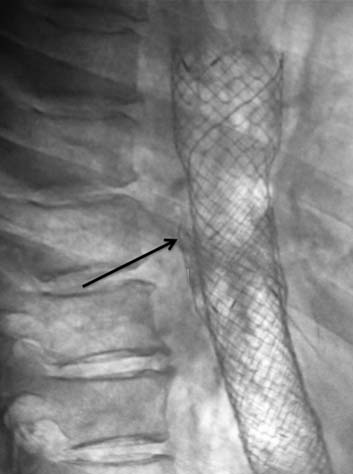
Stent in stent. Arrow is pointing towards proximal end of first stent.
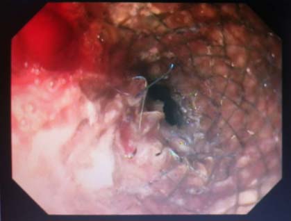
Food bolus impaction is also one of the main complications after stenting, but in various studies, it seems to be related not due to stent failure but lack of patient education [13,14]. In our study three patients had food bolus impaction which was managed by removing the bolus with the help of endoscopy. In one of our patient, stent wire was reason for food impaction. One of stent wire was hanging in the lumen, which was the cause of food impaction and it was corrected with biopsy forceps and balloon dilatation [Table/Fig-6]. This patient also underwent dilatation of obstruction due to proximal growth with SG dilator in the past and this may be the reason for stent wire to detach from stent. Dietary instructions should be given to patient and the relatives. Proper chewing of food, avoiding fibrous foods and increasing fluid intake and carbonated beverages and some time dilute solution of hydrogen peroxide helps to enhance the passage of solid food through stent lumen [13,14,18].
Stent wire hanging in the lumen, which caused food bolus impaction.
Inner sheath of stent can also detach from stent body causing luminal obstruction. This problem of membrane separation from the stent was taken care by laminating the membrane between two layers of wire mesh in a coaxial arrangement, which also increases the radial force exerted by the stent [13,18]. In our series only one patient had sheath detachment which caused stent obstruction. This patient surprisingly presented only 2 months after the index stenting and was managed by inserting another stent in the first stent.
Despite advancement in medical sciences, there is still lack of awareness among population regarding this disease. Patients usually come late at tertiary centre and at the time of presentation, they already have advanced disease. In our experience, patients came after 1-14 months of the symptoms with mean duration of 4.7 months after initiation of the symptoms. But now with the initiative of Government of Punjab, a mass public awareness door to door campaign was carried out regarding different alarming symptoms of various types of cancers and a separate Government fund has also been established for any person who is suffering from cancer [19-21]. Majority of the patients (more than 95%) in our series were stented free of cost with the help of this fund.
As patients came late, they have already reached advanced stage of disease, so mortality rate is also high. Out of 100 patients, 53 patients died after stenting and 36 were still alive (on radio and chemotherapy) with average time of death is 5.6 months after the stenting. Eleven patients were lost to follow-up.
Limitation
As this study was done by taking the retrospective data of only one institution and only a single surgeon’s experience, more centers and more doctors could be involved in future study to present representative data of the population. Also, well designed prospective trials can be done to ascertain in which group of patients of carcinoma of the esophagus, the success rate is best.
Conclusion
We conclude that, esophageal stenting is a relatively safe and easy procedure and it quickly relieves the dysphagia in a very sick patient and thus improves the patients’ symptoms very reliably. But a patient should be counseled properly regarding benefits and complications before doing the procedure. And also, the food habits and other precautions should be told before the discharge for successful outcome. As major and long term complications are very less, it is the best way of palliating the terminally ill cancer esophagus patient.