Morphological Variations of the Thyroid Gland among the People of Upper Assam Region of Northeast India: A Cadaveric Study
Anjan Jyoti Rajkonwar1, Giriraj Kusre2
1 Demonstrator, Department of Anatomy, Assam Medical College and Hospital, Dibrugarh, Assam, India.
2 Associate Professor, Department of Anatomy, Assam Medical College and Hospital, Dibrugarh, Assam, India.
NAME, ADDRESS, E-MAIL ID OF THE CORRESPONDING AUTHOR: Dr. Anjan Jyoti Rajkonwar, Department of Anatomy, Ground Floor, Basic Science Building, Assam Medical College and Hospital, Dibrugarh-786002, Assam, India.
E-mail: rajkonwaranjan@yahoo.com
Introduction
The morphological variations of the thyroid gland have been reported from different parts of the world. The variations are due to remnant or non-specific development of the parts of the thyroid gland. Surgical operation of the thyroid gland has been the treatment of choice in various thyroid pathologies. Prior knowledge of the morphological variation is important to ensure better results from these surgical operations.
Aim
To study the prevalence of morphological variations seen in the thyroid glands in the upper Assam region of Northeast India.
Materials and Methods
This was a hospital based cadaveric study. Total number of Thyroid glands dissected were 80. The thyroid gland was examined properly for the presence of pyramidal lobe, levator glandulae thyroideae and complete absence of isthmus. Statistical analysis was done by SPSS 21.0.
Results
It was found that 17 (21.25%) cadavers did not show an isthmus. The pyramidal lobe was present in 31(38.75%) cases and frequently arising from the left side (74.2%) of the isthmus. Levator glandulae thyroideae was found in 15 (18.75%) of the thyroid specimens. In all cases, it was extended from the apex of the pyramidal lobe to the hyoid bone.
Conclusion
Morphological variation of the thyroid gland is very common hence requires detection prior to any surgery on the thyroid gland.
Isthmus, Levator glandulae thyroideae, Pyramidal lobe
Introduction
The thyroid gland is a highly vascular gland placed anteriorly in the lower part of the neck consisting of two symmetrical lateral lobes connected by a midline isthmus [1]. The size of the thyroid gland varies considerably with age, sex, physiological state, race and geographical location and it is observed that the size is larger in female than in male [2]. The right lobe is slightly larger than the left one [3]. The thyroid gland appears as an epithelial proliferation in the floor of the pharynx between the tubercular impar and the copula in the form of bi-lobed diverticulum, called the thyroglossal duct. The lower part of the duct develops in to median isthmus and two lateral lobes. The upper connection of the duct with the floor of pharynx later disappears [4]. Agenesis of the thyroid isthmus is defined as the complete and congenital absence of the isthmus [5]. A high division of the thyroglossal duct generate two independent thyroid lobes with the absence of isthmus [6]. The embryological remnant of the caudal end of the thyroglossal duct commonly known as the pyramidal lobe often ascends from the isthmus or adjacent part of either lobe (more often from the left side) towards the hyoid bone [7]. The upper end of the pyramidal lobe continues as a fibro-muscular strand, called the Levator Glandulae Thyroideae (LGT), attached to the hyoid bone. When the pyramidal lobe is absent, LGT may attach to the upper part of the isthmus [8]. Thyroid diseases are among the most common endocrine disorders in India [9]. Most of the diseases, affecting the thyroid gland require medical and surgical intervention. The surgeons planning a total thyroidectomy operation must be aware about the morphological variations of the gland. Therefore, it is important to know the details regarding surgical anatomy of the thyroid gland. The North-eastern region of India is famous for its diverse ecology and distinct culture and lifestyle specific to different ethnic groups. Different ethnic groups existing in Assam are Ahoms, Bodos, Koch, Matak, Chutiya and different branches of Kacharis such as Dimasa, Sonowal and Thengal.
The prevalence of goitre remained high (42.2%) in Dibrugarh district [10]. Results of the morphological study of the thyroid gland in the people of this region are most likely to differ from those with adequate iodine intake, as iodine is very essential for the biological activity of the thyroid gland. The present study is an approach to popularise the information pool on the morphology of the thyroid gland and help the clinicians in their practice. Moreover, limited information is available regarding the morphological variations of the thyroid gland from the Upper Assam region of Northeast India. The present study is an approach to popularise the information pool on the morphology of the thyroid gland and help the clinicians in their practice.
Materials and Methods
A cross-sectional descriptive study was done in the Department of Anatomy, Assam Medical College and hospital, Dibrugarh for a period of one year from August 2014 to July 2015. A total of 80 thyroid glands (30 adult and 50 perinatal cadavers) were dissected and out of which 49 were male and 31 were female. Adult specimens (30) were collected from the cadavers provided for the dissection and perinatal specimens (50) were obtained from Department of Obstetrics & Gynaecology, Assam Medical College & Hospital.
After taking the approval from the institutional ethical committee, the cadavers were received in the Department of Anatomy. The particulars of the specimens were collected from the cadaver register. Cases with known thyroid diseases or crushing injury to the neck and those foetuses with gross congenital malformation were excluded from the study.
A vertical incision was given on the skin from the chin to the sternum in the mid line. The subcutaneous fat and deep fascia was exposed; the infrahyoid muscles were identified and reflected laterally. The pretracheal fascia was removed and the right and the left lobe of the gland were identified. The thyroid gland was examined carefully for the presence of isthmus, pyramidal lobe and LGT. When the pyramidal lobe was present, its position was observed. LGT if present, its extension and its relation with the pyramidal lobe were also observed.
Statistical Analysis
Statistical analysis was done using chi-square test by SPSS 21.0.
Results
A total 80 number of thyroid glands were collected from 49 male and 31 female cadavers. Presence of pyramidal lobe, levator glandulae thyroideae and absence of isthmus were the only morphological variations observed in the gland. Forty eight out of the total glands dissected had morphological variations the others had normal anatomy [Table/Fig-1]. Morphological variations were more common in males but the difference was not statistically significant (chi-square value=0.43, df=1; p-value = 0.67). All the major morphological variations observed in the study are summarized in [Table/Fig-2].
Morphological variations in either sex.
| Sex | Morphological variations | Normal anatomy |
|---|
| Male | 28 | 21 |
| Female | 20 | 11 |
| Total | 48 | 32 |
Different types of morphological variations and sex distribution of the cadavers dissected.
| Morphological variations | Male | Female | Total | Percentage |
|---|
| Pyramidal lobe (PL) | 18 | 13 | 31 | 38.75% |
| Absence of isthmus (ISTH) | 11 | 6 | 17 | 21.25% |
| Presence of Levator Glandulae Thyroideae (LGT) | 9 | 6 | 15 | 18.75% |
Note: More than one variations were seen in few specimens
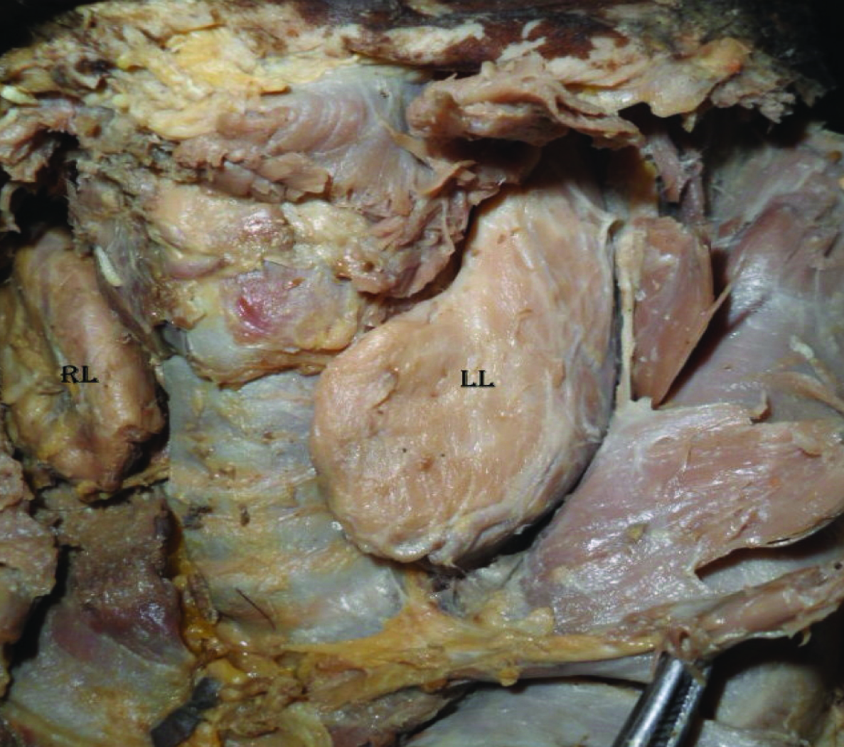
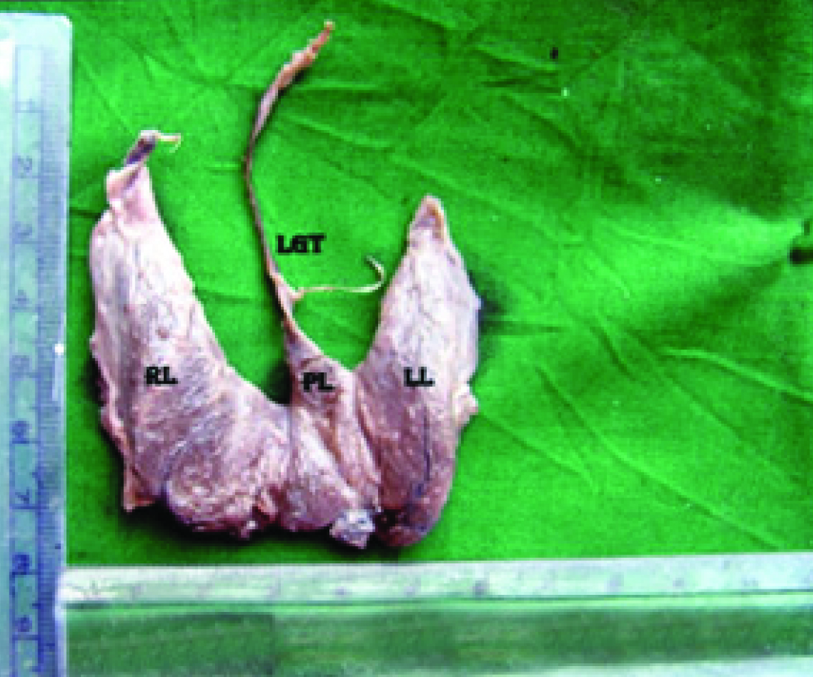
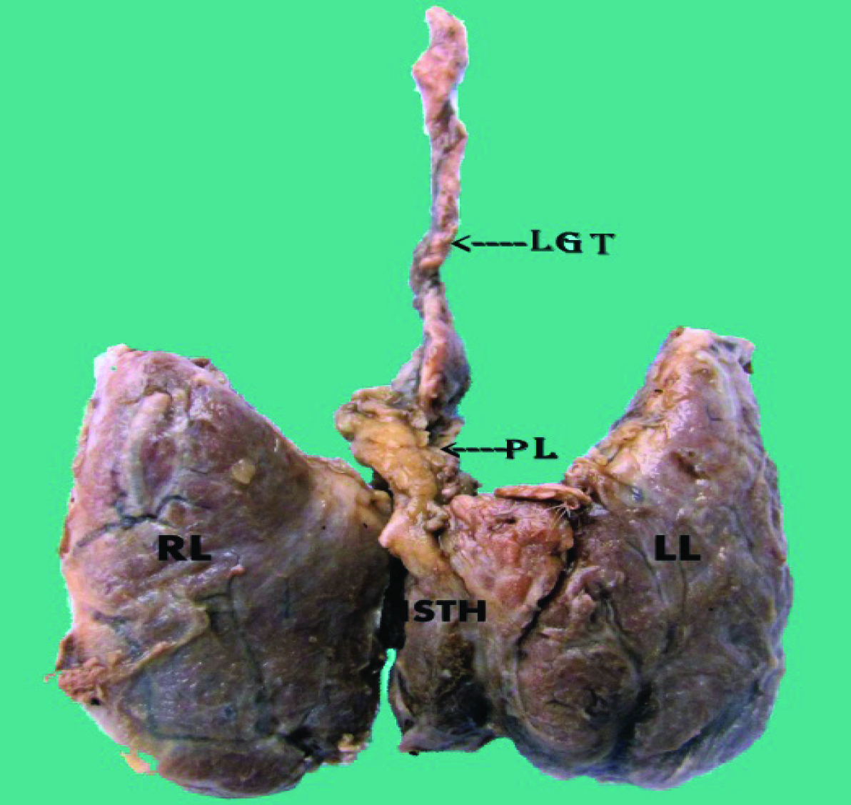
The isthmus was absent in 17 (21.25%) cases; of which 11 were male cadavers [Table/Fig-3]. The pyramidal lobe was observed in 31(38.75%) cases. Among them, pyramidal lobe was found more frequently in males (18). In 74.2% cadavers, the pyramidal lobe was attached to the left side of the isthmus [Table/Fig-4] and to the middle in 25.8% [Table/Fig-5]. According to the origin and location of pyramidal lobe, Milojevic B et al., classified five types of pyramidal lobe [11]. On the basis of that classification, we have found two types-Type III (74.2%) and Type I (25.8%) with the left sided being predominant [Table/Fig-6]. The LGT was present in 15(18.75%) cadavers [Table/Fig-5] and in all specimens LGT was extending from the apex of the pyramidal lobe to the hyoid bone.
Thyroid gland with absence of isthmus in an adult cadaver.
Note: LL - Left lobe; RL - Right lobe

Pyramidal lobe with levator glandulae thyroideae arising from the left half of isthmus.
Note: LL - Left lobe; RL - Right lobe; LGT - Levator Glandulae Thyroideae; PL - Pyramidal lobe

Pyramidal lobe with levator glandulae thyroideae arising from the middle of isthmus.
Note: LL - Left lobe; RL - Right lobe; LGT - Levator Glandulae Thyroideae; PL - Pyramidal lobe; ISTH - Isthmus

Classification of the pyramidal lobe on the basis of its attachment to the thyroid gland.
| Sex | Centralpart of theisthmus(Type I) | Junctionof the rightlobe withthe isthmus(Type II) | Junctionof theleft lobe withthe isthmus(Type III) | Left lobe(Type IV) | Right Lobe(Type V) | Total |
|---|
| Male | 5(16.1%) | 0 | 13 (41.9%) | 0 | 0 | 18 |
| Female | 3 (9.6%) | 0 | 10 (32.25%) | 0 | 0 | 13 |
| Total | 8 (25.8%) | 0 | 23 (74.2%) | 0 | 0 | 31 |
Discussion
The morphological variations of the thyroid gland found in present study were compared with other studies [Table/Fig-7] [12-18].
Morphological variations of the thyroid gland compared with other studies [12-18].
| Author | Sample Size | Year | Absent Isthmus | PyramidalLobe | LevatorGlandulaethyroideae |
|---|
| Ranade AV et al., [12] | 105 | 2008 | 33% | 58% | 49.5% |
| Nurunnabi ASM et al., [13] | 60 | 2008 | ----- | 41% | 20% |
| Begum M et al., [14] | 60 | 2009 | ------ | 26.7% | 15% |
| Joshi S D et al., [15] | 90 | 2010 | 16.7% | 37.8% | 30% |
| Kulkarni V et al., [16] | 20 | 2012 | 10% | ------ | 30% |
| Veerahanumaiah S et al., [17] | 89 | 2014 | 9% | 46% | 41% |
| Rathod S Mansingh et al., [18] | 76 | 2015 | 13% | 58% | ----- |
| Present Study | 80 | 2015 | 21.25% | 38.75% | 18.75% |
Incidence of absence of isthmus varies from 5% to 10% [5]. The absence of isthmus in the present study closely matched with the study by Joshi et al. (15 of 90) [15]. The differences between various studies were probably due to difference in sample selection or due to differences in the population studied.
The pyramidal lobe observed in the present study was comparable with that of (41%) [13] and (37.8%) [15]. In the present study, the pyramidal lobe was situated more on the left side of the gland which was also observed in various studies [11,14,15]. In studies based on the surgically removed thyroid gland after total thyroidectomy operation in Turkey and Serbia, 65.7% [19] and 61% [20] of thyroid glands respectively had pyramidal lobes. The conclusion of the above discussion is that presence of pyramidal lobe is quite common in all over the world.
The levator glandulae thyroidae observed in the present study was similar to observations of 20% [13] and 15% [14].
The morphological variation in the thyroid gland does not affect the function of the gland but presence of these variations may simulate some thyroid pathologies. Agenesis of isthmus is a differential pathological diagnosis for autonomous thyroid nodule, thyroiditis, primary carcinoma, neoplastic metastases and infiltrative diseases such as amyloidosis [21]. Agenesis of isthmus can be diagnosed via scintigraphy, ultrasonography, computed tomography and magnetic resonance imaging. Total or near total thyroidectomy is recommended for patients with ongoing thyroid cancer, those who refuse radio-ablation as a therapeutic procedure, or have a life threatening reaction to antithyroid drugs such as vasculitis, agranulocytosis and liver failure and is the operation of choice for patients undergoing surgical treatment for Grave’s disease [22]. Incomplete excision of thyroid gland in patients with auto-immune disorders may cause recurrence of the diseases. Anatomical variations of thyroid gland may cause many difficulties during complete removal of the gland. Removal of the pyramidal lobe and other accessory thyroid tissue has considerable beneficial effect on the total thyroidectomy operation [19].
In the present study, more than two-third of the thyroid glands dissected had one or more morphological variations hence presence of pyramidal lobe or other morphological variations should be looked for during total thyroidectomy operation to ensure complete removal of the gland tissue.
Limitation
This type of study can be done in all age groups; but present study focused only on adult and perinatal cadavers. We have only used dissection as a tool for the study but radiological techniques like MRI, CT scan and Ultrasonography can also be applied.
Conclusion
Precise and accurate knowledge of variations associated with the thyroid gland would be helpful for surgeons in performing total, subtotal & partial thyroidectomy and in the evaluation of Scintigraphic findings.
Note: More than one variations were seen in few specimens
[1]. Stranding S, Gray’s anatomy 2008 40th EdnChurchill LivingstoneElsevier:462 [Google Scholar]
[2]. Wood Jones, Buchanan’s Manual of Anatomy 1946 7th EdBailliore Tindall & Cox:1125 [Google Scholar]
[3]. Montgomery & Welbourn. Clinical Endocrinology for Surgeons 1963 1st EdEdward Arnold Pub:218 [Google Scholar]
[4]. Saddler TW, Langman’s Medical Embryology, Thyroid gland 2010 11th EdLippincott Williams and Wilkins:278 [Google Scholar]
[5]. Pastor VJF, Gil VJA, De Paz Fernández FJ, Cachorro MB, Agenesis of the thyroid isthmusEur J Anat 2006 10:83-84. [Google Scholar]
[6]. Dixit D, Shilpa MB, Harsh MP, Ravishankar MV, Agenesis of isthmus of thyroid gland in adult human cadavers-A case seriesCases Journal 2009 2:6640 [Google Scholar]
[7]. Sinnatamby CS, Last’s anatomy 2006 11th EdChurchill livingstoneElsevier:351-352. [Google Scholar]
[8]. Hamilton WJ, Textbook of Human Anatomy 1976 2nd EdThe McMillan Press Ltd:488 [Google Scholar]
[9]. Kochupillai N, Clinical endocrinology in IndiaCurrent Science 2000 79(8):106 [Google Scholar]
[10]. Patowary AC, Kumar S, Patowary S, Dhar P, Iodine deficiency disorders (IDD) and iodised salt in Assam: A few observationsIndian J Public Health 1995 39(4):135-40. [Google Scholar]
[11]. Milojevic B, Tosevski J, Milisavljevic M, Babic D, Malikovic A, Pyramidal lobe of the human thyroid gland- An anatomical study with clinical implicationsRom J Morphol Embryol 2013 54(2):285-89. [Google Scholar]
[12]. Ranade AV, Rai R, Pai M, Nayak SR, Prakash Krisnamurthy A, Anatomical variations of the thyroid gland-possible surgical implicationsSingapore Med J 2008 49(10):831-34. [Google Scholar]
[13]. Nurunnabi ASM, Alim A, Mahbub S, Segupta K, Begum M, Khatun M, Morphological and histological study of the pyramidal lobe of the thyroid gland in bangladeshi people-a postmortem studyBangladesh Journal of Anatomy 2009 7(2):94-100. [Google Scholar]
[14]. Begum M, Khatun M, Kishwara S, Ahmed R, Naushaba HA, Postmortem study of the pyramidal lobe of the thyroid gland in bangladeshi peopleJ Dhaka Med Coll 2009 18(2):120-23. [Google Scholar]
[15]. Joshi SD, Joshi SS, Daimi SR, Athavale SA, The thyroid gland and its variations-A cadaveric studyFolia Morphol 2010 69(1):47-50. [Google Scholar]
[16]. Kulkarni V, Sripadma S, Deshpande SK, Morphological variation of thyroid GlandMedica Innovatica 2012 1(2):36-38. [Google Scholar]
[17]. Veerahanumaiah S, Dakshayani KR, Menasinkai SB, Morphological variations of the thyroid glandInt J Res Med Sci 2015 3(1):53-57. [Google Scholar]
[18]. Mansingh RS, Mahesh ST, Santosh D, Morphological variations of thyroid glandInternational J. of Healthcare and Biomedical Research 2015 03(02):175-77. [Google Scholar]
[19]. Gurleyik E, Gurleyik G, Dogan S, Cobek U, Pyramidal lobe of the thyroid gland-surgical anatomy in patients undergoing total thyroidectomyAnatomy Research International 2015 2015:384148:55 [Google Scholar]
[20]. Zivic R, Radovanovic D, Vekic B, Surgical anatomy of the pyramidal lobe and its significance in thyroid surgerySAJS 2011 49(3):110-16. [Google Scholar]
[21]. Devi SK, Bhanu S, Susan P, Gajendra KPJ, Agenesis of isthmus of thyroid gland with bilateral levator glandulae thyroideaeInternational Journal of Anatomical Variations 2009 2:29-30. [Google Scholar]
[22]. Bojic T, Paunovic I, Diklic A, Zivaljevic V, Zoric G, Kalezic N, Total thyroidectomy as a method of choice in the treatment of Graves’disease-Analysis of 1432 patientsBMC Surg 2015 15:39 [Google Scholar]