Nasal Conidiobolomycosis- The Unknown Threat
Nikhil Arora1, Eishaan K. Bhargava2, Varun Rai2
1 Senior Resident, Department of Ear, Nose and Throat, Maulanaazad Medical College, New Delhi, India.
2 Senior Resident, Department of Ear, Nose and Throat, Maulanaazad Medical College, New Delhi, India.
3 Senior Resident, Department of Ear, Nose and Throat, Maulana Azad Medical College, New Delhi, India.
NAME, ADDRESS, E-MAIL ID OF THE CORRESPONDING AUTHOR: Dr. Nikhil Arora, D-55 Arya Nagar Apartments, I.P. Extension, Patparganj, New Delhi, India.
E-mail: for_nikhilarora@yahoo.com
Conidiobolus coronatus, Entompthoromycosis, Nasal obstruction
Nasal conidibolomycosis also called as entompthoromycosis is a rare fungal infection caused by Conidibolus coronatus. The fungus is present in soil and decaying vegetations [1]. It generally affects the upper respiratory tract, but rarely can affect the nose and subcutaneous tissue of face. Here, we report a case of farmer working in Orissa, who presented with the swelling of nose and after much dilemma was found to be suffering by Conidibolus infection.
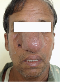
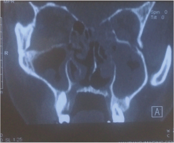
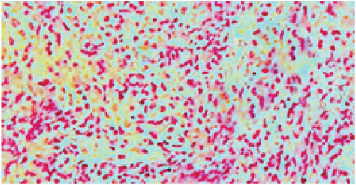
A 45-year-old male, farmer in Orissa presented to OPD with a progressive painless swelling over nose for duration of six months [Table/Fig-1]. Apart from right side nasal obstruction, there was no other specific complaint. Contrast Enhanced Computed Tomographic (CECT) scan of nose and paranasal sinuses revealed bilateral maxillary sinusitis [Table/Fig-2] and nasal endoscopic examination was found to be normal. Decision to take a skin biopsy was taken which showed superficial layer of keratinizing stratified squamous epithelium and dense neutrophilic infiltrate in dermis with thin wall fungal elements amidst granulomas surrounded by Splendore-Hoeppli phenomenon [Table/Fig-3] and after 72 hours growth on Sabouraud Dextrose Agar (SDA), the isolate of Conidiobolus coronatus was obtained. The diagnosis was further confirmed by light cycler PCR assay which accomplishes identification by PCR amplification and sequencing of DNA. Patient was started on itraconazole 200mg\day and potassium iodide 8 drops three times a day for a month and kept on regular follow-up with improvement seen as a reduction in size of the primary lesion.
Showing the clinical image of the patient.
Showing the CECT scan of nose and paranasal sinuses of patient having bilateral maxillary sinusitis.
Histopathologic image of the patient showing dense. neutrophilic infiltrate in dermis with fungal elements surrounded by splendore-hoeppli phenomenon.
Discussion
Conidiobolomycosis occurs mainly in tropical regions of Africa, South and Central America and South East Asia including India. It is generally seen in farmers and other outdoor occupations. The 3 species of the order Entomophthorales that cause conidiobolomycosis in humans and animals are C.coronatus, C. incongruus and C.lamprauges. Disease mainly affects the upper respiratory tract leading to a granulomatous reaction ultimately leading to formation of hard nodules over skin surface. The treatment of the disease is not well defined at present. A definitive diagnosis of rhinofacial entomophthoromycosis requires histopathologic demonstration of the etiologic agent and its isolation in culture [2,3]. The disease remains to be under reported due to the lack of clinical suspicion and lack of culture facilities. Our patient had responded well and is under regular follow-up with no systemic side effects of the treatment so far.
[1]. Pfaller MA, Diekema DJ, Unusual fungal and pseudofungal infections of humansJ Clin Microbiol 2005 43:1495-504. [Google Scholar]
[2]. Das P, Vijay MK, Joshi P, Yadav R, Singh G, Histological identification of Entomophthoromycosis in biopsy samples is requiredIndian J Pathol Microbiol 2014 57(3):514-16. [Google Scholar]
[3]. Chowdhary A, Randhawa HS, Khan ZU, Ahmad S, Khanna G, Gupta R, Rhinoentomophthoromycosis due to Conidiobolus coronatus. A case report and an overview of the disease in IndiaMedical Mycology 2010 48:870-79. [Google Scholar]