Forearm fractures are very common in children accounting for 45% of all fractures in childhood [1,2]. The incidence of fractures shaft forearm bones is more common in 6-16-year-old children, with higher incidence in children between 12-16 years of age [3]. As the fractures tend to occur in older children, their management becomes all the more difficult because of more proximal location of the fracture and hence more chances of re-displacement even after a successful closed reduction [4].
So, it becomes essential to manage these, with some kind of internal fixation so as to maintain a good fracture reduction and hence achieve satisfactory functional outcome. There are various methods of management of fractures in both bones of forearm in children. Historically, closed reduction and plaster cast application has been the gold standard in management of these fractures, however there are increased chances of re-displacement, particularly in older children [5]. As a result, there is a rising trend to fix most of these fractures. Fracture fixation may be done by extra-medullary devices like plates which have various disadvantages such as large incisions, more soft tissue dissection, more chances of infection and a re-surgery of almost similar magnitude for removal of implant. As suggested by Shoemaker et al., the ideal fixation mode should maintain alignment, be minimally invasive and should have least complications. This has led us to the use of intramedullary fixation devices. TENS (Titanium Elastic Nailing System) is a minimally invasive procedure that spares physis, provides 3 point fixation and hence mostly does not requires Plaster of Paris (POP) splint/cast, thereby allowing early mobilization to achieve excellent functional outcomes. Other devices for intramedullary fixation such as Kirschner wires/ pins/ nails lack these advantages and hence are inferior to TENS [6–10].
The aim of present study was to assess the clinical outcome of managing paediatric forearm fractures using TENS.
Materials and Methods
Among 549 patients who attended our tertiary care centre in Amritsar, for forearm diaphyseal fractures between February 2012 to January 2015, 87 patients with both bone fractures were treated with TENS. The study was approved by the ethical committee of the institute. The procedure was not being followed as a norm in all. Resources did not allow us to undertake comparative study.
Proper pre-operative anaesthetic checkup and investigations were conducted in all patients. Pre and post-operative cephalosporin antibiotics were administered for 3 days with dosage as per weight of patient.
Fifty patients were included in the present study with inclusion criteria of age 6–14 years, displaced fracture or grossly rotated fractures, failed closed manipulation and patients with minimum follow-up of six months. Patients with isolated forearm bone fracture, compound fractures or fracture with neurovascular injury were excluded.
Operative Technique- Under general anaesthesia, closed reduction was done under image intensifier. After achieving satisfactory reduction, ulna was fixed by antegrade nailing through the lateral surface of olecranon about 1.5 to 2 cm distal to physis. Radius was fixed by retrograde nailing through dorsal aspect of distal radius proximal to radial physis and just medial to lister’s tubercle. Special care was taken not to injure extensor tendons and superficial radial cutaneous nerve. The nail was prebent 30 degrees at the tip with additional gentle bend given so as the apex of bend overlaps with fracture site. The length and diameter of nails were varied as observed under image intensifier control. Wherever required, limited open reduction was carried out to achieve accurate reduction. The ends were bent and cut flush to the bone leaving enough length for subsequent removal and buried under the skin in all cases. During this period of study, we had same team of operating surgeons following the same technique as well as post-operative protocol. Post-operatively majority of patients required no external immobilization. However, depending on fracture stability, in some patients POP splint was given maximum up to 3 weeks in more comminuted fractures.
Early range of motion exercises were started and results were evaluated at 2, 4, 8, 12 and 24 weeks. Clinical results were evaluated as per scale developed by Price CT et al., for pain and range of motion of supination and pronation [Table/Fig-1] [11].
Price CT et al., criteria for evaluation of result [11].
| Outcomes | Symptoms | Loss of Forearm Rotation |
|---|
| Excellent | No complaints with strenuous activity | <15° |
| Good | Mild complaints with strenuous activity | 15°–30° |
| Fair | Mild complaints with daily activities | 31°–90° |
| Poor | All other results | >90° |
Results
On final evaluation at six months, there was no pain in all the patients. However some loss of forearm supination and pronation movements (between 15-30 degrees loss of motion) was seen in 4(8%) patients. Mean time for radiological bony union was 9.2 weeks (Range=6–13 weeks) [Table/Fig-2,3,4,5,6,7,8 and 9].
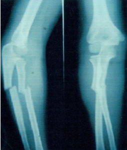
Pre-operative radiograph Antero-Posterior and lateral view of fractures in both bones of forearm.
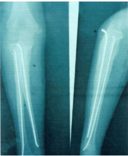
Post-operative radiograph Antero-posterior and lateral at 6 months showing bony union.
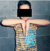
Clinical photograph showing full flexion at elbow.
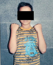
Clinical photograph showing full extension at elbow.
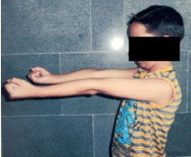
Clinical photograph showing full supination.
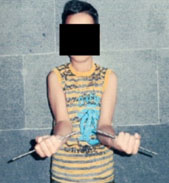
Clinical photograph showing full pronation.
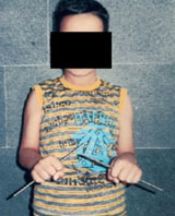
Clinical photograph showing full palmar flexion at wrist.
Clinical photograph showing full dorsiflexion at wrist.

According to Price CT et al., criteria, 46 patients (92%) had excellent results and 4 patients (8%) had good results. None of the patients had fair or poor results. [Table/Fig-10] depicts the excellent results obtained in majority of patients by means of TENS.
| S. No. | Criteria | Observations | Percentage |
|---|
| 1. | Age | | Mean age = 11.2 years |
| • 6-10 years | 22 | 44 |
| • 11-14 years | 28 | 56 |
| 2. | Sex |
| • Male | 43 | 86 |
| • Female | 7 | 14 |
| 3. | Type of injury |
| • Simple/ Closed | 46 | 92 |
| • Compound/ Open | 4 | 8 |
| 4. | Level of fracture in shaft of both bones |
| • Proximal 1/3rd | 20 | 40 |
| • Middle 1/3rd | 23 | 46 |
| • Distal 1/3rd | 7 | 14 |
| 5. | Type of Surgical Procedure |
| • CRIF | 47 | 94 |
| • ORIF | 3 | 6 |
| 6. | Results |
| • Excellent | 46 | 92 |
| • Good | 4 | 8 |
| • Fair | - | - |
| • Poor | - | - |
No significant complications were observed except for superficial pin tract infections at site of entry of nail in 3 (6%) patients. However, no deep infection, malunion, non-union, nerve palsy, refracture and nail migration were observed.
Discussion
Historically closed reduction and POP cast immobilisation has been the mainstay of treatment for fractures in both bones of forearm in children. However, fractures tend to redisplace especially in older children and when at more proximal location. How much malreduction is acceptable has always been a matter of great debate. As mentioned in literature, angular deformity >10° and complete displacement account for unacceptable reduction [6,12]. Also, younger children tend to tolerate greater deformity much better than older ones due to better remodelling potential [1,13–15].
In present study, majority of children were in age group of 11-14 years with mean age of 11.2 years. Similar observations were also made by Qidwai SA (11 years) and Garg NK et al., (11.8 years) [16,17]. So, mean age of incidence can inferred to be 11 years.
In present study, there were 46 patients with simple (closed) fractures constituting 92% of total patients and 4 patients with compound (open) fractures (Gustilo and Anderson grade I) constituting 8% of total patients. Kang SN et al., also mentioned in their study that 9% of their patients had open fracture and remaining (91%) were closed. This can be due to the fact that the injuries in children are low energy injuries [18].
In present study, there was fracture of shaft of forearm bones at proximal 1/3rd in 20 patients (40%) out of which 16 patients were among age group of 11-14 years. There was fracture forearm at middle 1/3rd in 23 (46%) and 15 among these were of age group of 11-14 years. Fracture forearm at distal 1/3rd in 7 (14%) who were in age group of 6-10 years. The incidence of proximal third fractures was similar in study conducted by Celebi L et al., in which mean age of patient was 10.6 which is similar to mean age group of our study (11.2 years). These findings are indicative of the fact that proximal fractures are more likely to occur in older children (>10 years) and distal fractures are more common in younger children (<10 years) [19].
We achieved closed reduction and intramedullary fixation in 47 patients (94%) under image intensifier guidance. However 3 patients required open reduction and intramedullary fixation (6%). This is in accordance to study conducted by Mohammed H et al., on 21 children with forearm fractures in which 4 patients (19%) had required open reduction and internal fixation with Elastic Stable Intramedullary Nailing (ESIN) and 19 were managed with Closed Reduction Internal Fixation (CRIF) [20].
On final follow-up at 24 weeks, 46 (92%) patients had loss of movement at forearm by less than 15 degree, 4 (8%) patients had loss of movement at forearm by 15-30 degrees and no patient had loss of movement at forearm more than 30 degrees. Similar results have been reported in literature in study by Kapoor V et al., in which 16% of patients had some loss of motion at forearm over a 24 weeks follow-up period [21].
Mean time for radiological bony union was 9.2 weeks (Range= 6–13 weeks) which is comparable to study done by Ali AM in which mean time for union was 10 weeks [22].
All 50 patients had excellent results in terms of fracture union. We had 46 patients (92%) with excellent results and 4 patients (8%) with good results according to Price criteria. The final result is in accordance with study conducted by Parajuli NP et al., in which 94 % patients had excellent results and 6% had good results [2].
Conclusion
Paediatric forearm fractures are quite common. However management tends to become difficult in more proximal fractures in older children (11-14 years) due to high incidence of redisplacement. Herein lies the importance of internal fixation. TENS is a modality which aids in the maintenance of radial bow and interosseous space between forearm bones while sparing the physis, thus achieving good functional results in terms of forearm movements. From the present study, we conclude that TENS is an effective and minimally invasive method of fixation of forearm fractures with excellent results in terms of bony union and functional outcomes with minimal complications. Therefore we strongly recommend its use in management of paediatric forearm fractures.